. 2011 Nov;35(11):1700-5.
doi: 10.1097/PAS.0b013e31822911e6.

Sarcina organisms in the gastrointestinal tract: a clinicopathologic and molecular study

Affiliations

Sarcina organisms in the gastrointestinal tract: a clinicopathologic and molecular study

Dora Lam-Himlin et al. Am J Surg Pathol. 2011 Nov.

Abstract

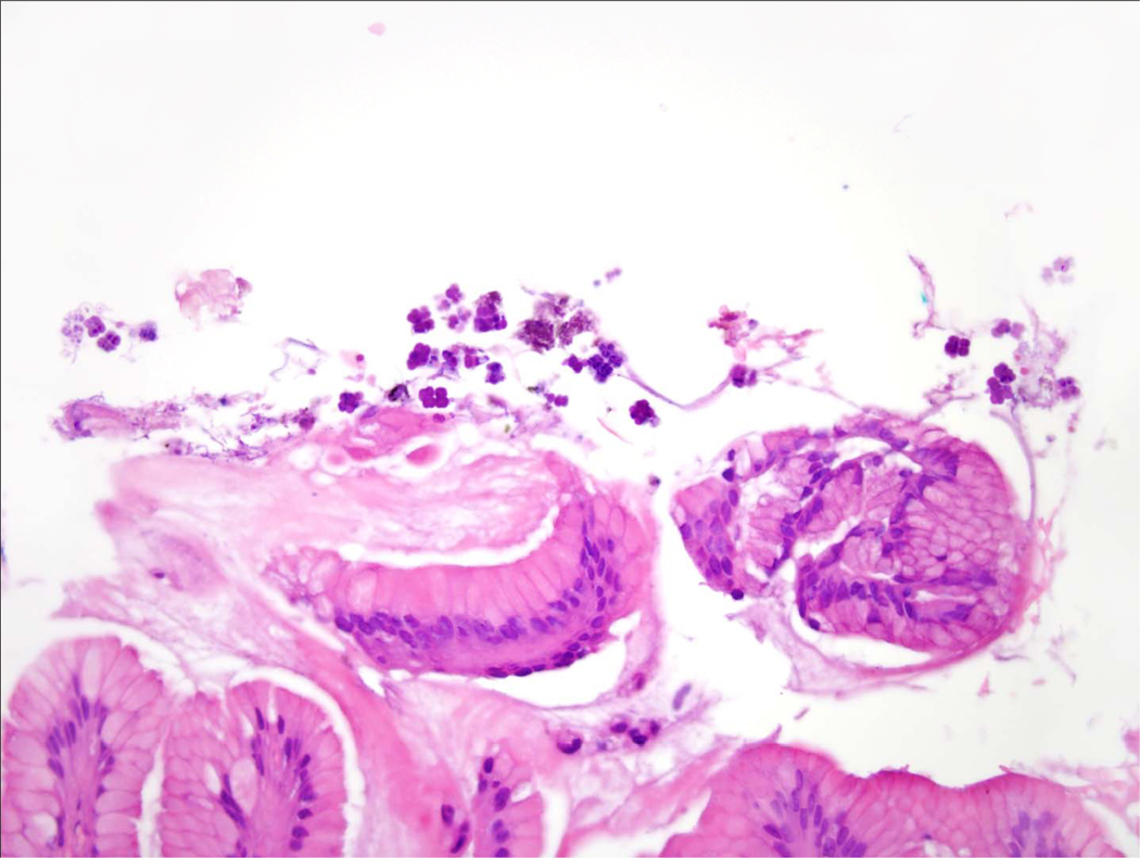
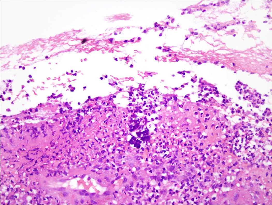
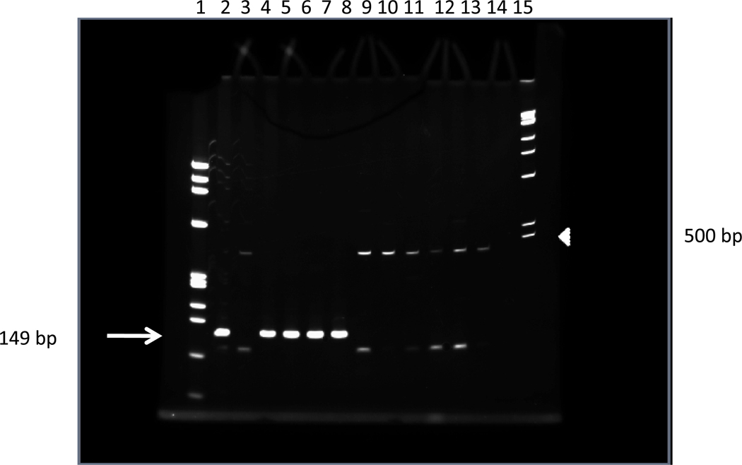
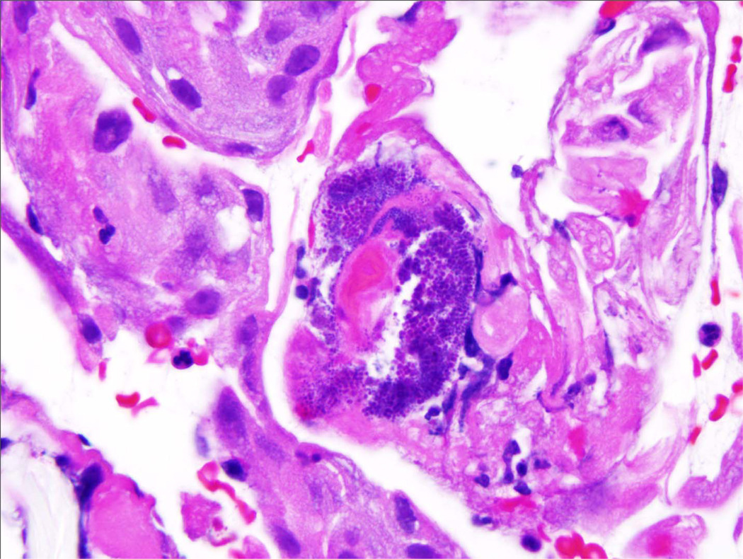
Sarcina organisms were first observed in and recorded from the stomach contents of a patient suffering from vomiting by John Goodsir in 1842. Since that time, their fine structure, phylogenetic classification, and biochemical characteristics have been described. Although numerous cases of fatal disease have been attributed to this organism in the veterinary literature, only a few human cases have been documented. As a result, whether this organism causes disease in humans has not been definitively established. We report the clinicopathologic findings in a series of 5 patients with Sarcina-like organisms identified in upper gastrointestinal endoscopic biopsies with molecular confirmation. Our findings have shown that the organism is most commonly found in patients with a history of gastric outlet obstruction or delayed gastric emptying. Although many of the patients do not demonstrate direct mucosal injury from the organism, the presence of a concurrent gastric ulcer puts the patient at increased risk for complications such as emphysematous gastritis or perforation. The finding of Sarcina organisms should prompt further investigation for functional causes of gastric outlet obstruction and delayed gastric emptying, such as occult malignancy.


Figures

Figure 1
Figure 1. Sarcina organisms in a biopsy of the gastric antrum
Note the characteristic packeting of tetrads or octets with individual bacterial cells measuring up to 3 microns; the resulting tetrads are comparable to the size of a lymphocyte nucleus. (Brown and Hopps, orginial magnification 100×, oil objective.)
Figure 2
Figure 2. Sarcina organisms in the gastric lumen
The distinctive octets are present in the superficial gastric mucin without apparent mucosal reaction or invasion. This was seen in 5 of the 6 biopsies, suggesting that Sarcina may be present in many cases without causing mucosal injury. (Hematoxylin and eosin, original magnification 100×, oil objective.)
Figure 3
Figure 3. Sarcina organisms embedded in a gastric ulcer
(Hematoxylin and eosin, original magnification 100×, oil objective.)
Figure 4
Figure 4. Summary of gel electrophoresis of PCR products targeting Sarcina PDC
(Lane 1) The φX174 DNA-HaeIII DNA ladder highlights DNA fragments between 72 and 1,353 base pairs. (Lane 15) The 1kb DNA ladder highlights DNA fragments between 500 (white arrowhead) and 10,000 base pairs. (Lanes 2–7) PCR amplification reactions from patient samples from histologically suspected cases of Sarcina. Lanes 2 and 4–7 show the 149 base pair PDC amplicon (white arrow). Patient 2 (Lane 3) does not show amplification of the PDC target. (Lanes 8–13) PCR amplification reactions from histologically unremarkable gastric biopsy sample serving as negative controls. None of the negative control reactions show amplification of the PDC target. In the absence of PDC amplification a non-specific amplicon of ~120 bp is seen. (Lane 14) The water template negative PCR control shows no amplification of PCR products.
Figure 5
Figure 5. Micrococcus spp
These organisms were identified in a gastric biopsy. While they have similar packet formation as Sarcina, the packets are more tightly clustered. In addition, the individual bacterial cells are much smaller, measuring about 0.5 microns. (Hematoxylin and eosin, original magnification 1000×, oil objective.)

Similar articles

Cited by

References

    1. Darwin Correspondence Project Database. Letter no. 4272. [Accessed 5 April 2011]; Available at: http://www.darwinproject.ac.uk/entry-4272.
    1. Beijerinck MW. An experiment with Sarcina ventriculi. Proceedings, Section of Sciences, Koninklijke Nederlandse Akademie van Wetenschappen. 1911;13:1234–1240.
    1. Burget GE. Note on the Flora of the Stomach. J Bacteriol. 1920;5:299–303. - PMC - PubMed
    1. Canale-Parola E, Wolfe RS. Studies on Sarcina ventriculi. I. Stock culture method. J Bacteriol. 1960;79:857–859. - PMC - PubMed
    1. Canale-Parola E, Wolfe RS. Studies on Sarcina ventriculi. II. Nutrition. J Bacteriol. 1960;79:860–862. - PMC - PubMed

Publication types

MeSH terms