. 2012 Jan;158(Pt 1):240-258.
doi: 10.1099/mic.0.051524-0. Epub 2011 Oct 13.

Blood-brain barrier invasion by Cryptococcus neoformans is enhanced by functional interactions with plasmin

Affiliations

Blood-brain barrier invasion by Cryptococcus neoformans is enhanced by functional interactions with plasmin

Jamal Stie et al. Microbiology (Reading). 2012 Jan.

Abstract

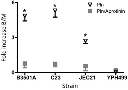
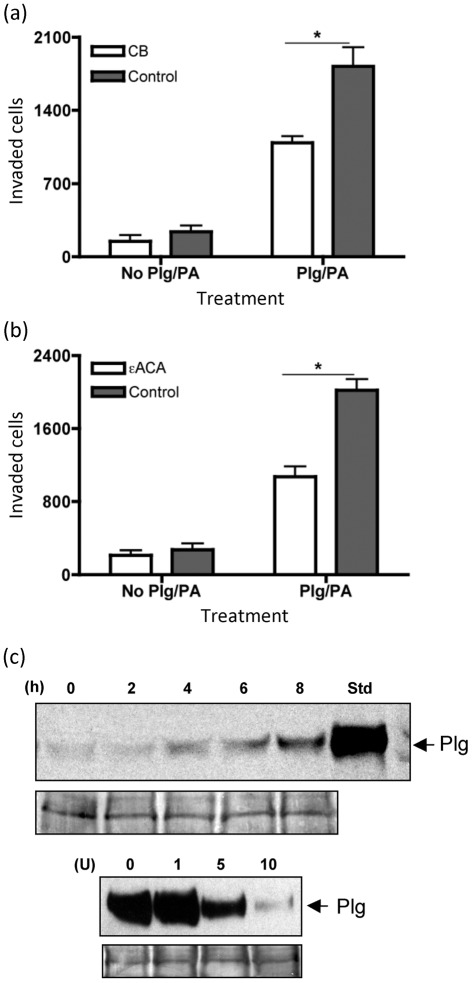
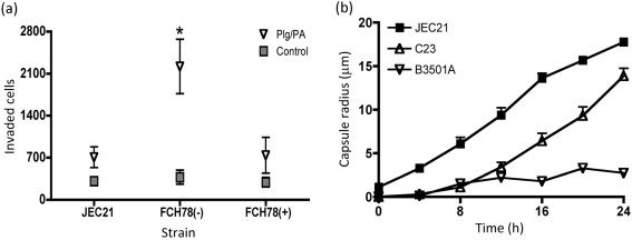
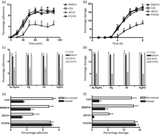
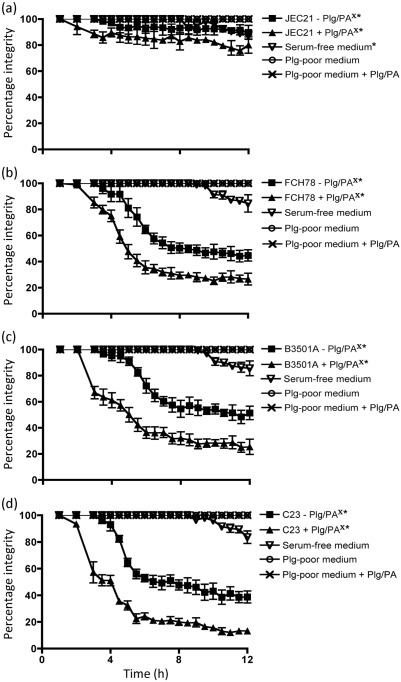
Cryptococcus neoformans can invade the central nervous system through diverse mechanisms. We examined a possible role for host plasma proteases in the neurotropic behaviour of this blood-borne fungal pathogen. Plasminogen is a plasma-enriched zymogen that can passively coat the surface of blood-borne pathogens and, upon conversion to the serine protease plasmin, facilitate pathogen dissemination by degrading vascular barriers. In this study, plasminogen-to-plasmin conversion on killed and viable hypoencapsulated strains of C. neoformans required the addition of plasminogen activator (PA), but this conversion occurred in the absence of supplemented PA when viable strains were cultured with brain microvascular endothelial cells (BMEC). Plasmin-coated C. neoformans showed an enhanced invasive ability in Matrigel invasion assays that was significantly augmented in the presence of BMEC. The invasive effect of plasmin required viable pathogen and correlated with rapid declines in BMEC barrier function. Plasmin-enhanced invasion was inhibited by aprotinin, carboxypeptidase B, the lysine analogue epsilon-aminocaproic acid, and by capsule development. C. neoformans caused plasminogen-independent declines in BMEC barrier function that were associated with pathogen-induced host damage; however, such declines were significantly delayed and less extensive than those observed with plasmin-coated pathogen. BMEC adhesion and damage by hypoencapsulated C. neoformans were diminished by capsule induction but unaltered by plasminogen and/or PA. We conclude that hypoencapsulated C. neoformans can invade BMEC by a plasmin-dependent mechanism, in vitro, and that small, or minimal, surface capsule expression during the blood-borne phase of cryptococcosis may promote virulence by means of plasmin(ogen) acquisition.


Figures

Fig. 1.
Fig. 1.
Fungal viability influences surface plasminogen activation in the presence of BMEC. Killed or viable C. neoformans strains B3501A, C23 and JEC21, and S. cerevisiae strain YPH499, were coated with plasminogen and incubated for 4 h with (+) or without (−) soluble PA or BMEC, as indicated. (a) The activation of surface-bound plasminogen to plasmin was demonstrated by the conversion of plasminogen (Plg) to plasmin heavy chain (PlaH). Twenty micrograms were loaded per lane, with protein loading controls located below each blot. Representative results from three separate experiments are shown. (b) Surface plasmin activity, with the plasmin substrate Chromogenix, on plasminogen-coated viable (left) and nonviable (right) strains after 4 h exposure to PA in the presence (+) or absence (−) of BMEC, as indicated. Background activity from fungal–BMEC control co-cultures without plasminogen and PA was subtracted from the results shown. Bars represent mean and sem from four separate experiments. *P<0.05 by t test for same-strain comparisons under the indicated incubation conditions. (c) Upper two panels: cleavage of the alpha (α), beta (β) and gamma (λ) bands of fibrinogen by plasminogen-coated viable and nonviable fungal strains after incubation with (+) or without (−) PA or BMEC, as indicated. The proteolysis of fibrinogen resulted in the concomitant appearance of a 40 kDa degradation product (*). Control lanes (−) include fibrinogen standard incubated in assay buffer with or without BMEC, as indicated. Lower panel: the fibrinogen cleavage activity of plasminogen-coated viable strains is shown after their co-incubation with BMEC, in the presence (+) or absence (−) of PA supplementation, as indicated. After incubation with BMEC and/or PA, fungal cells were suspended in a fibrinogen solution that was subsequently evaluated for fibrinogen proteolysis from 20 µg of total protein loaded per lane. The results shown are representative of four separate experiments.
Fig. 2.
Fig. 2.
The plasmin-enhanced invasion ability of C. neoformans strains is potentiated in the presence of BMEC. The effect of BMEC on the plasmin-enhanced invasion ability of C. neoformans was determined by comparing the invasion of plasmin-coated and non-coated C. neoformans strains B3501A, C23 and JEC21, and S. cerevisiae strain YPH499, in inserts layered with either BMEC–Matrigel or Matrigel alone after a 12 h incubation period. Invasion assays using plasmin-coated strains were conducted in the absence (Pln) or presence (Pln/Aprotinin) of the plasmin protease inhibitor aprotinin, as indicated, with fungal invasion quantified by YPD counts of fungal cells isolated from the lower chamber of transwell plates. The results were calculated using the formula presented in Methods and show the relative mean fold increases in plasmin-dependent invasion and sem, with the greater effect of plasmin on fungal invasion of BMEC–Matrigel (B) versus Matrigel alone (M) shown as the fold increase B/M (y axis). *P<0.05 by t test for plasmin pre-coated C. neoformans assayed in the presence or absence of aprotinin.
Fig. 3.
Fig. 3.
Inhibition of cryptococcal–plasminogen interactions reduces BMEC–Matrigel invasion. (a, b) BMEC–Matrigel invasion by strain B3501A was examined in the presence or absence of plasminogen and PA (Plg/PA) after: (a) removal of plasminogen-binding sites by pre-treatment with 10 U CB, or (b) the addition of a competitive inhibitor of plasminogen, the lysine analogue ϵACA, to the assay medium. The results shown for ϵACA treatment represent the mean inhibition with applied concentrations of 0.7–1.6 mM ϵACA. Bars represent the mean and sem from four separate experiments. *P<0.05 by t test for the datasets included under each horizontal line. (c) Upper panel: Western blot showing the re-expression of plasminogen-binding sites on strain B3501A under the assay conditions, after pretreatment with 10 U CB. Numbers represent the time (h) after the introduction of CB-pretreated fungi into the assay medium (0 h). Purified plasminogen is shown in the right-hand lane (Std). Lower panel: Western blot showing the concentration-dependent effect of CB on the plasminogen-binding activity of strain B3501A, with the number above each lane indicating the activity units (U) of CB applied. Twenty micrograms were loaded per lane, with protein loading controls located below each blot. The results shown are representative of three separate experiments.
Fig. 4.
Fig. 4.
Cryptococcal capsule induction during invasion assays and the effects of capsule development on plasmin-enhanced cryptococcal invasion of BMEC–Matrigel. (a) Invasive ability of the cap59 mutant FCH78, relative to the parental strain JEC21, before (−) or after (+) capsule transfer to FCH78. Plasminogen (Plg) and PA were either included (Plg/PA) or omitted (Control), as indicated. Values shown are mean±sem from five separate experiments. *P<0.05 by t test for comparisons between Plg/PA-treated strains. (b) Capsule growth on strains with a small, or minimal, initial capsule (0 h) was measured at various times over 24 h incubation in invasion assay medium at 37 °C with 5 % CO2. The radial thickness of capsules is shown in µm. Values shown are averaged values from two separate experiments.
Fig. 5.
Fig. 5.
Cryptococcal–BMEC adhesion and cytotoxicity. (a) Comparative adherence of C. neoformans strains B3501A, C23, JEC21 and FCH78 (cap59) to BMEC over time, measured as the percentage of adherent cells in relation to the total added fungi percentage adherence. (b) BMEC damage over time during exposure to C. neoformans, with percentage damage representing the amount of LDH activity detected in assay supernatants relative to the total BMEC LDH activity per well. (c, d) Fungal–BMEC adherence (c) and cytotoxicity (d) assays performed with plasminogen (Plg), plasminogen activator (PA), or both Plg and PA (Plg/PA) relative to Plg- and PA-negative controls (No Plg/PA) after 45 min (c) or 6 h (d) incubation. (e, f) Relative BMEC adherence (e) and damage (f) by fungal strains with a small capsule (Non-induced) or large capsule (Induced) after incubation times of 45 min (e) and 6 h (f). Bars represent mean and sem from a minimum of three separate experiments. *P<0.05 by t test for comparisons between strains in (a, b) or for same-strain comparisons in (e, f) under induced and non-induced conditions.
Fig. 6.
Fig. 6.
Effects of plasmin and cryptococcal–host damage on BMEC barrier resistance. BMEC barrier resistance was measured hourly by TEER during 12 h invasion assays. Strains JEC21, FCH78, B3501A and C23 were incubated with BMEC (a–d) with (+) or without (−) plasminogen (Plg) and PA. Controls showing the effect of plasminogen and PA on BMEC resistance in the absence of C. neoformans are indicated (Plg-poor/deficient medium+Plg/PA). BMEC were additionally examined in assay medium lacking both C. neoformans and plasminogen products (Plg-poor medium) or in medium lacking FCS (Serum-free medium). The 100 % TEER values measured in these experiments were 246.3±35.5 Ω cm2 and within the range of the maximal TEER values shown for confluent BMEC in Supplementary Fig. S2(b). Values shown are the mean±sem from at least three separate experiments. P<0.05 by ANOVA with Dunnett’s post-test for Xsame-strain comparisons (strain±Plg/PA), or *comparisons between the indicated datasets and Plg-poor medium controls.

Similar articles

Cited by

References

    1. Ambesi A., Klein R. M., Pumiglia K. M., McKeown-Longo P. J. (2005). Anastellin, a fragment of the first type III repeat of fibronectin, inhibits extracellular signal-regulated kinase and causes G1 arrest in human microvessel endothelial cells. Cancer Res 65, 148–156. - PubMed
    1. Attali C., Durmort C., Vernet T., Di Guilmi A. M. (2008). The interaction of Streptococcus pneumoniae with plasmin mediates transmigration across endothelial and epithelial monolayers by intercellular junction cleavage. Infect Immun 76, 5350–5356. 10.1128/IAI.00184-08 - DOI - PMC - PubMed
    1. Bennett J. E., Kwon-Chung K. J., Howard D. H. (1977). Epidemiologic differences among serotypes of Cryptococcus neoformans. Am J Epidemiol 105, 582–586. - PubMed
    1. Bergmann S., Hammerschmidt S. (2007). Fibrinolysis and host response in bacterial infections. Thromb Haemost 98, 512–520. - PubMed
    1. Bolaños B., Mitchell T. G. (1989). Phagocytosis and killing of Cryptococcus neoformans by rat alveolar macrophages in the absence of serum. J Leukoc Biol 46, 521–528. - PubMed

Publication types

LinkOut - more resources