

Rethinking in vitro embryo culture: new developments in culture platforms and potential to improve assisted reproductive technologies
- PMID: 21998170
- PMCID: PMC3316262
- DOI: 10.1095/biolreprod.111.095778
Rethinking in vitro embryo culture: new developments in culture platforms and potential to improve assisted reproductive technologies
Abstract
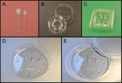
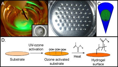
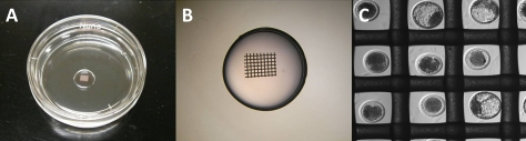
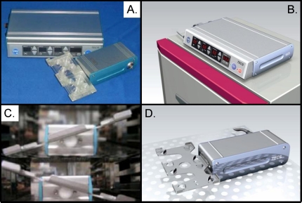
The preponderance of research toward improving embryo development in vitro has focused on manipulation of the chemical soluble environment, including altering basic salt composition, energy substrate concentration, amino acid makeup, and the effect of various growth factors or addition or subtraction of other supplements. In contrast, relatively little work has been done examining the physical requirements of preimplantation embryos and the role culture platforms or devices can play in influencing embryo development within the laboratory. The goal of this review is not to reevaluate the soluble composition of past and current embryo culture media, but rather to consider how other controlled and precise factors such as time, space, mechanical interactions, gradient diffusions, cell movement, and surface interactions might influence embryo development. Novel culture platforms are being developed as a result of interdisciplinary collaborations between biologists and biomedical, material, chemical, and mechanical engineers. These approaches are looking beyond the soluble media composition and examining issues such as media volume and embryo spacing. Furthermore, methods that permit precise and regulated dynamic embryo culture with fluid flow and embryo movement are now available, and novel culture surfaces are being developed and tested. While several factors remain to be investigated to optimize the efficiency of embryo production, manipulation of the embryo culture microenvironment through novel devices and platforms may offer a pathway toward improving embryo development within the laboratory of the future.
Figures
Similar articles
-
Advances in embryo culture systems.Semin Reprod Med. 2012 Jun;30(3):214-21. doi: 10.1055/s-0032-1311523. Epub 2012 May 14. Semin Reprod Med. 2012. PMID: 22585632
-
Advances in embryo culture platforms: novel approaches to improve preimplantation embryo development through modifications of the microenvironment.Hum Reprod Update. 2011 Jul-Aug;17(4):541-57. doi: 10.1093/humupd/dmr006. Epub 2011 Mar 31. Hum Reprod Update. 2011. PMID: 21454356 Review.
-
New culture devices in ART.Placenta. 2011 Sep;32 Suppl 3:S248-51. doi: 10.1016/j.placenta.2011.06.018. Epub 2011 Jul 18. Placenta. 2011. PMID: 21764448 Review.
-
Towards the use of microfluidics for individual embryo culture.Reprod Fertil Dev. 2010;22(1):32-9. doi: 10.1071/RD09219. Reprod Fertil Dev. 2010. PMID: 20003843 Review.
-
A microwell culture system that allows group culture and is compatible with human single media.J Assist Reprod Genet. 2018 Oct;35(10):1869-1880. doi: 10.1007/s10815-018-1252-z. Epub 2018 Jul 11. J Assist Reprod Genet. 2018. PMID: 29998386 Free PMC article.
Cited by
-
Uterine extracellular vesicles as multi-signal messengers during maternal recognition of pregnancy in the mare.Sci Rep. 2022 Sep 16;12(1):15616. doi: 10.1038/s41598-022-19958-z. Sci Rep. 2022. PMID: 36114358 Free PMC article.
-
Simple perfusion apparatus for manipulation, tracking, and study of oocytes and embryos.Fertil Steril. 2015 Jan;103(1):281-90.e5. doi: 10.1016/j.fertnstert.2014.09.039. Epub 2014 Nov 20. Fertil Steril. 2015. PMID: 25450296 Free PMC article.
-
Comparing blastocyst quality and live birth rates of intravaginal culture using INVOcell™ to traditional in vitro incubation in a randomized open-label prospective controlled trial.J Assist Reprod Genet. 2016 Apr;33(4):495-500. doi: 10.1007/s10815-016-0661-0. Epub 2016 Feb 3. J Assist Reprod Genet. 2016. PMID: 26843390 Free PMC article. Clinical Trial.
-
Dielectrophoretic Microfluidic Device for in Vitro Fertilization.Micromachines (Basel). 2018 Mar 20;9(3):135. doi: 10.3390/mi9030135. Micromachines (Basel). 2018. PMID: 30424069 Free PMC article.
-
Biology-inspired microphysiological system approaches to solve the prediction dilemma of substance testing.ALTEX. 2016;33(3):272-321. doi: 10.14573/altex.1603161. Epub 2016 May 15. ALTEX. 2016. PMID: 27180100 Free PMC article. Review.
References
-
- Lane M, Gardner DK. Embryo culture medium: which is the best? Best Pract Res Clin Obstet Gynaecol 2007; 21: 83 100 - PubMed
-
- Biggers JD, McGinnis LK, Lawitts JA. One-step versus two-step culture of mouse preimplantation embryos: is there a difference? Hum Reprod 2005; 20: 3376 3384 - PubMed
-
- Westrom L, Mardh PA, Mecklenburg CV, Hakansson CH. Studies on ciliated epithelia of the human genital tract. II. The mucociliary wave pattern of fallopian tube epithelium. Fertil Steril 1977; 28: 955 961 - PubMed
-
- Paltieli Y, Weichselbaum A, Hoffman N, Eibschitz I, Kam Z. Laser scattering instrument for real time in-vivo measurement of ciliary activity in human fallopian tubes. Hum Reprod 1995; 10: 1638 1641 - PubMed
-
- Fauci L, Dillon R. Biofluidmechanics of reproduction. Annu Rev Fluid Mech 2006; 38: 371 394
Publication types
MeSH terms
Grants and funding
LinkOut - more resources
Full Text Sources
Medical
Miscellaneous
