. 2011 Dec;22(24):4918-30.
doi: 10.1091/mbc.E11-09-0749. Epub 2011 Oct 12.

Defining pheromone-receptor signaling in Candida albicans and related asexual Candida species

Affiliations

Defining pheromone-receptor signaling in Candida albicans and related asexual Candida species

Ching-Hsuan Lin et al. Mol Biol Cell. 2011 Dec.

Erratum in

  • Mol Biol Cell. 2012 Mar;23(6):1140

Abstract

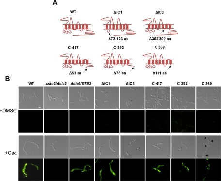
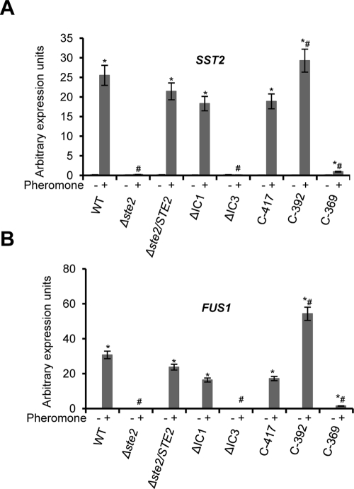
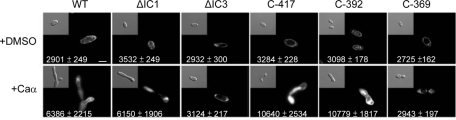
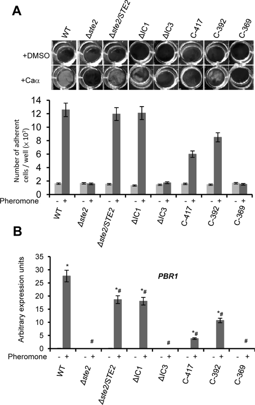
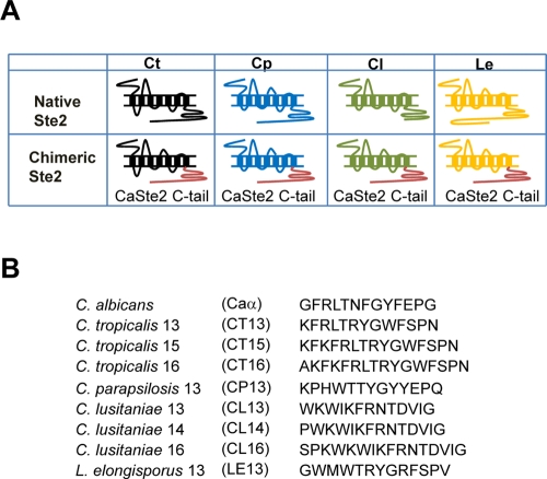
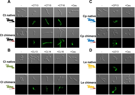
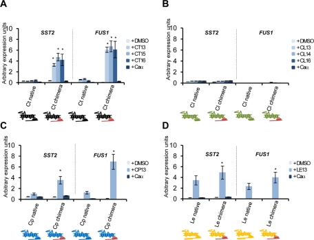
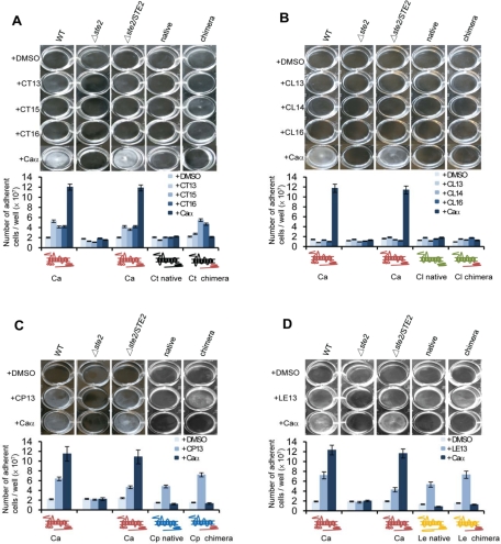
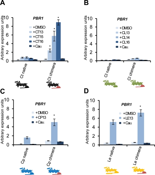
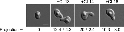
Candida albicans is an important human fungal pathogen in which sexual reproduction is under the control of the novel white-opaque switch. Opaque cells are the mating-competent form, whereas white cells do not mate but can still respond to pheromones, resulting in biofilm formation. In this study, we first define the domains of the α-pheromone receptor Ste2 that are necessary for signaling in both white and opaque forms. Both cell states require the IC loop 3 (IC3) and the C-terminal tail of Ste2 for the cellular response, whereas the first IC loop (IC1) of Ste2 is dispensable for signaling. To also address pheromone-receptor interactions in related species, including apparently asexual Candida species, Ste2 orthologues were heterologously expressed in Candida albicans. Ste2 receptors from multiple Candida clade species were functional when expressed in C. albicans, whereas the Ste2 receptor of Candida lusitaniae was nonfunctional. Significantly, however, expression of a chimeric C. lusitaniae Ste2 receptor containing the C-terminal tail of Ste2 from C. albicans generated a productive response to C. lusitaniae pheromone. This system has allowed us to characterize pheromones from multiple Candida species and indicates that functional pheromone-receptor couples exist in fungal species that have yet to be shown to undergo sexual mating.


Figures

FIGURE 1:
FIGURE 1:
Schematic of the C. albicans Ste2 receptor, including TM domains, IC and EC loops, and mutant receptors lacking these regions. Analysis of pheromone responses in C. albicans strains expressing mutant Ste2 receptors. (A) The Ste2 receptor has seven TM domains that result in three IC and three EC loops and an IC C-terminal cytoplasmic tail. The position and number of disrupted amino acids are indicated by arrows in the Ste2 receptors. (B) Response of wild-type and Ste2 mutant strains to pheromone. Images of mating projections (shmoos) in C. albicans opaque strains were taken after 24 h of α-pheromone treatment. Isolates are expressing a FUS1-GFP reporter construct that is expressed in opaque cells responding to pheromone. The ΔIC1, C-417, and C-392 derivatives of Ste2 form shmoos and activate FUS1 expression when challenged with α pheromone, while receptors lacking the IC3 region or the entire C-terminal tail (C-369) are defective in shmooing and FUS1 expression. Arrows in C-369 indicate very short mating projections formed in response to pheromone. Scale bar: 5 μm.
FIGURE 2:
FIGURE 2:
Quantitative RT-PCR indicating expression of the mating genes SST2 and FUS1 in response to pheromone. Deletion of the IC3 region of Ste2 or the entire C-terminal tail blocked pheromone induction of mating genes in opaque cells. Quantitative gene expression of SST2 (A) and FUS1 (B) was compared when each strain was treated with C. albicans α pheromone (+) or DMSO (−). *, p < 0.05 vs. DMSO. Expression in response to α pheromone was also compared between each mutant allele and wild-type STE2 (#p < 0.05 vs. wild-type). Each value is presented as the mean ± SD.
FIGURE 3:
FIGURE 3:
Analysis of Ste2-GFP fusion protein expression during the response to pheromone in strains expressing mutant Ste2 receptors. Microscopy was used to analyze Ste2-GFP expression in the presence or absence of C. albicans α pheromone. Ste2 localized to the cell surface without pheromone induction, while pheromone treatment stimulated both increased expression and internalization of wild-type, ΔIC1, C-417, and C-392 receptors, but not ΔIC3 or C-369. The GFP intensity is indicated at the bottom of each image. Values are given as the mean ± SD. Numbers are an average of five replicates. Scale bar: 5 μm.
FIGURE 4:
FIGURE 4:
Analysis of the pheromone response in C. albicans white cells expressing different STE2 alleles. We show that removal of the IC3 region or the entire cytoplasmic tail of Ste2 leads to a complete loss of pheromone-induced gene expression and biofilm development in white cells. (A) Images of white cells adhering to plastic when treated with pheromone. White MTLa cells were incubated in Lee's medium and treated with α pheromone in 12-well culture dishes for 24 h. Nonadherent cells were removed by washing, and the remaining adherent cells were photographed and quantitated. (B) Quantitative gene expression of PBR1, a gene induced in white cells in response to pheromone. Expression was analyzed and compared between α pheromone and the DMSO control (*, p < 0.05 vs. DMSO) or compared with the response in wild-type cells upon addition of pheromone (#, p < 0.05 vs. wild-type). Expression levels were normalized to the PAT1 gene. Values are the mean ± SD from two independent experiments with at least three replicates.
FIGURE 5:
FIGURE 5:
Schematic diagrams of native and chimeric Ste2 receptors from C. tropicalis (Ct), C. parapsilosis (Cp), C. lusitaniae (Cl), and L. elongisporus (Le), and the predicted sequences of α pheromones for these species. (A) Figure showing Ste2 receptors from different Candida clade species. Both native receptors and chimeric receptors were tested by expression in C. albicans, the latter made by replacing the C-terminal tail with that from C. albicans Ste2. (B) Sequences of the predicted α pheromones of related Candida clade species.
FIGURE 6:
FIGURE 6:
Images of shmoo formation in C. albicans strains expressing native and chimeric versions of Ste2 receptors from related Candida clade species. C. albicans Δste2 strains were engineered to express native or chimeric Ste2 receptors from C. tropicalis (A), C. lusitaniae (B), C. parapsilosis (C), or L. elongisporus (D). Each strain containing a FUS1-GFP reporter gene was challenged with either α pheromone for that species (see Figure 5B) or with C. albicans α pheromone (Caα) for 24 h. Top, native Ste2; bottom, chimeric Ste2. Scale bar: 5 μm.
FIGURE 7:
FIGURE 7:
Quantitative RT-PCR expression of mating genes in C. albicans strains expressing Ste2 receptors from alternative Candida clade species. Gene expression of SST2 and FUS1, two genes induced in mating opaque cells, was analyzed. Strains were treated with 10 μg/ml pheromone for 4 h in Spider medium. Strains expressed native or chimeric Ste2 from (A) C. tropicalis, (B) C. lusitaniae, (C) C. parapsilosis, or (D) L. elongisporus, in C. albicans. *, p < 0.05 native vs. chimeric Ste2. Expression levels were normalized to the PAT1 gene. Values are presented as the mean ± SD from two independent experiments with at least three replicates.
FIGURE 8:
FIGURE 8:
Pheromone induction of biofilm formation in C. albicans white cells expressing Ste2 receptors from other Candida clade species. Cell adhesion and biofilm development were analyzed in C. albicans white cells expressing Ste2 receptors from (A) C. tropicalis, (B) C. lusitaniae, (C) C. parapsilosis, or (D) L. elongisporus. Assays were performed in Lee's medium in 12-well plastic plates in the presence or absence of C. albicans α pheromone, or α pheromones from the respective species (see Figure 5B). Quantification of the number of adherent white cells is shown in the images. Each experiment was performed using at least two independent isolates and three experimental replicates. Values are represented as the mean ± SD.
FIGURE 9:
FIGURE 9:
Pheromone induction of PBR1 in white cells expressing Ste2 receptors from different Candida species. Expression of the PBR1 gene was analyzed in C. albicans white cells expressing native or chimeric Ste2 receptors from (A) C. tropicalis, (B) C. lusitaniae, (C) C. parapsilosis, or (D) L. elongisporus. PBR1 expression was quantitated after treatment of strains with 10 μg/ml pheromone for 4 h. The chimeric receptors significantly induced PBR1 gene expression over the native Ste2 receptors in three of the four species tested. *, p < 0.05 native vs. chimeric response. Expression levels were normalized to the PAT1 gene. Values are presented as the mean ± SD from two independent isolates with at least three experimental replicates.
FIGURE 10:
FIGURE 10:
Synthetic C. lusitaniae pheromones are able to induce a mating response in C. lusitaniae strains. The pheromone response of C. lusitaniae MTLa cells was tested by incubating cells on PDA together with the addition of synthetic pheromone. 13-mer, 14-mer, and 16-mer pheromones were tested, and each was found to induce shmoo formation. Images were taken after 24 h treatment. Scale bar: 2 μm.

Similar articles

Cited by

References

    1. Alby K, Bennett RJ. Sexual reproduction in the Candida clade: cryptic cycles, diverse mechanisms, and alternative functions. Cell Mol Life Sci. 2010;67:3275–3285. - PMC - PubMed
    1. Alby K, Bennett RJ. Interspecies pheromone signaling promotes biofilm formation and same-sex mating in Candida albicans. Proc Natl Acad Sci USA. 2011;108:2510–2515. - PMC - PubMed
    1. Alby K, Schaefer D, Bennett RJ. Homothallic and heterothallic mating in the opportunistic pathogen Candida albicans. Nature. 2009;460:890–893. - PMC - PubMed
    1. Bedell GW, Soll DR. Effects of low concentrations of zinc on the growth and dimorphism of Candida albicans: evidence for zinc-resistant and -sensitive pathways for mycelium formation. Infect Immun. 1979;26:348–354. - PMC - PubMed
    1. Bennett RJ. Coming of age–sexual reproduction in Candida species. PLoS Pathog. 2010;6:e1001155. - PMC - PubMed

Publication types

LinkOut - more resources