. 2011 Dec;22(23):4549-62.
doi: 10.1091/mbc.E11-05-0405. Epub 2011 Oct 12.

Hook2 is involved in the morphogenesis of the primary cilium

Affiliations

Hook2 is involved in the morphogenesis of the primary cilium

Carole L Baron Gaillard et al. Mol Biol Cell. 2011 Dec.

Abstract

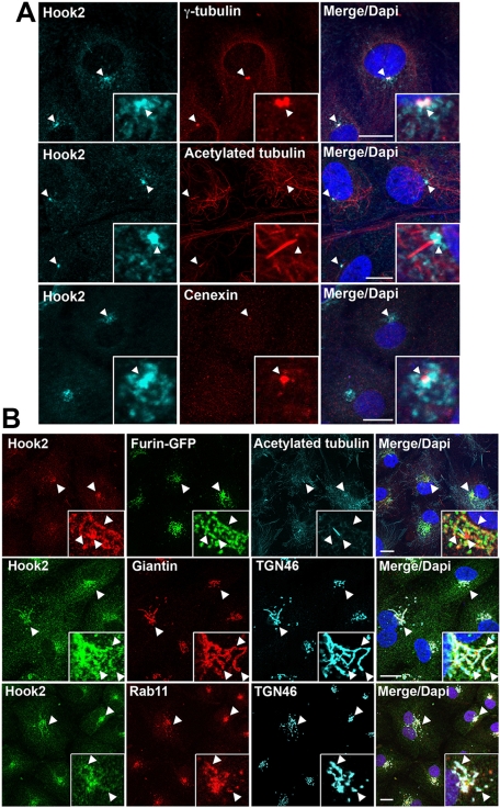
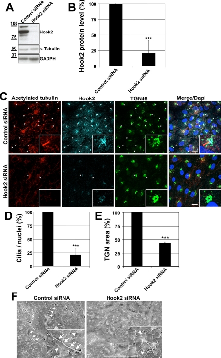
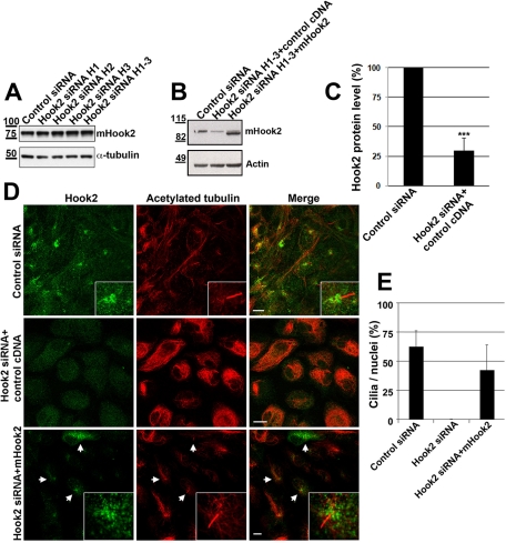
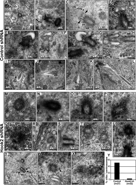
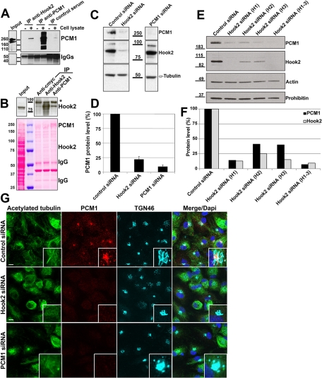
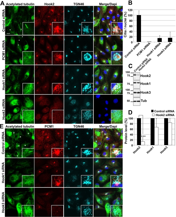
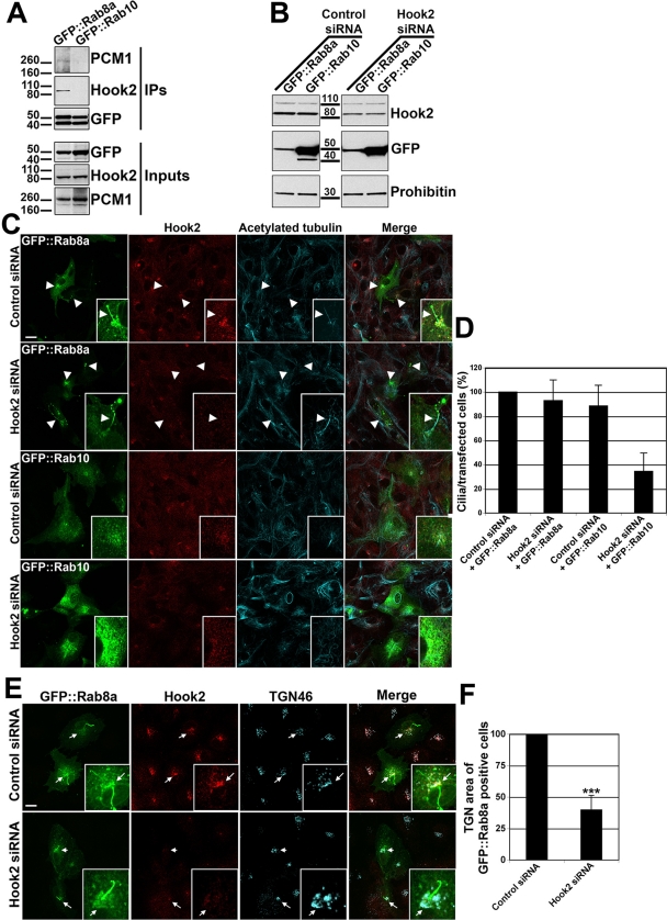
Primary cilia originate from the centrosome and play essential roles in several cellular, developmental, and pathological processes, but the underlying mechanisms of ciliogenesis are not fully understood. Given the involvement of the adaptor protein Hook2 in centrosomal homeostasis and protein transport to pericentrosomal aggresomes, we explored its role in ciliogenesis. We found that in human retinal epithelial cells, Hook2 localizes at the Golgi apparatus and centrosome/basal body, a strategic partitioning for ciliogenesis. Of importance, Hook2 depletion disrupts ciliogenesis at a stage before the formation of the ciliary vesicle at the distal tip of the mother centriole. Using two hybrid and immunoprecipitation assays and a small interfering RNA strategy, we found that Hook2 interacts with and stabilizes pericentriolar material protein 1 (PCM1), which was reported to be essential for the recruitment of Rab8a, a GTPase that is believed to be crucial for membrane transport to the primary cilium. Of interest, GFP::Rab8a coimmunoprecipitates with endogenous Hook2 and PCM1. Finally, GFP::Rab8a can overcome Hook2 depletion, demonstrating a functional interaction between Hook2 and these two important regulators of ciliogenesis. The data indicate that Hook2 interacts with PCM1 in a complex that also contains Rab8a and regulates a limiting step required for further initiation of ciliogenesis after centriole maturation.


Figures

FIGURE 1:
FIGURE 1:
Hook2 localizes to the centrosome, the basal body, and the Golgi apparatus. (A, B) Seven-day cultured ARPE19 cells were simultaneously fixed and permeabilized with methanol (A) or sequentially fixed in PFA and permeabilized with Tx100 (B). In A, the cells were then costained with antibodies raised against Hook2 and proteins localized either at the centrosome/basal body (γ-tubulin, top), the mother centriole (cenexin, bottom), or the primary cilium (acetylated tubulin, middle). In B, top, ARPE19 cells stably transfected with furin-GFP that localizes at the TGN were costained with antibodies staining Hook2 and acetylated tubulin. In B, middle and bottom, ARPE19 cells were costained with antibodies staining Hook2, as well as proteins that localize either in cis-medial Golgi compartments (giantin, middle) or in the TGN, the recycling endosomes, and the ciliary base (Rab11, bottom). Arrowheads indicate colocalizing markers. Bar, 10 μm; inset magnification, ×15. See also Supplemental Figure S1.
FIGURE 2:
FIGURE 2:
Hook2 depletion hinders ciliogenesis without Golgi breakdown in ARPE 19 cells. (A) A mix of 3 siRNAs (H1, H2, and H3) targeting human Hook2 mRNA was used to transiently decrease Hook2 expression for 7 d as measured by WB analysis of Hook2 levels in ARPE19 total cell lysates treated either with Hook2 siRNAs or luciferase siRNA (control siRNA). Antibodies against α-tubulin and GAPDH were used as loading controls. Molecular weight (MW) markers are indicated on the left in kilodaltons. (B) Quantification of Hook2 depletion. Error bars, SD; n = 20 and p < 0001. (C) SiRNA Hook2-depleted cells were fixed in PFA and compared with control siRNA-treated cells for their ability to promote ciliogenesis (acetylated tubulin staining) and maintain Golgi architecture (TGN46 staining) by immunofluorescence and confocal microscopy analysis. Bar, 10 μm; inset magnification, ×15. Asterisks indicate cilia. Note that Hook2 siRNA depletion does not induce breakdown but rather a compaction of the Golgi apparatus. (D) Quantification of ciliogenesis inhibition by Hook2 depletion from three independent experiments. Error bars, SD; n = 200 cells per condition and p < 0001. (E) Quantification of Golgi compaction by Hook2 depletion from three independent experiments. Error bars, SD; n = 100 cells per condition and p < 0001. (F) SEM analysis of control and Hook2 siRNA transiently depleted ARPE19 cells. Bar, 5 μm; inset magnification, ×2. Arrows indicate cilia in the control cells, and arrowheads mark some microvilli in Hook2-depleted cells. See also Supplemental Figure S2.
FIGURE 3:
FIGURE 3:
Exogenous mouse Hook2 rescues defect in ciliogenesis by Hook2 depletion in human ARPE19 cells. (A) 4T1 mouse cells were transiently transfected with control or different human Hook2 siRNA as indicated. After 7 d of incubation, the cells were lysed and tested by WB for Hook2 and α-tubulin production. Molecular weights are indicated on the left in kilodaltons. Note that human Hook2 siRNAs do not deplete mouse Hook2. (B–E) ARPE19 Hook2 siRNA transiently transfected cells were further transfected with an empty vector or mouse Hook2 cDNA and compared with control cells. After 7 d, cells were either processed for WB analysis of Hook2 and actin (B, and quantification of human Hook2 depletion in C) or fixed in PFA and compared by immunofluorescence and confocal microscopy analysis for their ability to promote ciliogenesis (D). Bar, 10 μm; inset magnification, ×15. Arrows indicate cells that express mouse Hook2 cDNA. (E) Quantification of ciliogenesis rescue. Bars represent the percentage of identified cilia per nuclei in each condition. Error bars, SD.
FIGURE 4:
FIGURE 4:
Hook2 is involved before the formation of the pericentriolar vesicle during the initiation of ciliogenesis. (A–X) Both control shRNA 2.3 and Hook2 shRNA 2.5 clones were assayed for primary cilium formation after 7 d in culture by TEM analysis. The different steps of ciliogenesis are shown for the control cells: (A) centriole duplication, (B) pericentriolar vesicle attachment, (C, D) vesicle flattening and axoneme growth, (E–H) sheath growth, (I, J) ciliary elongation, and (K, L) ciliary maturation. Only single centrioles (M–P), as well as centriole duplication (Q–U) and maturation (V–X), were observed for the Hook2-depleted cells. BB, basal body; BFC, basal foot and cap; C, cilium; Ce, centriole; DA, distal appendages; DC, daughter centriole; E, endosome; ER, endoplasmic reticulum; MC, mother centriole; G, Golgi apparatus; N, nucleus; PCS, pericentriolar satellite; PCV, pericentriolar vesicle; S, sheath; SA, subdistal appendages; SV, secondary vesicle; μ, microtubule. Long arrows in C depict a growing axoneme that flattens a pericentriolar vesicle to form the sheath at the distal tip of the mother centriole. The shorter arrows in G and I point at a secondary vesicle (SV) fusing with the ciliary sheath. (Y) Percentage of centrosomes that were associated with a pericentriolar vesicle or a cilium in control and Hook2-depleted cells. See also Supplemental Figure S3.
FIGURE 5:
FIGURE 5:
Hook2 interacts with and regulates PCM1. (A) Lysis buffer (−) or HEK cell lysate (+) were incubated overnight with rabbit anti-Hook2 antibodies, rabbit anti-PCM1 antibodies (positive control), or rabbit preimmune serum (negative control). The immune complexes were precipitated with protein A/G agarose bead mix, and the immunoprecipitates were analyzed by WB with anti-PCM1 (top) or goat anti-rabbit antibodies (bottom). Molecular weight markers are indicated in the middle in kilodaltons. IgGs, heavy chains of immunoglobulins. (B) The reverse experiment was repeated with HEK cell lysate (input) that was incubated with rabbit anti–c-Myc (negative control), rabbit anti-Hook2 (positive control), or rabbit anti-PCM1 antibodies for 12 h. The immune complexes were precipitated with protein A/G agarose bead mix, and the immunoprecipitates were analyzed by WB with anti-Hook2 antibodies (top). Molecular weight (MW) markers are indicated in the middle in kilodaltons. The Ponceau red staining of the membrane was sufficient to detect PCM1 in the IP PCM1, Hook2 in the Hook2 IP, and equal quantities of IgGs in each IP lane. The asterisk marks a 100-kDa cross-reacting band. (C, D) Control, Hook2, or PCM1 siRNA-transfected cells were compared by WB analysis (C) and further quantification (D) for PCM1 production. MW markers are indicated in the middle in kilodaltons; n = 3, p < 0001. Note that PCM1 siRNAs do not affect Hook2 level of expression. (E, F) ARPE19 cells were transfected with individual or a mix of the three siRNAs targeting Hook2 and compared with control siRNA-transfected cells after processing for WB analysis (B) and further quantification (C) for the production of Hook2 and PCM1. Actin and prohibitin were used as loading controls. MW markers are indicated on the left in kilodaltons; n = 2. (G) SiRNA Hook2–depleted cells were fixed in PFA and compared with control siRNA-treated cells by immunofluorescence and confocal microscopy analysis for their ability to promote ciliogenesis (acetylated tubulin staining), express PCM1 in the pericentriolar area (PCM1 staining), and maintain Golgi architecture (TGN46 staining). Bar, 10 μm; inset magnification, ×15. Note that Hook2 depletion highly decreases PCM1 levels of expression. See also Supplemental Figure S4.
FIGURE 6:
FIGURE 6:
Unlike Hook1 and Hook3, PCM1 phenocopies Hook2 depletion. (A) siRNA-treated cells were fixed in PFA after 7 d in culture and compared by immunofluorescence and confocal microscopy analysis for ciliogenesis (acetylated tubulin) and Hook2 localization as well as Golgi architecture (TGN46). Asterisks are next to cilia. Bar, 10 μm; inset magnification, ×15. Note that unlike PCM1 depletion, which phenocopies Hook2 depletion, Hook3 siRNA induces a Golgi breakdown, as well as a redistribution of Hook2 and consequently a ciliogenesis defect. (B) Quantification of the ciliogenesis defect induced by the depletion of Hook1 and Hook3 as compared with control cells or PCM1-depleted cells. (C, D) To confirm the specificity of Hook2 siRNA, ARPE19 cells were transfected with a mix of the three siRNAs targeting Hook2 and compared with control siRNA-transfected cells after processing for WB analysis (C) and further quantification (D) for the production of the three Hook isoforms. MW markers are indicated on the left in kilodaltons; n = 3, p < 0001. (E) siRNA-treated cells were fixed in PFA after 7 d in culture and compared by immunofluorescence and confocal microscopy analysis for ciliogenesis (acetylated tubulin) and PCM1 localization, as well as Golgi architecture (TGN46). Asterisks are next to cilia. Bar, 10 μm; inset magnification, ×15. See also Supplemental Figure S4.
FIGURE 7:
FIGURE 7:
Hook2 and PCM1 are in a complex with Rab8a. (A) Lysates from HEK cells transfected with GFP::Rab8a or GFP::Rab10 were lysed and incubated with G protein–coated beads coupled to mouse anti-GFP antibodies and processed for WB analysis with rabbit antibodies anti-PCM1, anti-Hook2, and anti-GFP and immunoglobulin light chain–specific secondary antibodies. MW markers are indicated on the left in kilodaltons. Note that Hook2 and PCM1 were coimmunoprecipitated with GFP::Rab8a but not with GFP::Rab10. (B, C) Control or Hook2 siRNA (H1–3) transiently depleted cells were further transfected with GFP::Rab8a or GFP::Rab10 cDNA in addition to control or Hook2 siRNA. After 7 d, the cells were processed for WB analyses (B) with the mitochondrial marker prohibitin as a loading control. In parallel, the cells were fixed in PFA, permeabilized with Tx100, stained with antibodies raised against Hook2 and acetylated tubulin, and compared by immunofluorescence and confocal microscopy analysis (C). Bars, 10 μm; inset magnification, ×15. Arrowheads indicate GFP::Rab8a–positive cilia, even in the absence of Hook2 (siRNA Hook2 panels). (D) The number of transfected cells with a cilium in each siRNA condition is quantified and normalized to 100% for the control cells (n = 5). (E) Control or Hook2 siRNA (H1–3) transiently depleted cells were further transfected with GFP::Rab8a in addition to control or Hook2 siRNA as in B and C. After 7 d, the cells were processed for confocal microscopy analysis after immunofluorescence staining with antibodies raised against Hook2 and TGN46. Bars, 10 μm; inset magnification, ×15. Arrows indicate the TGN area of transfected cells with a cilium. (F) The volume of the TGN was measured in GFP::Rab8a–transfected cells in control and Hook2 siRNA conditions (n = 3, p < 0001). Note that GFP::Rab8a restored the ciliogenesis defect but not the Golgi compaction induced by Hook2 depletion. See also Supplemental Figures S5 and S6.

References

    1. Alieva IB, Vorobjev IA. Vertebrate primary cilia: a sensory part of centrosomal complex in tissue cells, but a “sleeping beauty” in cultured cells? Cell Biol Int. 2004;28:139–150. - PubMed
    1. Dammermann A, Merdes A. Assembly of centrosomal proteins and microtubule organization depends on PCM-1. J Cell Biol. 2002;159:255–266. - PMC - PubMed
    1. den Hollander AI, et al. Mutations in LCA5, encoding the ciliary protein lebercilin, cause Leber congenital amaurosis. Nat Genet. 2007;39:889–895. - PubMed
    1. Dunn KC, Aotaki-Keen AE, Putkey FR, Hjelmeland LM. ARPE-19, a human retinal pigment epithelial cell line with differentiated properties. Exp Eye Res. 1996;62:155–169. - PubMed
    1. Ge X, Frank CL, Calderon de Anda F, Tsai LH. Hook3 interacts with PCM1 to regulate pericentriolar material assembly and the timing of neurogenesis. Neuron. 2010;65:191–203. - PMC - PubMed

Publication types

MeSH terms