. 2011 Dec;22(23):4575-87.
doi: 10.1091/mbc.E11-07-0616. Epub 2011 Oct 12.

Differential interactions of the formins INF2, mDia1, and mDia2 with microtubules

Affiliations

Differential interactions of the formins INF2, mDia1, and mDia2 with microtubules

Jeremie Gaillard et al. Mol Biol Cell. 2011 Dec.

Abstract

A number of cellular processes use both microtubules and actin filaments, but the molecular machinery linking these two cytoskeletal elements remains to be elucidated in detail. Formins are actin-binding proteins that have multiple effects on actin dynamics, and one formin, mDia2, has been shown to bind and stabilize microtubules through its formin homology 2 (FH2) domain. Here we show that three formins, INF2, mDia1, and mDia2, display important differences in their interactions with microtubules and actin. Constructs containing FH1, FH2, and C-terminal domains of all three formins bind microtubules with high affinity (K(d) < 100 nM). However, only mDia2 binds microtubules at 1:1 stoichiometry, with INF2 and mDia1 showing saturating binding at approximately 1:3 (formin dimer:tubulin dimer). INF2-FH1FH2C is a potent microtubule-bundling protein, an effect that results in a large reduction in catastrophe rate. In contrast, neither mDia1 nor mDia2 is a potent microtubule bundler. The C-termini of mDia2 and INF2 have different functions in microtubule interaction, with mDia2's C-terminus required for high-affinity binding and INF2's C-terminus required for bundling. mDia2's C-terminus directly binds microtubules with submicromolar affinity. These formins also differ in their abilities to bind actin and microtubules simultaneously. Microtubules strongly inhibit actin polymerization by mDia2, whereas they moderately inhibit mDia1 and have no effect on INF2. Conversely, actin monomers inhibit microtubule binding/bundling by INF2 but do not affect mDia1 or mDia2. These differences in interactions with microtubules and actin suggest differential function in cellular processes requiring both cytoskeletal elements.


Figures

FIGURE 1:
FIGURE 1:
Schematic diagrams of mDia1, mDia2, and INF2 primary structures. Domain boundaries are approximately to scale. For the domains relevant to this work, the boundaries are as follows: mDia1 (mouse): 1255 residues total; FH1, 559–747; FH2, 752–1148; DAD, 1182–1192. mDia2 (mouse): 1171 residues total; FH1, 554–596; FH2, 615–1007; DAD, 1041–1051. INF2 (human, CAAX splice variant): 1249 residues total; FH1, 421–520; FH2, 554–940; DAD/WH2, 973–985. The C-terminal regions of the proteins are indicated by brackets: mDia1, 1149–1255; mDia2, 1009–1171; INF2, 941–1249. Boundaries of the constructs used for these studies are given in Materials and Methods.
FIGURE 2:
FIGURE 2:
Microtubule binding by constructs of INF2, mDia1, and mDia2. Pelleting assays using 0.5 μM polymerized tubulin (dimer), Taxol stabilized. Formin concentrations represent monomer concentration. (A) FH1FH2C constructs: INF2, mDia1, and mDia2. (B) INF2 constructs: FH1FH2C and FH1FH2. (C) mDia2 constructs: FH1FH2C, FH1FH2, and GST-Cterm. (D) GST fusions of C-termini of INF2, mDia1, and mDia2. Representative Coomassie-stained SDS–PAGE gels of some of the data are shown in Supplemental Figure S1. (E) Stoichiometries of formin binding to microtubules at saturation, determined by densitometry vs. a standard curve for each protein. Stoichiometries reflect the dimers of both tubulins and formins.
FIGURE 3:
FIGURE 3:
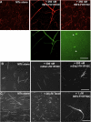
Fluorescence micrographs of microtubule bundling by INF2-FH1FH2C. (A) Fluorescence microscopy of Taxol-stabilized, Alexa 568–labeled microtubules (0.5 μM tubulin) in the absence or presence of fluorescein-labeled INF2-FH1FH2C or INF2-FH1FH2. Assays conducted in actin polymerization buffer. Arrows point to INF2-FH1FH2 labeling individual microtubules. (B) Fluorescence microscopy of Taxol-stabilized, Alexa 568–labeled microtubules in the absence or presence of 500 nM mDia1 or mDia2-FH1FH2C. Assays conducted in actin polymerization buffer. (C) Fluorescence micrographs of Alexa 568–tubulin (25 μM) polymerized in microtubule polymerization buffer for 45 min in the presence of the indicated additives. Formin monomer concentrations are given in all cases. Scale bars, 20 μm.
FIGURE 4:
FIGURE 4:
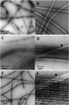
Negative-stained electron microscopy of microtubule bundles assembled by INF2-FH1FH2C. Taxol-stabilized microtubules (2 μM tubulin dimer) were incubated in the absence (E) or in the presence of 4 μM INF2-FH1FH2 (A, B) or 4 μM INF2-FH1FH2C (C, D, F). INF2 monomer concentrations are given. (F) High-magnification view of the MT bundle shown in D.
FIGURE 5:
FIGURE 5:
Microtubule dynamics in bundles induced by INF2-FH1FH2C. (A) Dynamic instability behavior of individual microtubules. The microtubule seeds (0.5 μM) are in red (Alexa 568–labeled tubulin), and elongating microtubule segments are in green (Alexa 488–labeled tubulin, 22 μM). (a–e) Growth and shortening at both microtubule ends observed in dual-view images from a time-lapse series (see Supplemental Movie S1). Time is in seconds; scale bar, 5 μm. The associated kymograph shows the microtubule elongation (red arrows) and catastrophe events (stars) of the microtubule at the center of the image. The microtubule seed is indicated by dotted lines. (B) Dual-view image series of the assembly of a microtubule bundle (see Supplemental Movie S2). Microtubule seed bundles were grown by adding Alexa 568–labeled tubulin (22 μM) in the presence of GFP-INF2 (0.5 μM monomer). Time is in seconds; bar, 5 μm. The corresponding kymograph indicates that the density of microtubules is high. Microtubules do not exhibit noticeable catastrophe and pause events, and microtubules grow progressively for nearly 15 min in this example. (C) Additional kymographs of microtubule bundles, showing examples of growth (red arrows), pauses (blue lines), and the only two catastrophe events observed in analyzing 11 bundles over an average of 15 min (stars). Scale bar, as in B. (D) Analysis of microtubule and bundle elongation in presence of INF2. (Left) Histogram of the mean elongation rates of microtubules during growth phases, when bundled by INF2 (n = 48). The average elongation rate is 1.54 μm/min. SD = 0.26, SE = 0.037. (Right) Histogram of the elongation rate of MT bundles (n = 11). The average elongation rate is 0.89 μm/min. SD = 0.22, SE = 0.066.
FIGURE 6:
FIGURE 6:
Microtubules inhibit actin polymerization by mDia1 and mDia2 but not by INF2. (A–C) Pyrene–actin polymerization assays using 1 μM actin (10% pyrene labeled) and 10 nM INF2-FH1FH2C (A), 5 nM mDia2-FH1FH2C (B), or 5 nM mDia1-FH1FH2C (C). Blue labels indicate nanomolar concentrations of MTs added in addition to the formin and actin. Inset in B shows expanded time course of polymerization (to 3600 s). (D) concentration curves of MT effects on the three formins (as well as mDia2-FH1FH2), plotted as T1/2 (time required to reach one-half of maximal actin polymerization). T1/2 for actin alone is 3122 s ± 152 (n = 8). (E) Pyrene–actin depolymerization assays in which 1.1 μM actin (10% pyrene labeled) is diluted to 1 μM in the presence or absence of INF2-FH1FH2C (250 nM) and/or microtubules (MTs, 750 nM). Formin monomer concentrations are given.
FIGURE 7:
FIGURE 7:
Actin monomers inhibit INF2-FH1FH2C binding to microtubules. (A–D) Fluorescence microscopy of Taxol-stabilized, Alexa 568–labeled microtubules (MT; 0.5 μM tubulin dimer) and 0.2 μM INF2-FH1FH2C alone (A), with 2 μM LatB-stabilized actin monomers (B; threefold molar excess of LatB), 2 μM profilin-bound actin monomers (C; threefold molar excess of profilin), or 2 μM phalloidin-stabilized actin filaments (D; 1.5-fold molar excess of phalloidin). Scale bar, 20 μm. Quantification of bundling is given in Table 1. (E) High-speed pelleting assays containing Taxol-stabilized microtubules (MT; 0.5 μM tubulin dimer) in the absence or presence of 0.5 μM FH1FH2C construct (INF2 or mDia2), 2.5 μM LatB-stabilized actin monomers (LBA; twofold molar excess of LatB), and 2.5 μM LatB-stabilized actin monomers with 7.5 μM profilin (LBAP). Assays are conducted in actin polymerization buffer. Pellet fractions are shown here. Formin monomer concentrations are given. (F) Concentration curve showing actin monomer inhibition of INF2-FH1FH2C binding to microtubules. INF2-FH1FH2C (monomer) and tubulin (dimer) concentrations are fixed at 0.5 μM. The y-axis reflects percentage of INF2-FH1FH2C bound, with 100% being the value without actin monomers present.
FIGURE 8:
FIGURE 8:
INF2 assembles coaggregates of microtubules and actin filaments. Fluorescence micrographs of Alexa 488–labeled microtubules (0.5 μM, Taxol stabilized, green) mixed with Alexa 568/phalloidin–stabilized actin filaments (1 μM, red) in the presence of 0.5 μM of the indicated FH1FH2C construct (monomer concentration) for 30 min at 23°C before imaging. Scale bar, 10 μm.

Similar articles

Cited by

References

    1. Al-Bassam J, Ozer RS, Safer D, Halpain S, Milligan RA. MAP2 and tau bind longitudinally along the outer ridges of microtubule protofilaments. J Cell Biol. 2002;157:1187–1196. - PMC - PubMed
    1. Al-Bassam J, Roger B, Halpain S, Milligan RA. Analysis of the weak interactions of ADP-Unc104 and ADP-kinesin with microtubules and their inhibition by MAP2c. Cell Motil Cytoskeleton. 2007;64:377–389. - PubMed
    1. Azoury J, Lee KW, Georget V, Rassinier P, Leader B, Verlhac MH. Spindle positioning in mouse oocytes relies on a dynamic meshwork of actin filaments. Curr Biol. 2008;18:1514–1519. - PubMed
    1. Bartolini F, Gundersen GG. Formins and microtubules. Biochim Biophys Acta. 2010;1803:164–173. - PMC - PubMed
    1. Bartolini F, Moseley JB, Schmoranzer J, Cassimeris L, Goode BL, Gundersen GG. The formin mDia2 stabilizes microtubules independently of its actin nucleation activity. J Cell Biol. 2008;181:523–536. - PMC - PubMed

Publication types

MeSH terms

LinkOut - more resources