. 2011 Nov 25;334(6059):1097-103.
doi: 10.1126/science.1213256. Epub 2011 Oct 13.

A potent and broad neutralizing antibody recognizes and penetrates the HIV glycan shield

Affiliations

A potent and broad neutralizing antibody recognizes and penetrates the HIV glycan shield

Robert Pejchal et al. Science. .

Abstract

The HIV envelope (Env) protein gp120 is protected from antibody recognition by a dense glycan shield. However, several of the recently identified PGT broadly neutralizing antibodies appear to interact directly with the HIV glycan coat. Crystal structures of antigen-binding fragments (Fabs) PGT 127 and 128 with Man(9) at 1.65 and 1.29 angstrom resolution, respectively, and glycan binding data delineate a specific high mannose-binding site. Fab PGT 128 complexed with a fully glycosylated gp120 outer domain at 3.25 angstroms reveals that the antibody penetrates the glycan shield and recognizes two conserved glycans as well as a short β-strand segment of the gp120 V3 loop, accounting for its high binding affinity and broad specificity. Furthermore, our data suggest that the high neutralization potency of PGT 127 and 128 immunoglobulin Gs may be mediated by cross-linking Env trimers on the viral surface.


Figures
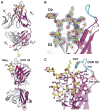
Fig. 1
Fig. 1
Unique binding mode of Man9 by antibody PGT 128 revealed by the high-resolution crystal structure of the complex. (A) Front (top) and side (bottom) views of PGT 128 Fab with bound Man9 glycan. The light and heavy chains are depicted as grey and magenta ribbons, respectively, and the glycan as yellow (carbons) and red (oxygen) ball-and sticks. (B) Close-up view of glycan binding site of PGT 128 showing electron density (2Fo-Fc) at 1.0 sigma for glycan and associated water molecules. Water molecules are shown as red spheres with the electron density colored red for waters that bridge mannose residues and green for waters in the glycan-antibody interface. (C) Detailed view of the interactions in the Man9 glycan binding site at the interface of CDRs H2, H3, L3 and FR2. Tryptophan (VH W52f, W56, W100e and VL W95) and Asn/Asp (VH N53, VL N94, D95a) residues from the Fab are enriched in the interface and dominate the interactions with the glycan. The D1 arm is bound by residues in the 6-amino acid CDR H2 insert and VH FR2. The D3 arm is bound by residues within CDR L3. Hydrogen bonds are shown as green dashes.
Fig. 2
Fig. 2
Crystal structure of PGT 128 Fab in complex with an engineered glycosylated gp120 outer domain (eODmV3). (A) Overall view of PGT 128/eODmV3. PGT 128 Fab heavy and light chains are depicted as in Fig. 1. The eODmV3 is shown in a green cartoon ribbon representation. Glycans are depicted in a ball-and-stick representation with carbons in yellow, oxygens in red and nitrogens in blue. PGT 128 binds the N332 glycan in the primary glycan binding site by interactions with the terminal mannose residues of the D1 and D3 arms. The mode of interaction and site of recognition is identical to that visualized in the high resolution Man9 complex. The secondary glycan binding site recognizes the N301 glycan. (B) Close up view of the secondary glycan interaction site and contacts made with N301 glycan. The mannose residues of the N301 glycan splay out around FR3 residues VH D72, T73, P74, and K75. The terminal mannose resides are not ordered in the electron density. (C) Close up view of V3 interactions with CDR H3. The C-terminus of V3, residues D325-Q328, makes van der Waals and hydrogen bonding contacts to one side of an extended β-strand region of PGT 128 CDR H3, which includes L100-D100d. The V3 base is intercalated between the apex of the CDR H2 insert (Y52e and W52f) and CDR H3.
Fig. 3
Fig. 3
Effect of PGT 128 paratope mutations in the individual glycan subsites on neutralization of HIV-1JR-FL and glycan binding. Binding of PGT 128 mutants to gp120 was tested by ELISA (left panel) or to glycans on the high mannose glycan microarray (right panel). (A) Mutation of select residues in the primary glycan binding site (Man8/9) that recognizes the N332 glycan. Residues (HC, heavy chain; LC, light chain) that disrupt the formation of the hydrophobic core of the binding site (VH K100gA, W100eA, and VL W95A) or disrupt hydrogen bonding to terminal mannose residues (VH H59A and VL D95aA) compromise neutralization (middle panel), as well as gp120 and glycan binding. (B) Mutation of select residues interacting with the secondary glycan binding site that recognizes the N301 glycan. Mutation of VH H52aA results in a decrease in gp120 binding and neutralization, while disruption of the CDR H1–H2 disulfide (VH C32A, C52bA, or double mutant) greatly compromises both gp120 binding and neutralization. There is much less effect on the glycan array which primarily reflects binding to the primary glycan binding site. A complete list of paratope mapping, as well as the effect on gp120 binding, is provided in Table S3. (C) Contribution of the 6-residue CDR H2 insert to neutralization and glycan binding. PGT 128 retains the ability to bind Man8/9 and neutralize to a lesser extent on deletion of the insert, whereas PGT 127 no longer neutralizes, although still has some ability to bind Man8/9. Swapping of the insert between 127 and 128 allows 128 to retain some binding and neutralization, but substantially reduces binding and abrogates neutralization when the PGT 128 H2 insert is transplanted onto PGT 127.
Fig. 4
Fig. 4
Negative stain reconstruction of partially-deglycosylated soluble 664G Env trimer in complex with PGT 128 Fab. Soluble (664G) Env trimer was complexed with Fab PGT 128 and treated with Endo H to remove non-protected glycans. (A) Coordinates of the 128/eODmV3 complex structure fitted into the reconstruction density (blue). Overhead (top) and side (bottom) views show the fit of the crystal structure to the EM density (see SOM). Fab 128, depicted as blue (heavy) and white (light), and eODmV3 (red) are depicted in schematic backbone representation with glycans shown as yellow sticks. (B) Reconstruction density overlayed with cryo-electron tomographic reconstruction of native, unliganded trimer (yellow) (30). The putative location of V1/V2 is indicated. V3, N301, and N332 are exposed on the surface of the outer domain and slightly below the trimer apex, which corresponds to location of the V1/V2 loops. The PGT 128 epitope is located approximately on the opposite side of gp120 from the CD4bs (fig. S13C).
Fig. 5
Fig. 5
Cell-surface binding and neutralization properties of PGT 127 and PGT 128 IgGs and Fabs. (A) (left) Binding of PGT 127 and PGT 128 Fabs and IgGs to HIV-1JR-FL trimers expressed on the surface of transfected 293T cells as determined by flow cytometry. (right) Neutralizing activity of PGT 127 and PGT 128 IgGs and Fabs against HIV-1JR-FL. 2G12 is included for comparison. Experiments were performed in duplicate and data are representative of at least two independent experiments. MFI, mean fluorescence intensity. (B) (top) Comparison of binding (EC50) and neutralization (IC50) for PGT 127 and PGT 128 Fabs and IgGs against HIV-1JR-FL. 2G12 is included for comparison. (bottom) Bar graph representation of Fab (IC50): IgG (IC50) ratios for PGT 127, PGT 128, b12, PG16, PGT 121, 2F5, and 4E10. 2G12 is not included as its two Fabs form a domain-swapped dimer (4). Ratios were calculated as IC50 of the Fab / IC50 of IgG.
Fig. 6
Fig. 6
Impact of PGT 127 and PGT 128 on viral infectivity decay. (A) Viral infectivity decay of HIV-1JR-FL was measured in the presence of PGT 127 and PGT 128 IgGs and Fabs. 2G12 is included for comparison. Data were fitted to a single-phase exponential decay to obtain half-life. Individual experiments were performed in triplicate, and error bars represent the standard error of two independent experiments. (B) The reduction in the half-life of HIV-1JR-FL (expressed as an x-fold decrease) in the presence of antibodies at concentrations providing 90% neutralization, compared to the absence of antibody. Error bars represent the standard error of two independent experiments.

Similar articles

Cited by

References

    1. Kwong PD, et al. Structure of an HIV gp120 envelope glycoprotein in complex with the CD4 receptor and a neutralizing human antibody. Nature. 1998;393:648–659. - PMC - PubMed
    1. Wyatt R, Sodroski J. The HIV-1 envelope glycoproteins: fusogens, antigens, and immunogens. Science. 1998;280:1884–1888. - PubMed
    1. Toone EJ. Structure and energetics of protein-carbohydrate complexes. Curr Opin Struct Biol. 1994;4:719–728.
    1. Calarese DA, et al. Antibody domain exchange is an immunological solution to carbohydrate cluster recognition. Science. 2003;300:2065–2071. - PubMed
    1. Walker LM, et al. Broad neutralization coverage of HIV by multiple highly potent antibodies. Nature. 2011;477:466–470. - PMC - PubMed

Publication types

MeSH terms