. 2012 Feb;40(3):1131-47.
doi: 10.1093/nar/gkr834. Epub 2011 Oct 13.

Genome sequencing of the lizard parasite Leishmania tarentolae reveals loss of genes associated to the intracellular stage of human pathogenic species

Affiliations

Genome sequencing of the lizard parasite Leishmania tarentolae reveals loss of genes associated to the intracellular stage of human pathogenic species

Frédéric Raymond et al. Nucleic Acids Res. 2012 Feb.

Abstract

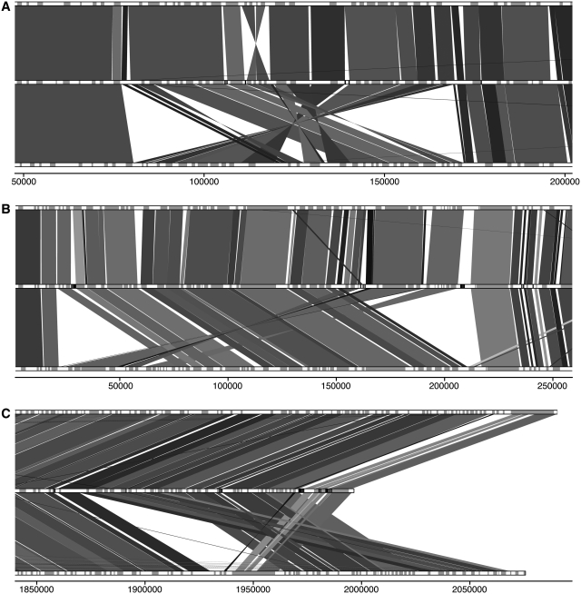
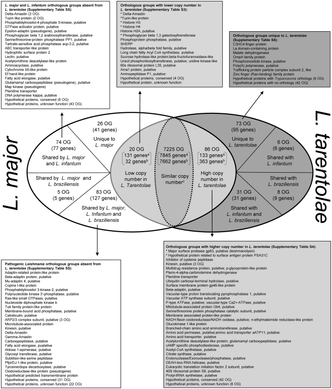
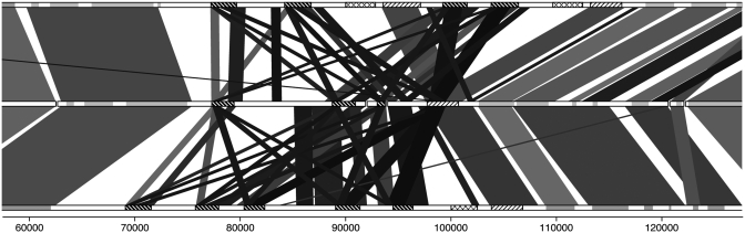
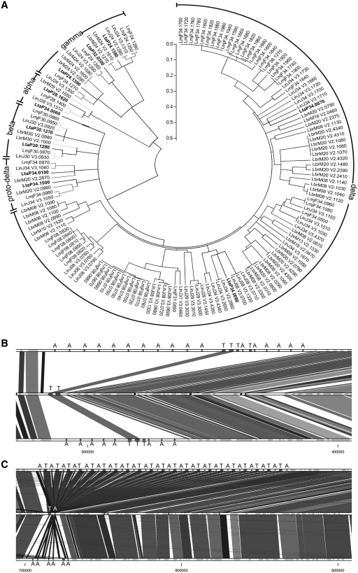
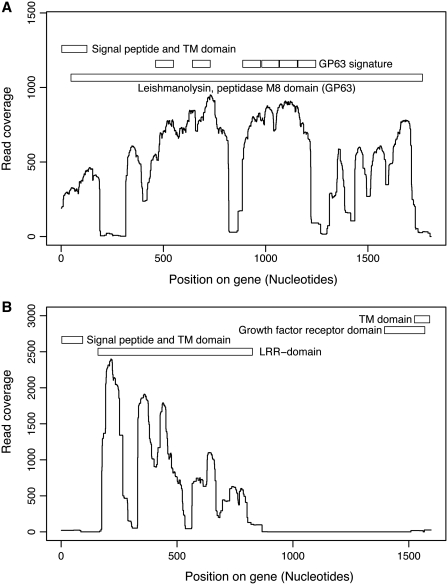
The Leishmania tarentolae Parrot-TarII strain genome sequence was resolved to an average 16-fold mean coverage by next-generation DNA sequencing technologies. This is the first non-pathogenic to humans kinetoplastid protozoan genome to be described thus providing an opportunity for comparison with the completed genomes of pathogenic Leishmania species. A high synteny was observed between all sequenced Leishmania species. A limited number of chromosomal regions diverged between L. tarentolae and L. infantum, while remaining syntenic to L. major. Globally, >90% of the L. tarentolae gene content was shared with the other Leishmania species. We identified 95 predicted coding sequences unique to L. tarentolae and 250 genes that were absent from L. tarentolae. Interestingly, many of the latter genes were expressed in the intracellular amastigote stage of pathogenic species. In addition, genes coding for products involved in antioxidant defence or participating in vesicular-mediated protein transport were underrepresented in L. tarentolae. In contrast to other Leishmania genomes, two gene families were expanded in L. tarentolae, namely the zinc metallo-peptidase surface glycoprotein GP63 and the promastigote surface antigen PSA31C. Overall, L. tarentolae's gene content appears better adapted to the promastigote insect stage rather than the amastigote mammalian stage.


Figures

Figure 1.
Figure 1.
Synteny map of L. tarentolae (middle) compared to L. major (top) and L. infantum (bottom). Genes on chromosome tracks are grey. Leishmania tarentolae contig delimitation is indicated in black in the middle lane. Shade of synteny blocks is proportional to sequence identity, the darker the more similar are the sequences. The scale represents nucleotide position on the chromosome. (A) 5′ region of chromosome 28. (B) 5′ region of chromosome 7. (C) 3′-end of chromosome 35.
Figure 2.
Figure 2.
Differential distribution of genes and OGs of genes between L. tarentolae and L. major. aGene counts referring to L. major. bGene counts referring to L. tarentolae. Lists include the description of OGs of genes that have differential distribution between L. tarentolae and the three sequenced Leishmania pathogenic species. Counts of genes within the different OG are in parenthesis. The complete list of genes and OGs for selected categories is shown in Supplementary Tables S3–S6. Asterisk indicates genes with the highest copy number variability in L. tarentolae.
Figure 3.
Figure 3.
Differential distribution of genes involved in lipophosphoglycan and phosphoglycan modification in L. tarentolae (middle) as compared to L. major (top) and L. infantum (bottom). Phosphoglycan β 1,2 arabinosyltransferase are shaded with crosses, a first group of phosphoglycan β 1,3 galactosyltransferase is shaded with thin lines and a second group with bold lines. The other genes on chromosome tracks are grey. Leishmania tarentolae contig delimitation is in black in the middle lane. Shade of synteny blocks is proportional to sequence identity, the darker the more similar are the sequences. The scale represents nucleotide position on chromosome 2.
Figure 4.
Figure 4.
Analysis of amastin-coding genes in pathogenic and non-pathogenic Leishmania spp. (A) Phylogenetic tree of the amastin genes in L. tarentolae, L. major, L. infantum and L. braziliensis. Labels refer to amastin subfamilies. L. tarentolae amastins are in bold. The evolutionary history was inferred using the Neighbor-Joining method, with a bootstrap test of 500 replicates. Phylogenetic analyses were conducted in MEGA4 (29). (B) Synteny of L. major (top), L. tarentolae (middle) and L. infantum (bottom) amastin/tuzin cluster located on chromosome 8. Amastins are marked with the letter A and tuzins with the letter T. (C) Synteny of L. major (top), L. tarentolae (middle) and L. infantum (bottom) amastin/tuzin cluster located on chromosome 34.
Figure 5.
Figure 5.
Density of read coverage for genes present in high copy number in L. tarentolae. For each position of the reference L. major genes, the number of corresponding reads were counted and plotted on the graph. Protein domains are indicated on the upper portion of each graph. (A) Leishmanolysin (GP63) gene; LmjF10.0480 is used as a reference. (B) Promastigote surface antigen PSA31C gene; LmjF31.1440 is used as a reference.
Figure 6.
Figure 6.
Protease activity of GP63 in six Leishmania species. (A) Western blot using monoclonal antibody targetting GP63 shows the quantity of this protease in each sample. (B) Gelatin zymography assay determining the protease activity of GP63. No signal was observed for L. tarentolae, suggesting the absence of GP63 activity in this species. Lane 1, L. mexicana; lane 2, L. major; lane 3, L. donovani; lane 4, L. infantum; lane 5, L. amazonensis; and lane 6, L. tarentolae.
Figure 7.
Figure 7.
Southern blot hybridization of genes whose copy number varies between L. tarentolae and the other Leishmania pathogenic species. Total genomic DNA of Leishmania isolates was digested with XhoI, run on agarose gels, blotted and hybridized with a combination of PCR-specific probes derived from each species (see Supplementary Table S2 for primer sequences and probe details). (A) Delta-amastin; (B) Proto-delta-amastin (shared by the four species) as a control; (C) Phosphoglycan β 1,3 galactosyltransferase; (D) Phosphoglycan β 1,2 arabinosyltransferase; (E) GP63; and (F) Surface antigen protein PSA31C. DNA loading was estimated by hybridizing the same blot with the single copy gene PTR1 (shown in the lower portion of each panel). Lanes 1, L. tarentolae Parrott-TarII; lane 2, L. tarentolae S125; lane 3, L. major Friedlin; lane 4, L. infantum JPCM5; lane 5, L. braziliensis WHOM/BR/75/M2904.

Similar articles

Cited by

References

    1. Murray HW, Berman JD, Davies CR, Saravia NG. Advances in leishmaniasis. Lancet. 2005;366:1561–1577. - PubMed
    1. Bates PA. Transmission of Leishmania metacyclic promastigotes by phlebotomine sand flies. Int. J. Parasitol. 2007;37:1097–1106. - PMC - PubMed
    1. Noyes HA, Chance ML, Croan DG, Ellis JT. Leishmania (sauroleishmania): A comment on classification. Parasitol. Today. 1998;14:167. - PubMed
    1. Simpson L, Holz G. The status of Leishmania tarentolae/Trypanosoma platydactyli. Parasitol. Today. 1988;4:115–118. - PubMed
    1. Elwasila M. Leishmania tarentolae Wenyon, 1921 from the gecko Tarentola annularis in the Sudan. Parasitol. Res. 1988;74:591–592. - PubMed

Publication types