. 2011 Dec 16;286(50):43259-71.
doi: 10.1074/jbc.M111.264580. Epub 2011 Oct 13.

The orchestration of mammalian tissue morphogenesis through a series of coherent feed-forward loops

Affiliations

The orchestration of mammalian tissue morphogenesis through a series of coherent feed-forward loops

Qing Xie et al. J Biol Chem. .

Abstract

Tissue morphogenesis requires intricate temporal and spatial control of gene expression that is executed through specific gene regulatory networks (GRNs). GRNs are comprised from individual subcircuits of different levels of complexity. An important question is to elucidate the mutual relationship between those genes encoding DNA-binding factors that trigger the subcircuit with those that play major "later" roles during terminal differentiation via expression of specific genes that constitute the phenotype of individual tissues. The ocular lens is a classical model system to study tissue morphogenesis. Pax6 is essential for both lens placode formation and subsequent stages of lens morphogenesis, whereas c-Maf controls terminal differentiation of lens fibers, including regulation of crystallins, key lens structural proteins required for its transparency and refraction. Here, we show that Pax6 directly regulates c-Maf expression during lens development. A 1.3-kb c-Maf promoter with a 1.6-kb upstream enhancer (CR1) recapitulated the endogenous c-Maf expression pattern in lens and retinal pigmented epithelium. ChIP assays revealed binding of Pax6 and c-Maf to multiple regions of the c-Maf locus in lens chromatin. To predict functional Pax6-binding sites, nine novel variants of Pax6 DNA-binding motifs were identified and characterized. Two of these motifs predicted a pair of Pax6-binding sites in the CR1. Mutagenesis of these Pax6-binding sites inactivated transgenic expression in the lens but not in retinal pigmented epithelium. These data establish a novel regulatory role for Pax6 during lens development, link together the Pax6/c-Maf/crystallin regulatory network, and suggest a novel type of GRN subcircuit that controls a major part of embryonic lens development.


Figures
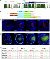
FIGURE 1.
FIGURE 1.
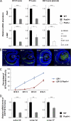
Identification and functional characterization of a novel 5′ lens-preferred enhancer in the c-Maf locus. A, evolutionarily conserved regions (CR1, 2, and 3) in the 12-kb region of the mouse c-Maf locus. Mammal Cons displays the conservation of DNA in multiple mammalian species shown in the University of California at Santa Cruz genome browser. B, a diagrammatic summary of four EGFP reporter constructs tested in transgenic mice. The individual conserved regions CR1 (1.6 kb), CR2 (2.5 kb), and CR3 (1.8 kb) were tested in combination with a 1.3 kb c-Maf promoter (-500 to +800). C, expression of c-Maf in the mouse embryonic lens detected by immunofluorescence. D, expression of EGFP driven by CR1/1.3 kb c-Maf promoter in the transgenic mouse eye (line 10). Expression of c-Maf in RPE by in situ hybridizations and using the knocked-in lacZ marker were described elsewhere (23, 24). LP, lens pit; LV, lens vesicle; 1oLF, primary lens fibers; RE, retina; RPE, retinal pigmented epithelium. Scale bars = 100 μm (C and D). Blue, DAPI; red, anti-c-Maf; green, anti-GFP. E, quantitative analysis of c-Maf- and EGFP-expressing cells in three independent transgenic lines (lines 10, 14, and 17; see C and D and supplemental Fig. S1). The percentages of c-Maf- and EGFP-positive cells were calculated as described under “Experimental Procedures.”
FIGURE 2.
FIGURE 2.
Temporal and spatial patterns of c-Maf expression in the lens are changed as a result of Pax6 haploinsufficiency. A, quantitative RT-PCR analysis of Pax6 and c-Maf expression in E15.5 and P1 wild-type Pax6+/− lenses and wild-type and E9.5 Pax6−/− lens placodes. B, EGFP expression driven by CR1/1.3kb c-Maf promoter in Pax6+/− eye. LP, lens pit; LV, lens vesicle; 1oLF, primary lens fibers; RE, retina; RPE, retinal pigmented epithelium. Scale bars = 100 μm (C and D). Blue, DAPI; green, anti-GFP. C, quantitative analysis of EGFP-positive cells generated in wild-type and Pax6+/− transgenic lenses by ImageJ. Data from three different transgenic lines were combined. D, quantitative analysis of EGFP mRNA levels of three individual transgenic lines (10, 14, and 17) in the wild type and in Pax6+/− transgenic lenses by quantitative RT-PCR.
FIGURE 3.
FIGURE 3.
Pax6 and c-Maf regulate expression of c-Maf. A, distribution of Pax6 and c-Maf detected by qChIP analysis over the 12-kb c-Maf locus in lens chromatin. The relative enrichments are shown as 1% of the input. Localization of two Pax6-binding sites (P6, see Fig. 5A) in CR1 corresponds to a ChIP-positive signal (asterisk). Specific Pax6 and c-Maf enrichments above the background noise, calculated as described under “Experimental Procedures,” are shown by vertical arrows. Pax6 and c-Maf binding (Cryaa promoter) and no-binding (Cryaa +6kb) regions are shown (right side of the diagram) as positive and negative controls, respectively (50). The noise levels (0.148 and 0.112) for Pax6 and c-Maf ChIP data, respectively, are shown by horizontal gray lines. B, transient cotransfection studies of the c-Maf promoter and CR1/c-Maf promoter. Each transfection was performed twice in triplicates. Relative promoter activities were calculated using the “empty vector” value set as 1. Mean ± S.D. of individual data and corresponding p values are indicated.
FIGURE 4.
FIGURE 4.
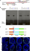
Identification and functional characterization of novel Pax6 DNA-binding site variants. A, a schematic diagram of Pax6 and Pax6(5a) proteins and luciferase reporter constructs. β, β-turns; PAI, N-terminal subdomain of PD; L, linker region; RED, C-terminal subdomain of PD; P/S/T, transcriptional activation domain rich on serine, threonine, and proline residues. 6x motifs represent individual Pax6-binding variants. E4 TATA is a minimal promoter. B, a summary of novel variants of Pax6-binding sites. Motifs 1-1, 2-1, and 3-1 were found for Pax6. Motif 4-1 was found for Pax6(5a). Motifs 1-2, 1-3, 2-2, 3-2, and 3-3 were generated from known/validated Pax6-binding sites (supplemental Fig. S4). In vitro binding of Pax6 and Pax6(5a) to individual motifs was tested by EMSAs (supplemental Fig. S5). Cotransfection assays were performed in P19 cells, and the data are expressed as relative fold changes elicited in the presence of Pax6 or Pax6(5a) compared with the changes found with the empty vector.
FIGURE 5.
FIGURE 5.
Pax6 is essential for CR1 enhancer function through a tandem of sites. A, a prediction of two Pax6-binding sites that resemble motifs 2-1 (site 2) and 2-2 (site 1) in CR1 of the mouse c-Maf locus. B, EMSA confirmation of Pax6 binding to sites 1 and 2. Five point mutations in these Pax6 binding sites were also tested as specific competitors. C, a diagrammatic summary of wild-type CR1 and two mutants, CR1-Mut and CR1-Del, in EGFP reporter constructs. D, temporal and spatial analysis of EGFP expression in the presence of two Pax6 mutants (C) in CR1 in transgenic mice. Analysis of additional CR1-Mut and CR1-Del transgenic lines is shown in supplemental Fig. S6. LP, lens pit; LV, lens vesicle; 1oLF, primary lens fibers; RE, retina; RPE, retinal pigmented epithelium. Scale bars = 100 μm (C and D). Blue, DAPI; green, anti-GFP.
FIGURE 6.
FIGURE 6.
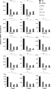
Analysis of crystallin gene expression in Pax6 heterozygous lenses. Significant decrease of transcripts encoding two α-, six β-, and six γ-crystallin genes was found in E15.5 Pax6+/− lenses by quantitative RT-PCR experiments. The relative mRNA abundance is calculated using the reference gene β2 microglobulin (B2M) as described under “Experimental Procedures.” Samples from one wild–type and three Pax6+/− embryos (#1, #2, and #3) were tested in each independent biological replicate. Each reaction was performed in triplicate, and experiments were replicated three times. Mean ± S.D. of individual data and corresponding p values are indicated.
FIGURE 7.
FIGURE 7.
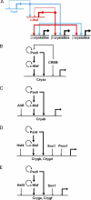
A potent GRN subcircuit comprised of Pax6 and c-Maf is used to control crystallin gene expression in the lens. A, the core regulatory subcircuit. The feed-forward loop is shown in boldface. B, GRN for αA-crystallin gene expression. The core module is expanded by utilization of the cAMP response element-binding protein that regulates Pax6 expression (81) and binds both the αA-crystallin promoter and its 3′-distal enhancer, DCR3 (50). C, GRN for αB-crystallin gene expression. The core module is expanded by utilization of AhR that binds the 5′ distal enhancer (71). D, regulation of γB- and γD-crystallins by the Pax6/c-Maf core module in combination with Hsf4, Prox1, and Sox1 (–74). E, regulation of γE- and γF-crystallins by the Pax6/c-Maf core module in combination with Hsf4 and Sox1 (72, 73). The Pax6/c-Maf core module is shown in boldface. α-crystallins, αA and αB; β-crystallins, βA3/A1, βA2, βA4, βB1, βB2, and βB3; γ-crystallins, γA, γB, γC, γD, γE, and γF; Cryaa, αA-crystallin; Cryab, αA-crystallins; Crygb, γB-crystallin; Crygd, γd-crystallin; Cryge, γE-crystallin; Crygf, γF-crystallin.

Similar articles

Cited by

References

    1. Ali R. R., Sowden J. C. (2011) Nature 472, 42–43 - PubMed
    1. Eiraku M., Takata N., Ishibashi H., Kawada M., Sakakura E., Okuda S., Sekiguchi K., Adachi T., Sasai Y. (2011) Nature 472, 51–56 - PubMed
    1. Yang C., Yang Y., Brennan L., Bouhassira E. E., Kantorow M., Cvekl A. (2010) FASEB J. 24, 3274–3283 - PMC - PubMed
    1. Davidson E. H. (2010) Nature 468, 911–920 - PMC - PubMed
    1. Peter I. S., Davidson E. H. (2011) Cell 144, 970–985 - PMC - PubMed

Publication types

MeSH terms