. 2011 Dec 9;286(49):42725-42735.
doi: 10.1074/jbc.M111.265207. Epub 2011 Oct 13.

Functional characterization of alternative splicing in the C terminus of L-type CaV1.3 channels

Affiliations

Functional characterization of alternative splicing in the C terminus of L-type CaV1.3 channels

Bao Zhen Tan et al. J Biol Chem. .

Abstract

Ca(V)1.3 channels are unique among the high voltage-activated Ca(2+) channel family because they activate at the most negative potentials and display very rapid calcium-dependent inactivation. Both properties are of crucial importance in neurons of the suprachiasmatic nucleus and substantia nigra, where the influx of Ca(2+) ions at subthreshold membrane voltages supports pacemaking function. Previously, alternative splicing in the Ca(V)1.3 C terminus gives rise to a long (Ca(V)1.3(42)) and a short form (Ca(V)1.3(42A)), resulting in a pronounced activation at more negative voltages and faster inactivation in the latter. It was further shown that the C-terminal modulator in the Ca(V)1.3(42) isoforms modulates calmodulin binding to the IQ domain. Using splice variant-specific antibodies, we determined that protein localization of both splice variants in different brain regions were similar. Using the transcript-scanning method, we further identified alternative splicing at four loci in the C terminus of Ca(V)1.3 channels. Alternative splicing of exon 41 removes the IQ motif, resulting in a truncated Ca(V)1.3 protein with diminished inactivation. Splicing of exon 43 causes a frameshift and exhibits a robust inactivation of similar intensity to Ca(V)1.3(42A). Alternative splicing of exons 44 and 48 are in-frame, altering interaction of the distal modulator with the IQ domain and tapering inactivation slightly. Thus, alternative splicing in the C terminus of Ca(V)1.3 channels modulates its electrophysiological properties, which could in turn alter neuronal firing properties and functions.


Figures

FIGURE 1.
FIGURE 1.
Characterization of pAb_CaV1.342 and pAb_CaV1.342A specific antibodies Western blot and immunostaining. A, left, schematic of CaV1.3 channel pore-forming α1 subunit (α1D), with hot spots for CaM/channel regulation in the carboxyl terminus (EF, EF-hand (32); pre-IQ and IQ domains (33, 34). Right, diagrammatic representation of two C-terminal splice variants of the CaV1.3 α1 subunits. Incorporation of exon 42A instead of exon 42 results in a truncated C terminus 6 amino acids after exon 41. Rabbit polyclonal antibodies were raised specifically against these two splice variants. The peptide sequences utilized are in red and italics, namely for pAb_CaV1.342, CCEDDSSPTWSRQNYSYYNRYPGSSMD, and pAb_CaV1.342A, CLQMLERL. B, Western blot of total membrane protein extracted from wild type and CaV1.3−/− knock-out mice. Left blot was probed with pAb_CaV1.342 antibody. A single band of ∼250 kDa was detected in a wild type mouse but absent in the knock-out mouse. Middle blot was probed with pAb_CaV1.342A antibody. A single band of ∼150 kDa was detected in a wild type mouse but absent in the knock-out mouse. Right blot was probed with commercial pAb_CaV1.3 antibody. C, double labeling of the CA3 region of wild type mouse dorsal hippocampus with bassoon (red) and pAb_CaV1.342 or pAb_CaV1.342A (green). Positive staining for presynaptic vesicle proteins (bassoon) is restricted to the stratum lucidum (SL), a mossy fiber recipient layer of the CA3 subfield of the hippocampus. Strong staining of pAb_CaV1.342 (top) and pAb_CaV1.342A (bottom) was observed in the stratum pyramidale (SP) of the region. No co-localization was observed between the bassoon and pAb_CaV1.342 or pAb_CaV1.342A. Scale bar, 50 μm.
FIGURE 2.
FIGURE 2.
Similar localization of CaV1.342 and CaV1.342A proteins in many regions of brain and spinal cord. Wild type mouse brain and spinal cord sections are immunostained with pAb_CaV1.342 and pAb_CaV1.342A antibodies, using 3,3′-diaminobenzidine as the chromogen. Left panel, brain and spinal cord sections were stained for CaV1.342 channels. Right panel, brain and spinal cord sections were stained for CaV1.342A channels. Similar staining patterns were observed for both CaV1.342 and CaV1.342A proteins in many regions of the brain and spinal cord.
FIGURE 3.
FIGURE 3.
Correction of cloning error in the original CaV1.342 clone to CaV1.3A2123V. A, amino acid alignments of the CTM region for long variants of human, mouse, and rat CaV1.3 (GenBank accession numbers: human, NM000720; mouse, NM028981; rat, NM017298). Positions of exons 48 and 49 are given. Highlighted green residues mark the differences between these variants. Highlighted purple residues mark the position whereby alanine is observed in position 2123 in rat CaV1.342 clone (GenBank accession number D38101) instead of valine. B, top, direct sequencing results of rat clone CaV1.342 clone. The peptide combination is listed in black; bottom, direct sequencing results of RT-PCR amplified from rat brain. Highlighted purple residues mark the position of cloning error. C, representative IBa (gray) and ICa (black) traces during depolarization to 10 mV. The IBa and ICa traces were scaled to enable comparison between the two profiles. Current scales were drawn for both IBa (gray) and ICa (black). The time scales for each IBa and ICa pair are the same. D, normalized I-V plots for IBa (top) and ICa (bottom) of CaV1.3A2123V and CaV1.342A. The curves were fitted with the equation described under “Experimental Procedures.” E, calcium-dependent inactivation of current through CaV1.3A2123V and CaV1.342A. The fraction of the peak current, Ipeak, remained at time intervals of 300 ms upon depolarization to the indicated voltages for CaV1.3A2123V channels. The difference between the remaining current for IBa and ICa, f value, indicates the strength of CDI. The presence of CDI is also marked by “U”-shaped dependence of ICa on voltage (i.e. CaV1.342A, black curves). The curves are visual fits of the values plotted to facilitate comparison. The number of cells recorded are given in parentheses.
FIGURE 4.
FIGURE 4.
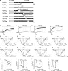
Current-voltage relationships of CaV1.3 alternatively spliced variants. A, schematic representation of alternatively spliced CaV1.3 channel constructs. The channel backbone consists of CaV1.3 (GenBank accession number: D38101, white box), whereas the cytosolic tail consists of CaV1.3 long form (CaV1.342), or alternatively spliced variants IQΔ, Δ41, 43S, 43S-2, Δ44, and 48S (black). The stop codons for IQΔ, Δ41, 43S, and 43S-2 are indicated by black and white filled circles. Numbering follows the CaV1.3 amino acid sequence. B–E, representative IBa (gray) and ICa (black) traces during depolarization to 10 mV for alternatively spliced constructs Δ41, 43S-2, Δ44, and 48S. The IBa and ICa traces were scaled to enable comparison between the two profiles. Current scales were drawn for both IBa (gray) and ICa (black). The time scales for each IBa and ICa pair are the same. F–I, normalized I-V plots for IBa of alternatively spliced constructs Δ41, 43S-2, Δ44, and 48S. The curves were fitted with the equation described under “Experimental Procedures.” In parentheses are the number of cells recorded. J-M, same as F-I, but for ICa. N-Q, calcium-dependent inactivation of current through alternatively spliced variants Δ41, 43S-2, Δ44, and 48S. The fraction of peak current, Ipeak, remained at time intervals of 300 ms upon depolarization for IBa and ICa. f value indicates the strength of the CDI. The curves are visual fits of the values plotted to facilitate comparison. The number of cells recorded are given in parentheses.
FIGURE 5.
FIGURE 5.
Voltage-dependent electrophysiological properties of splice variants CaV1.3Δ41, CaV1.343S-2, CaV1.3Δ44, and CaV1.348S. A–D, activation and steady-state inactivation properties. Normalized plots for activation (dashed lines) and steady-state inactivation (solid lines) for long form (CaV1.3A2123V) and alternatively spliced constructs Δ41, 43S-2, Δ44, and 48S. For steady-state inactivation, peak currents obtained after the 15-s inactivating pulse were normalized to that obtained before inactivation and plotted against voltage. The curves are fitted with the Boltzmann relationship. For the activation plots, the peak of the tail currents (G) were normalized against the largest peak and plotted against voltage. The curves were fitted using the equation given under “Experimental Procedures.” The number of cells recorded are given in parentheses, “(/)” for steady-state inactivation and “[/]” for activation. Plots of the long form (CaV1.3A2123V) were redrawn in each graph for comparison. E–G, recovery from inactivation. Fractional recovery was plotted as a function of ΔT for CaV1.3A2123V and splice variants Δ41, 43S-2, Δ44, and 48S, respectively. The curve for CaV1.3A2123V was redrawn for all plots for comparison. Curves were fitted as described under “Experimental Procedures.” I–L, density of Ba2+ currents through CaV1.3A2123V and splice variants Δ41, 43S-2, Δ44, and 48S. The peak of tail currents measured at the end of short depolarizing pulses evoked at different potentials were normalized against the membrane capacitance (Cm) of the recorded cell to obtain current density. The number of cells are indicated in the parentheses. ***, p < 0.001 (compared with CaV1.3A2123V) (Kruskal-Wallis test followed by Dunn's multiple comparison post test).

Similar articles

Cited by

References

    1. Dunlap K., Luebke J. I., Turner T. J. (1995) Trends Neurosci. 18, 89–98 - PubMed
    1. Hell J. W., Westenbroek R. E., Warner C., Ahlijanian M. K., Prystay W., Gilbert M. M., Snutch T. P., Catterall W. A. (1993) J. Cell Biol. 123, 949–962 - PMC - PubMed
    1. Lipscombe D., Helton T. D., Xu W. (2004) J. Neurophysiol. 92, 2633–2641 - PubMed
    1. Xu W., Lipscombe D. (2001) J. Neurosci. 21, 5944–5951 - PMC - PubMed
    1. Koschak A., Reimer D., Huber I., Grabner M., Glossmann H., Engel J., Striessnig J. (2001) J. Biol. Chem. 276, 22100–22106 - PubMed

Publication types

LinkOut - more resources