. 2011 Nov 11;109(11):1290-301.
doi: 10.1161/CIRCRESAHA.111.250167. Epub 2011 Oct 13.

CD4+ T cells and complement independently mediate graft ischemia in the rejection of mouse orthotopic tracheal transplants

Affiliations

CD4+ T cells and complement independently mediate graft ischemia in the rejection of mouse orthotopic tracheal transplants

Mohammad A Khan et al. Circ Res. .

Abstract

Rationale: While microvascular injury is associated with chronic rejection, the cause of tissue ischemia during alloimmune injury is not yet elucidated.

Objective: We investigated the contribution of T lymphocytes and complement to microvascular injury-associated ischemia during acute rejection of mouse tracheal transplants.

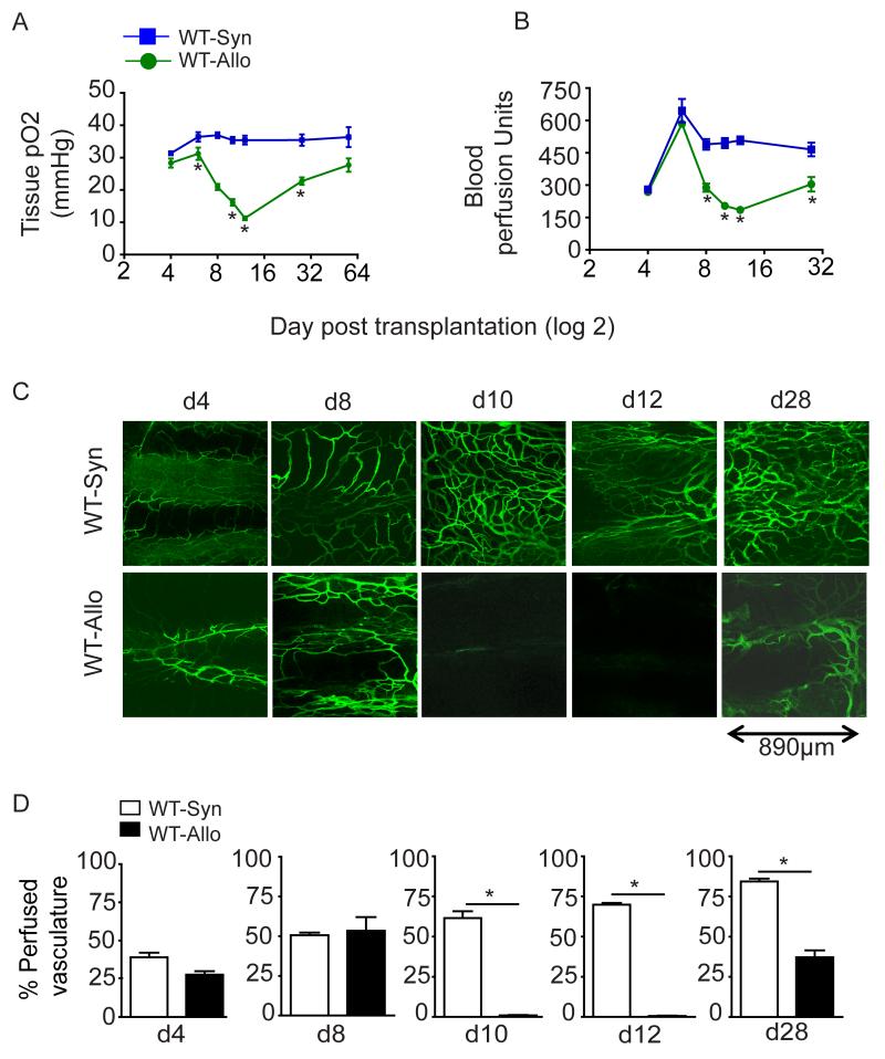
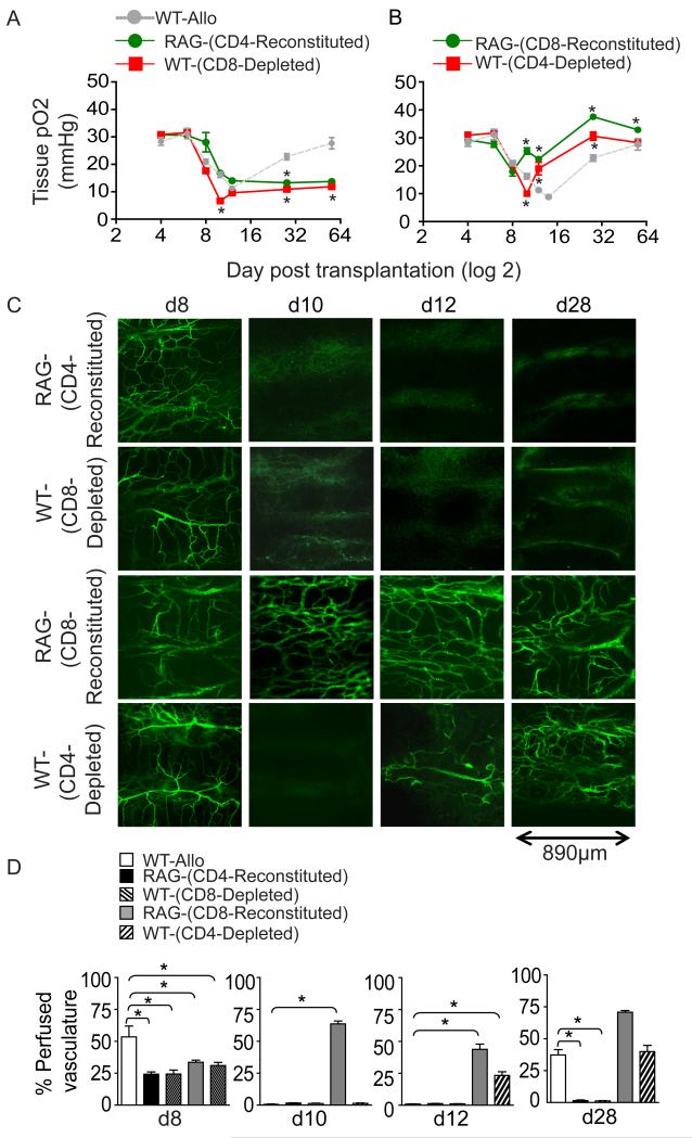
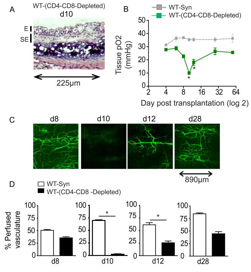
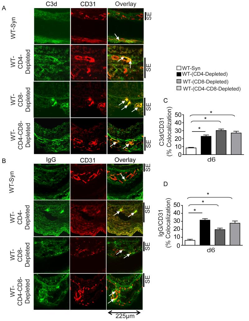
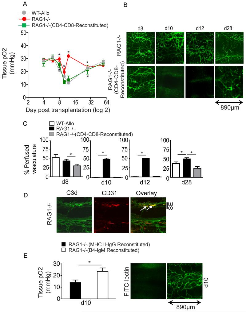
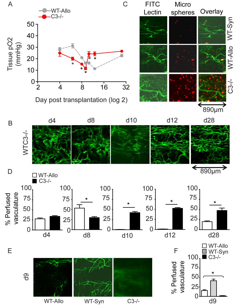
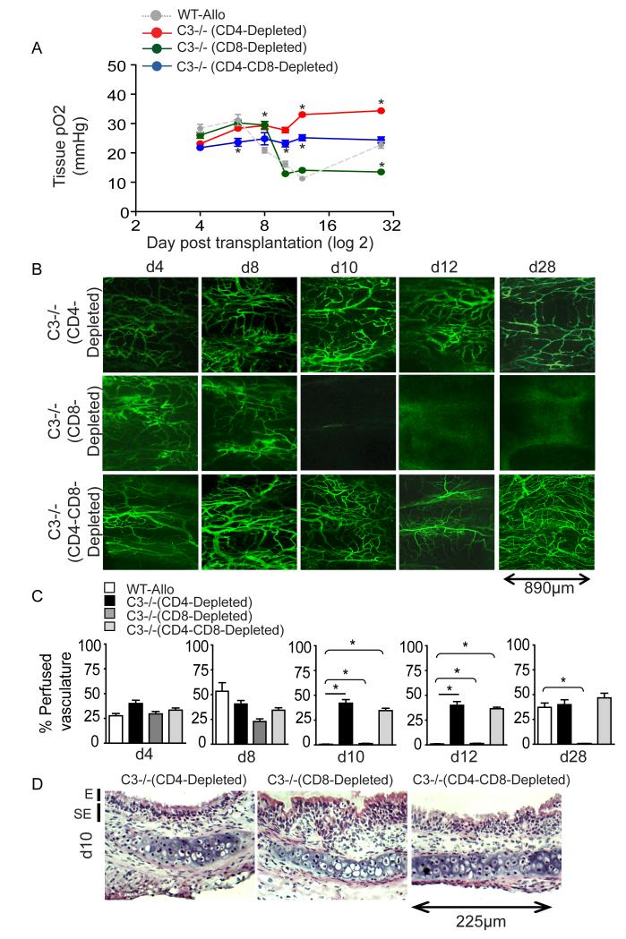
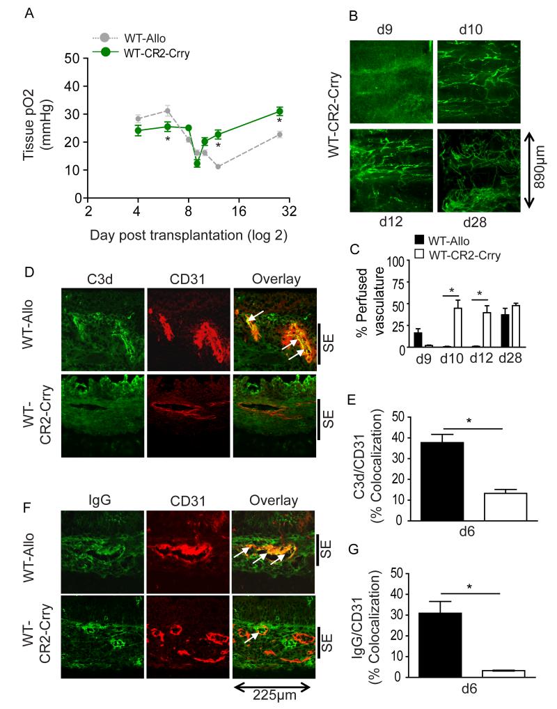
Methods and results: Using novel techniques to assess microvascular integrity and function, we evaluated how lymphocyte subsets and complement specifically affect microvascular perfusion and tissue oxygenation in MHC-mismatched transplants. To characterize T cell effects on microvessel loss and recovery, we transplanted functional airway grafts in the presence and absence of CD4(+) and CD8(+) T cells. To establish the contribution of complement-mediated injury to the allograft microcirculation, we transplanted C3-deficient and C3-inhibited recipients. We demonstrated that CD4(+) T cells and complement are independently sufficient to cause graft ischemia. CD8(+) T cells were required for airway neovascularization to occur following CD4-mediated rejection. Activation of antibody-dependent complement pathways mediated tissue ischemia even in the absence of cellular rejection. Complement inhibition by CR2-Crry attenuated graft hypoxia, complement/antibody deposition on vascular endothelium and promoted vascular perfusion by enhanced angiogenesis. Finally, there was a clear relationship between the burden of tissue hypoxia (ischemia×time duration) and the development of subsequent airway remodeling.

Conclusions: These studies demonstrated that CD4(+) T cells and complement operate independently to cause transplant ischemia during acute rejection and that sustained ischemia is a precursor to chronic rejection.


Figures

Figure 1
Figure 1. The progressive hypoxia of acutely rejecting allografts is reversed as perfusion is restored during chronic rejection
(A) Tissue pO2 (Mean±SE, mmHg) was plotted over different time points (d4 - d56), (n=4-6 animals/time point). Rejecting allografts become progressively hypoxic over time beginning with early acute rejection responses observed in the first week following transplantation. Airway pO2 nadirs on d14 and increases during chronic rejection. B) Blood perfusion (Mean±SE, units) was plotted over different time points (d4-d28), (n=4-6 animals/time points).Rejecting allografts demonstrate less blood perfusion as compare to syngrafts during acute rejection. (C) FITC-lectin perfusion profile of whole mounts tracheal grafts from d4 to d28 illustrates that falling airway pO2s correlate with the loss and restoration of perfusion during allograft rejection. (D) Morphometric assessments of perfused vasculature (FITC-lectin perfusing vessels/unit area) in tracheal grafts at different time points following transplantation demonstrate neovascularization after 28 days of rejection. Data are shown as means with SEM for five independent experiments. *, p<0.05. Original magnification, X10.
Figure 2
Figure 2. Effects of CD4+ and CD8+ T cells on kinetics of tissue pO2 and perfusion
(A) Tissue pO2 kinetics, in rejecting CD4-reconstituted/CD8-deficient RAG1−/− or CD4-replete/CD8-depleted WT recipients, were serially evaluated between d4 and d56 (n=4-6 animals/time point). CD4+ cells are sufficient to induce sustained graft hypoxia. Absence of CD8+ cells is correlated with persistent graft hypoxia relative to CD8-replete control rejection responses (See Fig. 1a). (B) Tissue pO2 kinetics, in rejecting CD8-reconstituted/CD4-deficient RAG1−/− or CD8-replete/CD4-depleted WT recipients, were serially evaluated between d4 and d56 (n=4-6 animals/time point). CD8+ reconstitution does not result in significant hypoxia in RAG1−/− recipients. (C) FITC-lectin perfusion profiles were serially assessed in whole mounts from d8 to d28 and demonstrated that perfusion was lost in CD4-reconstituted/CD8-deficient RAG1−/− and CD4-replete/CD8-depleted WT recipients. By contrast, CD8-reconstituted/CD4-deficient RAG1−/− recipients were not ischemic and CD8-replete/CD4-depleted WT recipients demonstrated an accelerated restoration of blood flow by d12, which was earlier than the WT response illustrated in Fig. 1c. (D) Morphometric assessment of perfused vasculature in tracheal graft at different time points. Data are shown as means with SEM of four independent experiments. *, p<0.05. Original magnification, X10.
Figure 3
Figure 3. Combined CD4+ and CD8+ T cell depletion prevents acute rejection, but grafts become ischemic
(A) H&E of BALB/c →B6 tracheal transplants 10 days after transplantation shows no acute mononuclear cell infiltration (H&E, Original magnification, X40). (B) CD4/CD8-depleted recipients exhibit significant hypoxia similar to rejecting non-depleted WT recipients (n=4-6 animals/time point). (C) Reperfusing vessels in pan T-depleted recipients are detected on d12. (D) % perfused vasculature is significantly decreased on d10 and d12 relative to syngrafts. Data are shown as means with SEM. *, p<0.05. (FITC-lectin, Original magnification, X10).
Figure 4
Figure 4. Increased microvascular deposition of C3d and IgG identifies grafts that will lose perfusion
(A) Immunofluorescent staining demonstrates increased vascular C3d on post-operative day 6 in the T-cell depleted allografts relative to syngrafts; these allograft groups lose perfusion on d10. Increased C3d/IgG endothelial deposition is noted even the combined CD4/CD8 T cell depleted graft recipients that do not exhibit acute rejection. Arrows highlighted overlaid C3d on CD31+ cells. A small amount of endothelial C3d expression is noted in the adventitia of the syngrafts (n=8/group). (B) A similar pattern of vascular IgG deposition was noted, with greater deposition in the allograft groups than the syngraft group. (C) Morphometric assessments of C3d/CD31 colocalization revealed that all allograft groups demonstrated significantly more endothelial complement deposition. (D) Morphometric assessments for IgG/CD31 colocalization similarly showed increased immunoglobulin deposition in allografts. Data are shown as means with SEM and representative images of at least four different experiments. *, p<0.05. Original magnification, X40. SE symbolizes sub epithelial area in grafts.
Figure 5
Figure 5. Effects of absence of T and B-lymphocytes on kinetics of tissue pO2 and loss of microvascular flow
(A) Tissue pO2 values of Balb/c allografts in non-reconstituted and CD4+/CD8+ reconstituted RAG1−/− recipients were plotted over different time points (d10-d58), (n=4-6 animals/time point). B, C) FITC-lectin perfusion profile and % perfused vasculature show that perfusion is maintained over time in RAG1−/− recipients and lost in reconstituted animals. (FITC-lectin, Original magnification, X10). D) Immunofluorescence colocalization of C3d in CD31+ vascular endothelial cells in RAG1−/− mice at day 6 of transplantation. E) Tissue pO2 and FITC-lectin perfusion in donor-specific MHC-II IgG2 and B4-IgM reconstituted RAG1−/− allografts at d10 post transplantation. Adoptive transfer of donor-specific antibodies causes ischemia whereas administration of B4-IgM antibodies (specific to annexin IV) did not cause graft hypoxia or ischemia. Original magnification, X40. Data are shown as means with SEM. *, p<0.05. Data are shown as means with SEM.
Figure 6
Figure 6. Absence of complement in the allograft recipient limits graft hypoxia and ischemia during rejection
(A) BALB/c→B6 C3−/− allografts were assessed for tissue pO2 over different time points (d4-d28), (n=4-6 animals/time point) and demonstrated a brief period of hypoxia on d9. (B) FITC-lectin perfusion profile of whole mounts tracheal grafts from d4 to d28 illustrate rapid recovery of microvascular flow from d10 of perfusion during allograft rejection. D4 microvessels appear dilated in C3−/− recipients. (C) Significant microvascular permeability was noted in d4 tracheal C3−/− recipients by using FITC-lectin in conjunction with R50 Fluromax red microsphere infusion. (D) Morphometric assessments of perfused vasculature (FITC-lectin perfusing vessels/unit area) in tracheal grafts at different time points. (E) FITC-lectin perfusion studies in C3−/− recipients demonstrate a brief period of ischemia on d9. (F) Morphometric assessment of perfused vessels on d9. Data are shown as means with SEM of four different experiments. *, p<0.05. Original magnification, X10.
Figure 7
Figure 7. CD4+ T cell depletion synergizes with C3 deficiency to prevent airway hypoxia and maintain perfusion in rejection
(A) Lymphocyte subset-depleted Balb/c→B6 C3−/− recipients were serially assessed for graft pO2 between d4-d28 (n=4-6 animals/time points). While CD4-depleted C3−/− recipients exhibited a pO2 profile similar to syngrafts, CD8-depleted C3−/− recipients reveal prolonged and unremitting graft hypoxia. (B, C) FITC-lectin perfusion profiles and morphometric assessments of whole mount tracheal grafts from d4 to d28 demonstrate preserved perfusion in CD4-depleted and CD4/CD8 depleted allografts over time in C3−/− recipients (FITC-lectin, original magnification, X10. (D) Tracheal transplants harvested on d10 post-transplant in C3−/− recipients subjected to CD4+ and/or CD8+ cell depletion (H&E, original magnification, X40). Data are shown as means with SEM of four different experiments.*, p<0.05. SE symbolizes sub epithelial area in grafts.
Figure 8
Figure 8. Effects of CR2-Crry treatment on kinetics of tissue hypoxia and loss of microvascular flow in rejecting allografts
(A) Allograft tissue pO2 in CR2-Crry treated recipients (n=4-6 animals/time point). (B, C) FITC-lectin perfusion profile and % perfused vasculature (per unit area) during course of treatment from d10 - d28 in WT-allografts (FITC-lectin, original magnification, X10. (D-G) Immunofluorescent staining for C3d, IgG and colocalization of C3d and IgG on vascular endothelial cells. Arrows indicate colocalization of C3d and IgG on CD31+ cells. (Image representative of n= 6). Data are shown as means with SEM. *, p<0.05. Original magnification, X40.

Similar articles

Cited by

References

    1. Libby P, Pober JS. Chronic rejection. Immunity. 2001;14(4):387–397. - PubMed
    1. Ozdemir BH, Demirhan B, Ozdemir FN, Dalgic A, Haberal M. The role of microvascular injury on steroid and OKT3 response in renal allograft rejection. Transplantation. 2004;78(5):734–740. - PubMed
    1. Babu AN, Murakawa T, Thurman JM, Miller EJ, Henson PM, Zamora MR, Voelkel NF, Nicolls MR. Microvascular destruction identifies murine allografts that cannot be rescued from airway fibrosis. J Clin Invest. 2007;117(12):3774–3785. - PMC - PubMed
    1. Luckraz H, Goddard M, McNeil K, Atkinson C, Charman SC, Stewart S, Wallwork J. Microvascular changes in small airways predispose to obliterative bronchiolitis after lung transplantation. J Heart Lung Transplant. 2004;23(5):527–531. - PubMed
    1. Luckraz H, Goddard M, McNeil K, Atkinson C, Sharples LD, Wallwork J. Is obliterative bronchiolitis in lung transplantation associated with microvascular damage to small airways? Ann Thorac Surg. 2006;82(4):1212–1218. - PubMed

Publication types

MeSH terms