. 2011 Dec;60(12):3159-68.
doi: 10.2337/db10-1805. Epub 2011 Oct 13.

Deficiency for costimulatory receptor 4-1BB protects against obesity-induced inflammation and metabolic disorders

Affiliations

Deficiency for costimulatory receptor 4-1BB protects against obesity-induced inflammation and metabolic disorders

Chu-Sook Kim et al. Diabetes. 2011 Dec.

Abstract

Objective: Inflammation is an important factor in the development of insulin resistance, type 2 diabetes, and fatty liver disease. As a member of the tumor necrosis factor receptor superfamily (TNFRSF9) expressed on immune cells, 4-1BB/CD137 provides a bidirectional inflammatory signal through binding to its ligand 4-1BBL. Both 4-1BB and 4-1BBL have been shown to play an important role in the pathogenesis of various inflammatory diseases.

Research design and methods: Eight-week-old male 4-1BB-deficient and wild-type (WT) mice were fed a high-fat diet (HFD) or a regular diet for 9 weeks.

Results: We demonstrate that 4-1BB deficiency protects against HFD-induced obesity, glucose intolerance, and fatty liver disease. The 4-1BB-deficient mice fed an HFD showed less body weight gain, adiposity, adipose infiltration of macrophages/T cells, and tissue levels of inflammatory cytokines (e.g., TNF-α, interleukin-6, and monocyte chemoattractant protein-1 [MCP-1]) compared with HFD-fed control mice. HFD-induced glucose intolerance/insulin resistance and fatty liver were also markedly attenuated in the 4-1BB-deficient mice.

Conclusions: These findings suggest that 4-1BB and 4-1BBL may be useful therapeutic targets for combating obesity-induced inflammation and metabolic disorders.


Figures
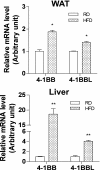
FIG. 1.
FIG. 1.
HFD upregulates 4-1BB and 4-1BBL gene expression in adipose tissue and liver. C57BL/6 mice were fed an HFD or RD for 9 weeks. Levels of the mRNAs of 4-1BB and 4-1BBL in the epididymal adipose tissue (upper) and liver (lower) of mice fed an RD or HFD. Levels of mRNA were estimated by quantitative PCR. Results are means ± SEM (n = 4 mice per group). *P < 0.05, **P < 0.01 compared with RD. WAT, white adipose tissue.
FIG. 2.
FIG. 2.
Body weight change and adiposity in HFD-fed 4-1BB–deficient mice. WT and 4-1BB–deficient mice were fed an HFD for 9 weeks. A: Expression of 4-1BB mRNA in epididymal adipose tissue, liver, and skeletal muscle. Body weight changes and gross morphology of mice (B), energy intake (C), and adipose tissue weight (Ep, epididymal; Re, retroperitoneal; Me, mesenteric; and Sc, subcutaneous) of WT (n = 8) and 4-1BB–deficient mice (n = 8) fed an RD or HFD, and gross morphology of adipose tissues (D). Results are means ± SEM. *P < 0.05, **P < 0.01, #P < 0.005 compared with WT mice fed an HFD. E: Histological analysis of epididymal adipose tissue and size distribution of adipocytes from WT and 4-1BB–deficient mice fed an HFD. Sections were stained with hematoxylin-eosin. Hypertrophied adipocytes are indicated by asterisks. Original magnification is ×200 (scale bar = 50 μm). The sizes of adipocytes in randomly chosen fields were measured with a microscope (magnification ×200) and calculated using Axiovision AC software. #P < 0.005 compared with WT mice fed an HFD. WAT, white adipose tissue. (A high-quality color representation of this figure is available in the online issue.)
FIG. 3.
FIG. 3.
Energy expenditure, locomotor activity, and body temperature in 4-1BB–deficient mice. A: Energy expenditure (EE) was measured in WT (n = 4) and 4-1BB–deficient mice (n = 4) fed an HFD. Locomotor activity (B) and body temperature (C) were measured in WT (n = 10–11) and 4-1BB–deficient mice (n = 7) fed an RD or HFD. Results are means ± SEM. *P < 0.05, ##P < 0.001 compared with WT mice fed an HFD. D: Levels of UCP-1 protein in BAT. Levels of protein were determined by Western blotting. The intensities of UCP-1 protein were normalized to those of β-actin and are expressed as means ± SEM of 4 mice per group. *P < 0.05 compared with WT mice fed an HFD. E: Histological analysis of BAT from WT and 4-1BB–deficient mice fed an HFD (hematoxylin-eosin). Original magnification is ×200 (scale bar = 50 μm). (A high-quality color representation of this figure is available in the online issue.)
FIG. 4.
FIG. 4.
Adipose tissue macrophages and T cells in HFD-fed 4-1BB–deficient mice. A: Histological analysis of epididymal adipose tissue from WT and 4-1BB–deficient mice fed an HFD. Sections were stained with hematoxylin-eosin (H&E) in epididymal adipose tissue from WT and 4-1BB–deficient mice fed an HFD. Stained cells are indicated by arrows. Original magnifications are ×200 (upper) and ×400 (lower) (scale bar = 50 μm). FACS quantification of immune cell population and numbers in visceral adipose SVF from WT and 4-1BB–deficient mice fed an HFD. SVFs were double stained with fluorescein isothiocyanate–conjugated phycoerythrin-conjugated anti-CD4 (helper T cell)/anti-CD8 (cytotoxic T cell), anti-F4/80/CD11b (macrophage), anti-CD4 (or CD8)/CD44high (activated T cell), and anti-CD4 (or CD8)/CD62Llow (activated T cell). B: The values in the panels indicate the percentages of each cell population. C: The total immune cell numbers in adipose tissue. Results are mean ± SEM. *P < 0.05, **P < 0.01 compared with WT mice fed an HFD. (A high-quality color representation of this figure is available in the online issue.)
FIG. 5.
FIG. 5.
Adipose tissue inflammatory responses in HFD-fed 4-1BB–deficient mice. Concentrations of inflammatory proteins (TNF-α, IL-6, and MCP-1) (A) and an anti-inflammatory protein (adiponectin) (B) in adipose tissue from WT (n = 8) and 4-1BB–deficient mice (n = 8) fed an HFD. Adipose tissue (0.5 g) was homogenized with 1 mL of 100 mmol/L Tris-HCl and 250 mmol/L sucrose buffer (pH 7.4) supplemented with protease inhibitors. Lipids were removed by centrifugation at 10,000g for 10 min. Levels of cytokines/adipokines in homogenates were measured by enzyme-linked immunosorbent assay and normalized for protein content. Levels of HMW adiponectin were assessed in plasma samples from WT (n = 8) and 4-1BB–deficient mice (n = 8). Results are mean ± SEM. *P < 0.05, #P < 0.005 compared with WT mice fed an HFD. C: NF-κB activation in adipose tissue was determined using the p65 TransAM assay as described in research design and methods. Results are mean ± SEM (n = 6 mice per group). ##P < 0.001 compared with WT mice fed an HFD.
FIG. 6.
FIG. 6.
Deficiency of 4-1BB ameliorates insulin resistance and improves insulin signaling in mice fed an HFD. A: Fasting glucose and insulin levels. B: Plasma TG and total cholesterol levels in WT and 4-1BB–deficient mice fed an RD or HFD. Results are means ± SEM (n = 5–6 mice per group). *P < 0.05, **P < 0.01 compared with WT mice fed an HFD. C: Glucose tolerance tests. Mice fed an HFD for 7 weeks were fasted for 5 h before receiving an oral administration of 20% glucose solution at a dose of 2 g/kg, and blood samples were taken at the indicated times (n = 5). D: Insulin tolerance tests. Mice fed an HFD for 7 weeks were fasted for 5 h before receiving an intraperitoneal injection of 0.75 units/kg insulin, and blood samples were taken at the indicated times (n = 5). Results are means ± SEM. *P < 0.05, **P < 0.01, #P < 0.005 compared with WT mice fed an HFD. E: Western blots of phosphorylated Akt (p-Akt) and total Akt in adipose tissue, liver, and skeletal muscle from WT (n = 4) and 4-1BB–deficient mice (n = 4) fed an HFD. Mice were fasted for 5 h before receiving a 10 mU/g i.p. insulin injection and killed 4 min later, and tissues were collected for Western blotting. WAT, white adipose tissue. (A high-quality color representation of this figure is available in the online issue.)
FIG. 7.
FIG. 7.
Deficiency of 4-1BB ameliorates hepatic steatosis. A: Gross morphology and histological analysis (hematoxylin-eosin) of livers from WT and 4-1BB–deficient mice fed an HFD. Original magnification is ×200 (scale bar = 50 μm). B: Levels of TG content of livers from WT (n = 4) and 4-1BB–deficient mice (n = 4) fed an HFD. Results are means ± SEM. ##P < 0.001 compared with WT mice fed an HFD. C: Expression of lipogenic genes (SREBP-1, ACC1, and FAS) in livers from WT (n = 4) and 4-1BB–deficient mice (n = 4) fed an HFD. Levels of mRNA were estimated by quantitative PCR. Results are means ± SEM. *P < 0.05 compared with WT mice fed an HFD. D: Western blots of phosphorylated AMPK (p-AMPK), acetyl-CoA carboxylase (ACC), and PPAR-α in livers from WT and 4-1BB–deficient mice fed an HFD. The intensity of the bands was quantified by densitometry and is expressed as means ± SEM (n = 4 mice per group). **P < 0.05 compared with WT mice fed an HFD. E: Levels of inflammatory proteins (TNF-α, IL-6, and MCP-1) in livers from WT (n = 8) and 4-1BB–deficient mice (n = 8) fed an HFD. Levels of cytokines/chemokines in liver were measured by enzyme-linked immunosorbent assay. Results are means ± SEM. *P < 0.05, **P < 0.01 compared with WT mice fed an HFD. (A high-quality color representation of this figure is available in the online issue.)

Comment in

Similar articles

Cited by

References

    1. Hotamisligil GS, Shargill NS, Spiegelman BM. Adipose expression of tumor necrosis factor-alpha: direct role in obesity-linked insulin resistance. Science 1993;259:87–91 - PubMed
    1. Shoelson SE, Lee J, Goldfine AB. Inflammation and insulin resistance. J Clin Invest 2006;116:1793–1801 - PMC - PubMed
    1. Hotamisligil GS. Inflammation and metabolic disorders. Nature 2006;444:860–867 - PubMed
    1. Guilherme A, Virbasius JV, Puri V, Czech MP. Adipocyte dysfunctions linking obesity to insulin resistance and type 2 diabetes. Nat Rev Mol Cell Biol 2008;9:367–377 - PMC - PubMed
    1. Weisberg SP, McCann D, Desai M, Rosenbaum M, Leibel RL, Ferrante AW., Jr Obesity is associated with macrophage accumulation in adipose tissue. J Clin Invest 2003;112:1796–1808 - PMC - PubMed

Publication types

MeSH terms