. 2011 Oct 12;3(104):104ra100.
doi: 10.1126/scitranslmed.3002949.

Human TH17 cells are long-lived effector memory cells

Affiliations

Human TH17 cells are long-lived effector memory cells

Ilona Kryczek et al. Sci Transl Med. .

Abstract

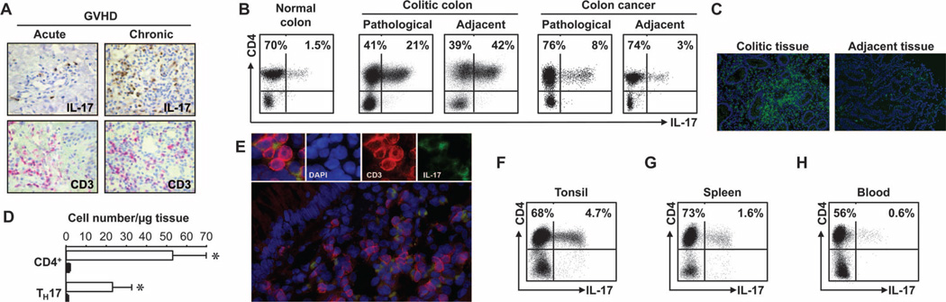
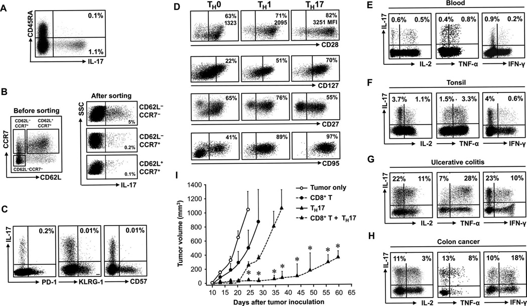
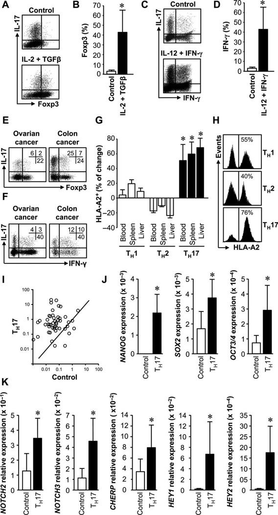
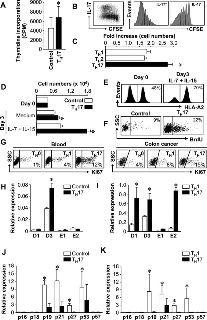
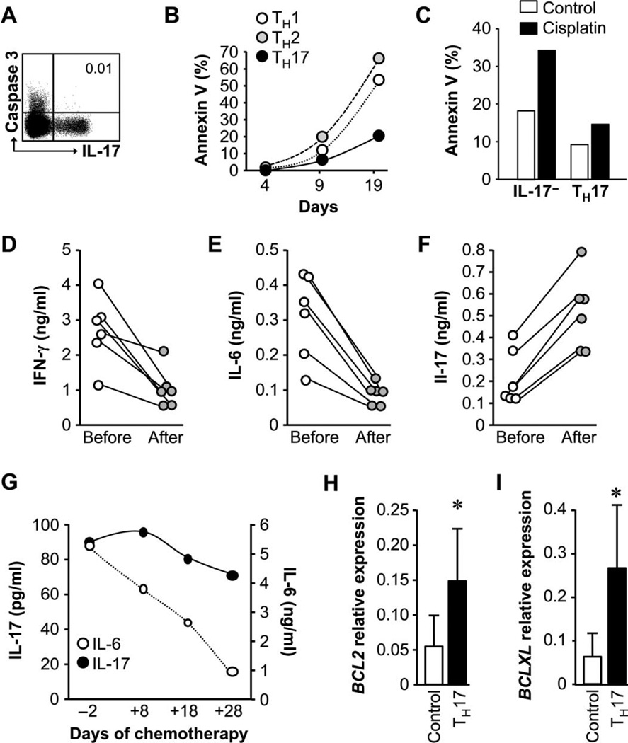
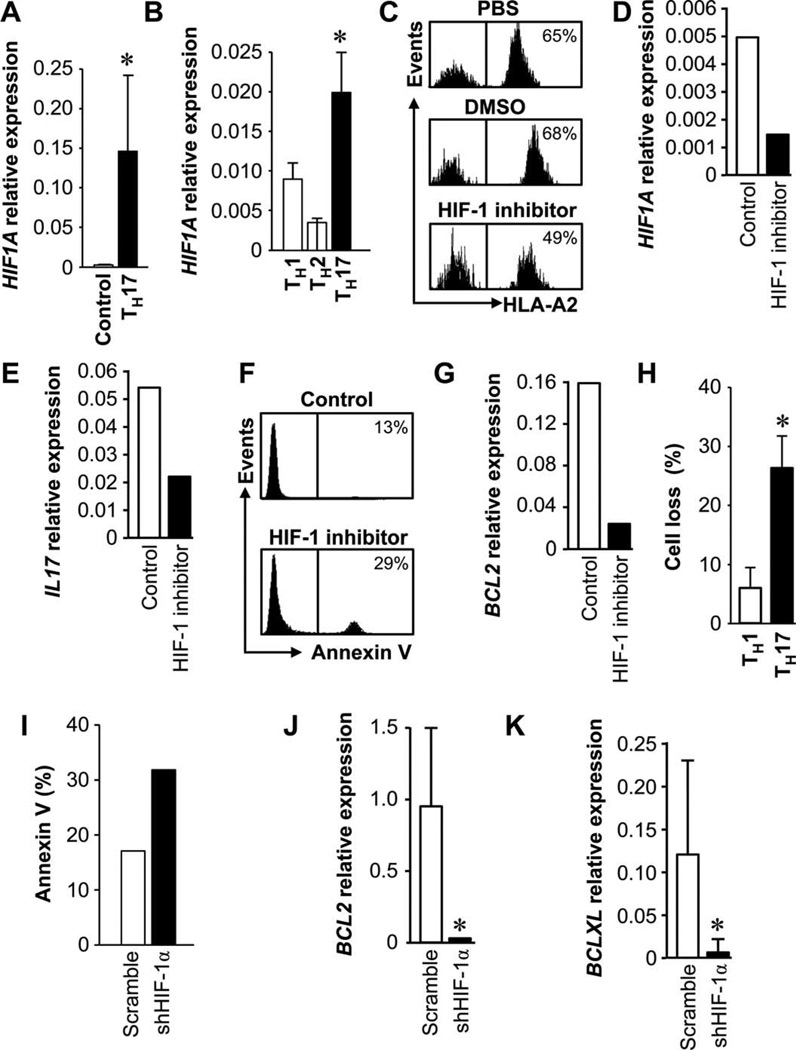
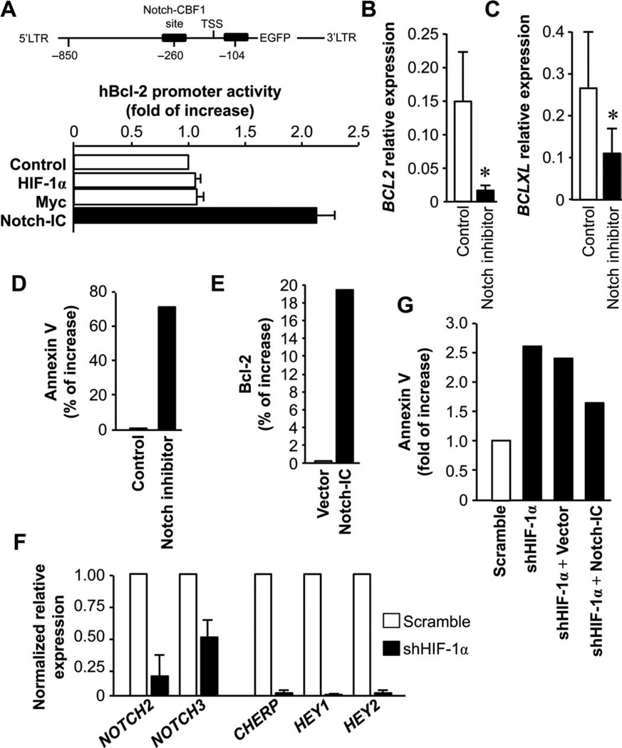
T helper 17 (TH17) cells have been shown to contribute to multiple disease systems. However, the functional phenotype and survival pattern of TH17 cells as well as the underlying mechanisms that control TH17 cells have been poorly investigated in humans, significantly hampering the clinical targeting of these cells. Here, we studied human TH17 cells in the pathological microenvironments of graft-versus-host disease, ulcerative colitis, and cancer; TH17 cell numbers were increased in the chronic phase of these diseases. Human TH17 cells phenotypically resembled terminally differentiated memory T cells but were distinct from central memory, exhausted, and senescent T cells. Despite their phenotypic markers of terminal differentiation, TH17 cells mediated and promoted long-term antitumor immunity in in vivo adoptive transfer experiments. Furthermore, TH17 cells had a high capacity for proliferative self-renewal, potent persistence, and apoptotic resistance in vivo, as well as plasticity-converting into other types of TH cells. These cells expressed a relatively specific gene signature including abundant antiapoptotic genes. We found that hypoxia-inducible factor-1α and Notch collaboratively controlled key antiapoptosis Bcl-2 family gene expression and function in TH17 cells. Together, these data indicate that human TH17 cells may be a long-lived proliferating effector memory T cell population with unique genetic and functional characteristics. Targeting TH17-associated signaling pathway would be therapeutically meaningful for treating patients with autoimmune disease and advanced tumor.


Figures

Fig. 1
Fig. 1
High amounts of TH17 cells are found in sites of chronic diseases. Case numbers: blood (31), oral mucosa (6), spleen (5), tonsil (6), colon cancer (21), and colitis (12). (A) High numbers of IL-17+ cells were detected in oral mucosa tissues in patients with GVHD. Consecutive sections of oral mucosa biopsies were stained for IL-17 (brown) or CD3 (red). P < 0.001, acute versus chronic. (B) High percentages of TH17 cells were found in colitic colon and colon cancer tissues. Tissue single cells were stained for TH17 cell markers, analyzed by flow cytometry, and gated on CD3+ cells. (C and D) CD4+ and TH17 cells were found in colitic colon tissues. Colon tissues were stained for CD3 (green) (C). Absolute numbers of CD4+ and TH17 cells were quantified based on flow cytometry analysis. Results are expressed as the absolute numbers of CD4+ or TH17 per microgram tissue ± SEM (D). P < 0.001, colitic lesion (empty bars) versus adjacent tissue (filled bars). (E) Colon tissues were stained for CD3 and IL-17, and analyzed with a fluorescence microscope. Red, CD3; green, IL-17. DAPI, 4′,6-diamidino-2-phenylindole. (F to H) TH17 cells were found in different organs. Single cells were stained for TH17 cell markers and analyzed by flow cytometry. P < 0.001, tonsil and spleen as compared with blood.
Fig. 2
Fig. 2
Primary TH17 cells exhibit a terminally differentiated phenotype but mediate potent antitumor immunity. (A to D) Tissue or blood cells were stained for the indicated markers and cytokines and analyzed by flow cytometry. Results are expressed as the percentage of the specific subset of CD4+ T cells. n = 10 to 25. (A) TH17 cells were in the CD45RO+CD45RA memory T subset. (B) Relationship between TH17 cells and expression of CCR7 and CD62L. Three CD4+ T cell subsets were sorted and stained for IL-17. (C) Relationship between TH17 cells and expression of CD57, KLRG-1, and PD-1. (D) Expression of the described markers on primary TH0, TH1, and TH17 cells. P < 0.001, for CD28 and CD95 expression on TH17 as compared with TH1 and TH0 cells, and P < 0.05, for CD127 and CD27 expression on TH17 as compared with TH1 and TH0 cells. (E to H) Effector cytokine profile of primary TH17 cells in different tissues gated on CD4+ T cells. (I) TH17 cells mediate and promote tumor regression in vivo. Ovarian cancer–bearing NSG mice (16, 17) received PBS, TH17, and/or CD8+ T cells. Mean ± SEM of tumor volumes are shown (n = 4 to 6 mice per group). *P < 0.05.
Fig. 3
Fig. 3
TH17 cells have functional and genetic stem cell–like features. (A to D) TH17 cells gave rise to other TH cells. Primary TH17 cells were stimulated with the cytokine milieu for Treg (A and B) or TH1 (C and D) induction for 3 days and analyzed by flow cytometry. Results are expressed as the percentage of Foxp3+ (B) or IFN-g+ cells (D) in TH17 cells. n = 5. *P < 0.05. (E and F) IL-17+Foxp3+(E) and IL-17+IFN-γ+(F) T cells in cancers. Colon (n = 9) and ovarian cancer (n = 25) T cells were analyzed by flow cytometry. (G and H) TH17 cells had superior in vivo persistence. HLA-A2+ TH subsets were mixed with HLA-A2IL-17CD4+ T cells and transfused to NSG mice. On day 5, human T cells were analyzed. (I) The percentage of changes between the initial and the recovered ratios of HLA-A2+ and HLA-A2 cells was calculated. Three paired donors. *P < 0.001. (J) Representative histogram shows different T subsets (HLA-A2+) in spleen. (I to K) TH17 cells expressed stem cell genes. SuperArray was performed in primary TH17 and controls (I). Selective stem cell genes were quantified (J and K). n = 8. P < 0.05.
Fig. 4
Fig. 4
TH17 cells have increased proliferative capacity. (A to C) TH17 cells had potent expansion capacity. Primary (A and B) and polarized TH subsets (C) were stimulated with TCR for 3 (A and C) or 14 (B) days. Cell proliferation/expansion was detected by [3H]thymidine incorporation (A), CFSE dilution (B), or increased cell numbers (C). n = 3. *P < 0.05. (D and E) Cytokines stimulated TH17 expansion. Primary HLA-A2+ TH17 and HLA-A2IL-17 control T cells were separately cultured (D) or initially mixed and cocultured (E) for 3 days with IL-7 plus IL-15. The absolute cell numbers (D) or the percentage (E) of TH17 cells was determined by flow cytometry. n = 3. *P < 0.05. (F) TH17 cells were in S phase. NSG mice received TH17 or control cells and BrdU. Cell cycling phase was analyzed on day 5 by flow cytometry to determine BrdU+ human T cells in mouse spleen. n = 2. (G) TH17 cells expressed increased Ki67 expression in blood and colon cancer. Eight to 10 donors. P < 0.05, TH17 as compared with TH1/TH0. (H to K) TH17 cells expressed different amounts of cell cycling genes. Expression of multiple cyclin genes (H and I) and CDK inhibitors (J and K) was quantified in primary (H and J) and polarized TH17 cells (I and K). n = 8. *P < 0.05.
Fig. 5
Fig. 5
TH17 cells are resistant to apoptosis. (A) TH17 cells did not express caspase 3. The expression of caspase 3 in T cells was analyzed by flow cytometry. n = 4. (B) TH17 cells were resistant to apoptosis induced by TCR activation. T subsets were stimulated with anti-CD3, and cell apoptosis was analyzed with annexin V staining. n = 5. P < 0.01, TH17 as compared with TH1/TH2 on day 19. (C) TH17 cells were resistant to apoptosis induced by in vitro cisplatin. Primary TH17 and control cells were treated with cisplatin for 48 hours. Annexin V+ cells were analyzed. n = 4. P < 0.01. (D to F) Chemotherapy increased IL-17 production. Ovarian cancer patients received one cycle of cisplatin therapy. Blood mononuclear cells were activated, and cytokines were measured in the supernatants with enzyme-linked immunosorbent assay (ELISA). n = 6. P < 0.001. (G) Chemotherapy reduced IL-6 but not IL-17 production in ovarian cancer ascites. Ovarian cancer patient was treated with cisplatin. Cytokines were measured in the ascites fluid by ELISA. Left scale shows IL-17 (filled circles), and right scale shows IL-6 (empty circles). (H and I) TH17 cells spontaneously expressed high levels of BCL2 family genes. Real-time PCR was performed in primary TH17 and control cells for BCL2 (H) and BCLXL (I). n = 5. *P < 0.001.
Fig. 6
Fig. 6
HIF-1α regulates TH17 cell apoptosis and persistence. (A and B) TH17 cells expressed HIF1A. HIF1A expression was quantified by real-time PCR in primary TH17 (A) and polarized T cell subsets (B). n = 5 to 8. *P < 0.05. (C to E) HIF-1 blockade reduced TH17 persistence in vivo. Primary HLA-A2+ TH17 cells and HLA-A2IL-17CD4+ T cells were mixed and pretreated with PBS, DMSO, and the HIF-1 inhibitor echinomycin for 48 hours and transfused to NSG mice. Human T cells were stained and analyzed for HLA-A2 and human CD5 (C), and human HIF1A (D) and IL17A (E) were quantified in the spleen. n = 5. (F and G) HIF-1 blockade increased TH17 apoptosis in vivo. Primary TH17 cells were pretreated with HIF-1 inhibitor or DMSO and transferred to NSG mice. After 2 days, annexin V expression (F) and BCL2 gene expression (G) were analyzed in human T cells in mouse spleen. n = 6. P < 0.05. (H) HIF-1 blockade increased TH17 apoptosis in vitro. Enriched primary TH17 cells were transfected with lentiviral vector encoding shHIF-1α and then activated with TCR for 3 days. TH17 and TH1 cell numbers were counted, and the percentage of cell loss was calculated. n = 3. P < 0.05. (I to K) HIF-1 blockade increased TH17 apoptosis induced by chemotherapy. Primary TH17 cells were transfected with lentiviral vector encoding shHIF-1 or scramble and cultured with cisplatin for 48 hours. Cell apoptosis was analyzed with annexin V expression (I). BCL2 (J) and BCLXL (K) genes were quantified. n = 7. P < 0.01.
Fig. 7
Fig. 7
Notch and Bcl-2 family regulate the stem cell-like feature of the TH17 cells. (A) Notch bound to and activated the Bcl-2 promoter. Black boxes show Notch binding sites in the BCL2 promoter (upper panel). Human embryonic kidney (HEK) 293 cells were cotransfected with the hBCL2-EGFP (enhanced green fluorescent protein) promoter with the indicated plasmids for 38 hours. The intensity of EGFP was measured with flow cytometry. LTR, long terminal repeat; TSS, transcription start site. (B and C) Notch blockade suppressed BCL2 and BCLXL expression. Primary T cells were treated with Notch inhibitor for 24 hours. BCL2 (B) and BCLXL (C) expression was quantified by real-time PCR. n = 3. P < 0.05. (D) Notch blockade increased TH17 cell apoptosis. Primary TH17 cells were cultured with the Notch inhibitor for 3 days. Annexin V expression was analyzed by flow cytometry. n = 3. (E) Notch activation increased BCL2 expression on TH17 cells. Primary TH17 cells were transfected with Notch-IC. After 24 hours, Bcl-2 expression was analyzed by flow cytometry. n = 3. (F) HIF blockade reduced Notch signaling gene expression in TH17 cells. Primary TH17 cells were transfected with shHIF-1α. After 24 hours, Notch signaling gene expression was quantified. n = 3. (G) Notch activation rescued TH17 cell apoptosis induced by HIF blockade. Primary TH17 cells were transfected with lentiviral vectors encoding shHIF-1α and/or Notch-IC. After 24 hours, annexin V expression was analyzed by flow cytometry. n = 3.

Similar articles

Cited by

References

    1. Dong C. Diversification of T-helper-cell lineages: Finding the family root of IL-17-producing cells. Nat. Rev. Immunol. 2006;6:329–333. - PubMed
    1. Harrington LE, Hatton RD, Mangan PR, Turner H, Murphy TL, Murphy KM, Weaver CT. Interleukin 17–producing CD4+ effector T cells develop via a lineage distinct from the T helper type 1 and 2 lineages. Nat. Immunol. 2005;6:1123–1132. - PubMed
    1. Kolls JK, Lindén A. Interleukin-17 family members and inflammation. Immunity. 2004;21:467–476. - PubMed
    1. Veldhoen M, Hocking RJ, Atkins CJ, Locksley RM, Stockinger B. TGFβ in the context of an inflammatory cytokine milieu supports de novo differentiation of IL-17-producing T cells. Immunity. 2006;24:179–189. - PubMed
    1. Kleinschek MA, Boniface K, Sadekova S, Grein J, Murphy EE, Turner SP, Raskin L, Desai B, Faubion WA, de Waal Malefyt R, Pierce RH, McClanahan T, Kastelein RA. Circulating and gut-resident human Th17 cells express CD161 and promote intestinal inflammation. J. Exp. Med. 2009;206:525–534. - PMC - PubMed

Publication types

Substances

LinkOut - more resources