. 2011 Oct;7(10):e1002179.
doi: 10.1371/journal.pcbi.1002179. Epub 2011 Oct 6.

Computational modeling of allosteric communication reveals organizing principles of mutation-induced signaling in ABL and EGFR kinases

Affiliations

Computational modeling of allosteric communication reveals organizing principles of mutation-induced signaling in ABL and EGFR kinases

Anshuman Dixit et al. PLoS Comput Biol. 2011 Oct.

Abstract

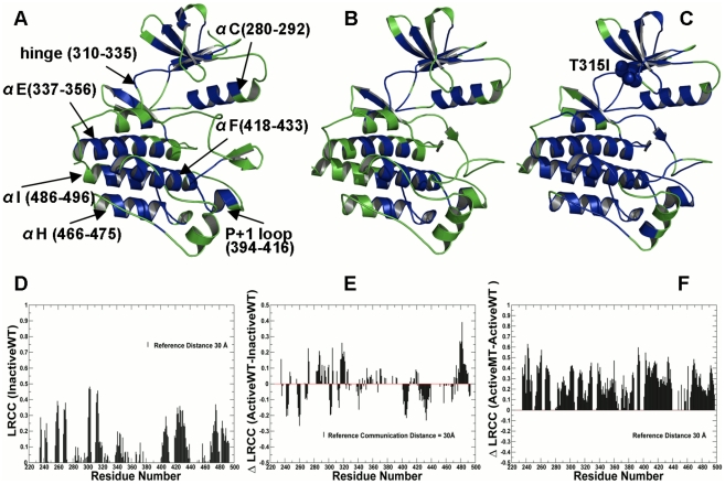
The emerging structural information about allosteric kinase complexes and the growing number of allosteric inhibitors call for a systematic strategy to delineate and classify mechanisms of allosteric regulation and long-range communication that control kinase activity. In this work, we have investigated mechanistic aspects of long-range communications in ABL and EGFR kinases based on the results of multiscale simulations of regulatory complexes and computational modeling of signal propagation in proteins. These approaches have been systematically employed to elucidate organizing molecular principles of allosteric signaling in the ABL and EGFR multi-domain regulatory complexes and analyze allosteric signatures of the gate-keeper cancer mutations. We have presented evidence that mechanisms of allosteric activation may have universally evolved in the ABL and EGFR regulatory complexes as a product of a functional cross-talk between the organizing αF-helix and conformationally adaptive αI-helix and αC-helix. These structural elements form a dynamic network of efficiently communicated clusters that may control the long-range interdomain coupling and allosteric activation. The results of this study have unveiled a unifying effect of the gate-keeper cancer mutations as catalysts of kinase activation, leading to the enhanced long-range communication among allosterically coupled segments and stabilization of the active kinase form. The results of this study can reconcile recent experimental studies of allosteric inhibition and long-range cooperativity between binding sites in protein kinases. The presented study offers a novel molecular insight into mechanistic aspects of allosteric kinase signaling and provides a quantitative picture of activation mechanisms in protein kinases at the atomic level.


Conflict of interest statement

The authors have declared that no competing interests exist.

Figures

Figure 1
Figure 1. Structural Mapping of Allosteric Communication Profiles in the ABL Kinase Catalytic Domain.
Structural mapping of residues involved in long-range communication in different functional states of the ABL catalytic domain: (A) the inactive ABL-WT structure (PDB ID 1IEP); (B) the active ABL-WT structure (PDB ID 1M52); (C) the active form of the ABL-T315I mutant (PDB ID 2Z60). The catalytic core is shown in green. The highlighted in blue allosterically coupled clusters correspond to the peaks in the residue-based LRCC profile computed with the reference communication threshold of 30 Å. The kinase segments and corresponding residue ranges are indicated by respective arrows. (D) The absolute LRCC values of the inactive ABL-WT form. (E) The relative LRCC values between the active and inactive ABL-WT forms. (F) The relative LRCC values between the active ABL-T315I and active ABL-WT. Each bin refers to a residue and shows the fraction of residues that efficiently communicate with this particular residue at distances greater the reference communication threshold of 30 Å.
Figure 2
Figure 2. Structural Mapping of Allosteric Communication Profiles in the ABL Regulatory Complexes.
The crystal structures of the ABL-SH2-SH3 complex in the inactive, autoinhibited form (PDB ID 2FO0, colored in green) (A) and the active form (PDB ID 1OPL, colored in green) (B). Structural mapping of the catalytic domain regions involved in the allosteric communication (in blue) correspond to the peaks in the residue-based LRCC profile computed with the reference communication threshold of 30 Å. (C) The relative LRCC between the inactive ABL-T334I and the inactive ABL-WT. (D) The relative LRCC between the active ABL-T334I and the active ABL-WT.
Figure 3
Figure 3. Structure-Functional Analysis of the Inter-Domain Interface in the ABL Regulatory Complexes.
(A) An overview of the inter-domain interface in the crystal structure of the inactive ABL complex. The crystal structure is colored according to the protein flexibility (light blue corresponds to more flexible regions, dark blue corresponds to structurally rigid regions). The residues involved in the interdomain interface are shown in green sticks. (B) A close-up view of the interdomain interface between the kinase catalytic core and the SH2 domain. The critical residues are shown in green sticks. (C) The occupancy of the interdomain interface contacts in the inactive, down-regulated form of the ABL-SH2-SH3 complex. (D) The occupancy of the interdomain interface contacts in the active form of the ABL-SH2-SH3 complex. The occupancies of the interdomain interface contacts are shown in blue filled bars for the ABL-WT and in red filled bars for the ABL-T334I mutant.
Figure 4
Figure 4. Structural Mapping of Allosteric Communication Profiles in the EGFR Kinase Catalytic Domain.
Structural mapping of residues involved in long-range communication in different functional states of the EGFR catalytic domain: (A) the inactive EGFR-WT structure (PDB ID 1XKK); (B) the active EGFR-WT structure (PDB ID 2J6M); (C) the active form of the EGFR-T790M mutant (PDB ID 2JIT). The catalytic core is shown in green. The highlighted in blue are allosterically coupled clusters correspond to the peaks in the LRCC profile computed with the reference communication threshold of 30 Å. The kinase segments and corresponding residue ranges are indicated by respective arrows. (D) The absolute LRCC values of the inactive ABL-WT form. (E) The relative LRCC values between the active and inactive EGFR-WT forms. (F) The relative LRCC values between the active EGFR-T790M mutant and inactive EGFR-WT forms.
Figure 5
Figure 5. MD Simulations of the EGFR Regulatory Dimers.
20 ns MD simulations were performed for both a symmetric EGFR and asymmetric dimers in the WT and mutant forms. Upper Panel: The RMSD fluctuations of the Cα atoms (A) and the RMSF values of the Cα atoms (B) obtained from MD simulations of a symmetric EGFR dimer (EGFR-WT shown in blue, EGFR-T766M shown in red). Lower Panel: The RMSD fluctuations of the Cα atoms (A) and the RMSF values of the Cα atoms (B) obtained from MD simulations of an asymmetric active EGFR dimer (EGFR-WT shown in blue, EGFR-T766M shown in red).
Figure 6
Figure 6. Allosteric signaling in the EGFR Kinase Regulatory Dimers: The Intra-monomer Communications.
Structural mapping of the kinase residues involved in the efficient long-range communications at the reference communication distance of 30 Å is shown for an asymmetric dimer (A) and a symmetric dimer (B). The reference communication distance of 30 Å was used to analyze primarily long-range intra—domain (intra-monomer) communications and interfacial inter-monomer communications. The depicted mapping and analysis of allosteric communications is based on simulations of the crystal structure of an asymmetric EGFR dimer (PDB ID 2GS6) and inactive symmetric dimer (PDB ID 2GS7). The crystal structures include the monomer A (receiver molecule, colored in green) and the monomer B (activator molecule, colored in pink). The highlighted in blue are allosterically coupled regions. The kinase segments are pointed to by respective arrows. (C) The relative LRCC values computed between an asymmetric EGFR-WT dimer and a symmetric EGFR-WT. (D) The relative LRCC values computed between an asymmetric EGFR-T766M dimer and a symmetric EGFR-WT. The LRCC values were plotted as respective bars depicted for the monomer A in blue and the monomer B in red.
Figure 7
Figure 7. Mutation-Induced Allosteric Signaling in the EGFR Kinase Regulatory Dimers: Analysis of Long-Range Communications between EGFR Monomers.
Structural mapping of the kinase residues involved in the efficient long-range communications at the reference communication distance of 60 Å is shown for an asymmetric dimer (A) and a symmetric dimer (B). We adopted a reference distance of 60 Å to highlight the effect and contribution of the inter-monomer communications. The highlighted in blue are allosterically coupled regions. A schematic representation of allosteric coupling between monomers in an asymmetric EGFR dimer is depicted in blue spheres. The crystal structures include the monomer A (receiver colored in green) and the monomer B (activator colored in pink). (C) The relative LRCC values computed between an asymmetric EGFR-WT dimer and a symmetric EGFR-WT (D) The relative LRCC values computed between an asymmetric EGFR-T766M dimer and a symmetric EGFR-WT. The LRCC values were plotted as respective bars depicted for the monomer A (receiver molecule, in blue) and the monomer B (activator molecule, in red).
Figure 8
Figure 8. Structural Analysis of the Inter-monomer Interface in Functional Asymmetric EGFR Dimer.
(A) An overview of the inter-monomer interface in the crystal structure of an asymmetric EGFR dimer (PDB ID 2GS6). The monomer A (receiver molecule) is shown in green and the monomer B (activator molecule) is shown in pink. The mutational site T766M is in green spheres. The interdomain interface is indicated by a rectangular. (B) A detailed close-up view of the inter-monomer interface between the N-terminal αC-helix of the receiver monomer A (in green) and the C-terminal αH-helix and αI-helix of the activator monomer B (in pink). (C) The occupancy of the critical interdomain interface contacts in an asymmetric EGFR dimer. The occupancies for the EGFR-WT are shown in blue bars and for the EGFR-T766M mutant in red bars.
Figure 9
Figure 9. Structural Analysis of the Inter-monomer Interface in a Symmetric EGFR Dimer.
(A) An overview of the inter-monomer interface in the crystal structure of a symmetric inactive EGFR dimer (PDB ID 2GS7). The monomer A (receiver molecule) is shown in green and the monomer B (activator molecule) is shown in pink. The primary interdomain interface is indicated by a rectangular. (B) An exploded view of the electrostatic inter-monomer interface formed between the N-terminal lobe of the receiver monomer A (shown in green), residues from the “electrostatic hook (shown in magenta) and the C-terminal αI-helix and the αE-helix of the activator monomer B (in pink). (C) The occupancy of the critical interdomain interface contacts in a symmetric EGFR dimer. The occupancies for the EGFR-WT are shown in blue filled bars and for the EGFR-T766M mutant in red filled bars.
Figure 10
Figure 10. Allosteric Communications and Stabilizing Interactions in an Extended Structure of a Symmetric EGFR Dimer.
Structural mapping of the kinase regions involved in long-range communications (highlighted in blue) at the reference communication distance of 30 Å (A) and 60 Å (B). The monomer A (receiver molecule) is shown in green and the monomer B (activator molecule) is shown in pink. The extended crystal structure of the inactive symmetric dimer (PDB ID 3GT8) was used in simulations and allosteric analysis. An overview of the inter-monomer interface is indicated by a rectangular in (B). (C) The occupancy of the critical interdomain interface contacts in the symmetric EGFR dimer. The occupancies for the EGFR-WT are shown in blue bars and for the EGFR-T766M mutant in red bars. (D) An exploded view of the electrostatic hook formed between the C-terminal tail residues 979–990 (shown in pink) and the hinge region in the receiver kinase domain (shown in green).

Similar articles

Cited by

References

    1. Koshland DE, Némethy G, Filmer D. Comparison of experimental binding data and theoretical models in proteins containing subunits. Biochemistry. 1966;5:365–385. - PubMed
    1. Monod J, Wyman J, Changeux JP. On the nature of allosteric transitions: A plausible model. J Mol Biol. 1965;12:88–118. - PubMed
    1. Cui Q, Karplus M. Allostery and cooperativity revisited. Protein Sci. 2008;17:1295–1307. - PMC - PubMed
    1. Goodey NM, Benkovic SJ. Allosteric regulation and catalysis emerge via a common route. Nat Chem Biol. 2008;4:474–482. - PubMed
    1. Tsai CJ, Sol AD, Nussinov R. Allostery: absence of a change in shape does not imply that allostery is not at play. J Mol Biol. 2008;378:1–11. - PMC - PubMed

Publication types

MeSH terms

Substances