. 2011 Oct;7(10):e1002288.
doi: 10.1371/journal.ppat.1002288. Epub 2011 Oct 6.

Homeostatic proliferation fails to efficiently reactivate HIV-1 latently infected central memory CD4+ T cells

Affiliations

Homeostatic proliferation fails to efficiently reactivate HIV-1 latently infected central memory CD4+ T cells

Alberto Bosque et al. PLoS Pathog. 2011 Oct.

Abstract

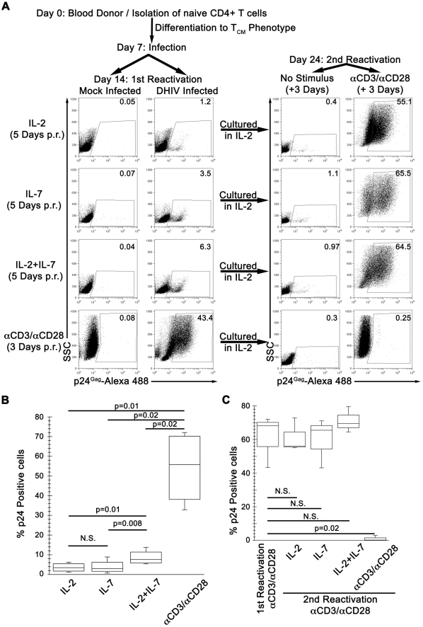
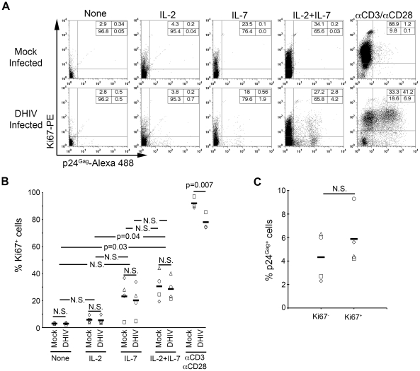
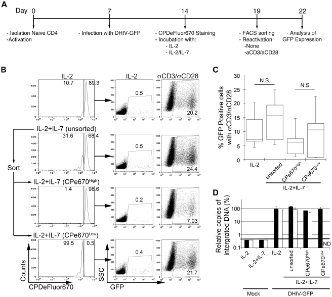
Homeostatic proliferation ensures the longevity of central memory T-cells by inducing cell proliferation in the absence of cellular differentiation or activation. This process is governed mainly by IL-7. Central memory T-cells can also be stimulated via engagement of the T-cell receptor, leading to cell proliferation but also activation and differentiation. Using an in vitro model of HIV-1 latency, we have examined in detail the effects of homeostatic proliferation on latently infected central memory T cells. We have also used antigenic stimulation via anti-CD3/anti-CD28 antibodies and established a comparison with a homeostatic proliferation stimulus, to evaluate potential differences in how either treatment affects the dynamics of latent virus populations. First, we show that homeostatic proliferation, as induced by a combination of IL-2 plus IL-7, leads to partial reactivation of latent HIV-1 but is unable to reduce the size of the reservoir in vitro. Second, latently infected cells are able to homeostatically proliferate in the absence of viral reactivation or cell differentiation. These results indicate that IL-2 plus IL-7 may induce a detrimental effect by favoring the maintenance of the latent HIV-1 reservoir. On the other hand, antigenic stimulation efficiently reactivated latent HIV-1 in cultured central memory cells and led to depletion of the latently infected cells via virus-induced cell death.


Conflict of interest statement

The authors have declared that no competing interests exist.

Figures

Figure 1
Figure 1. IL-7 induces partial reactivation of latent HIV-1 in cultured TCM.
(A) Mock or DHIV latently infected, cultured TCM were incubated in the presence of IL-2, IL-7 or a combination of IL-2 and IL-7 (IL-2+IL-7) or costimulated with antibodies to CD3 and CD28 (αCD3/αCD28) and assessed for intracellular p24Gag by flow cytometry (1st Reactivation). The percentage of p24Gag positive cells is indicated in each panel. After reactivation, cells were kept in culture for an additional 7-day period in the presence of IL-2. At day 24, cells were incubated in the presence of IL-2 or costimulated with antibodies to CD3 and CD28 (αCD3/αCD28) and assessed for intracellular p24Gag by flow cytometry (2nd Reactivation). Data is representative of 4 donors for 1st reactivation and 3 donors for 2nd reactivation (p.r. post reactivation). (B) Box-plots corresponding to the 4 donors analyzed as in A, left panels. Horizontal lines indicate median values; statistical significance was assessed by 2-tailed paired-sample t test analysis (P values provided, N.S. not significant). (C) 3 of these previous donors were analyzed as in A (right panels, 2nd Reactivation), and compared with the first reactivation with αCD3/αCD28. Horizontal lines indicate median values; statistical significance was assessed by 2-tailed paired-sample t test analysis (P values provided, N.S. not significant).
Figure 2
Figure 2. IL-7 induces cell cycle entry of cultured TCM.
(A) Mock or DHIV latently infected, cultured TCM were left in the absence of cytokines (None) or in the presence of IL-2, IL-7 or a combination of IL-2 and IL-7 (IL-2+IL-7) or costimulated with antibodies to CD3 and CD28 (αCD3/αCD28) and assessed for intracellular p24Gag and Ki67 expression by flow cytometry. Numbers in boxes indicate percentages. (B) Analysis corresponding to 4 different donors analyzed as shown in panel A; each donor is represented with a different symbol and horizontal lines indicate media values. Statistical significance was calculated by 2-tailed paired-sample t test analysis (P values provided, N.S. not significant). (C) Analysis of the percentage of p24Gag+ cells in each compartment (Ki67 or Ki67+) after IL-2+IL-7 treatment in the four donors analyzed in B. Data was normalized as indicated in the text. Horizontal lines indicate media values. Statistical significance was calculated by 2-tailed paired-sample t test analysis (N.S. not significant).
Figure 3
Figure 3. Most latently infected cells can proliferate in response to IL-2 plus IL-7 without inducing viral reactivation.
(A) Timeline of the experiment. (B) At day 19, cells treated with IL-2 plus IL-7 (unsorted) were subjected to cell sorting into two populations: CPe670High (undivided cells) or CPe670Low (divided cells). The percentages of CPe670High and CPe670Low cells are indicated in the histograms. After sorting, cells were incubated in the presence of IL-2 or costimulated with antibodies to CD3 and CD28 (αCD3/αCD28) and assessed for GFP expression by flow cytometry. The three groups of cells treated with IL-2+IL-7 were compared with the same cells treated with IL-2 alone (IL-2). The percentages of GFP positive cells are indicated in each panel. (C) Box-plots corresponding to the 5 donors analyzed as in B. Horizontal lines indicate median values; statistical significance was assessed by 2-tailed paired-sample t test analysis (N.S. not significant). (D) Integrated HIV-1 DNA was analyzed by Alu-LTR PCR in duplicates in two donors from panel C. Results were normalized for each donor relative to the levels of integration in cells treated with IL-2. Black bars correspond to a donor with 6.5% of GFP positive cells after reactivation with αCD3/αCD28 of cells treated with IL-2. White bars correspond to a donor with 4.2% of GFP positive cells after reactivation with αCD3/αCD28 of cells treated with IL-2. Mock infected cells retrieved a value below the threshold of detection of the technique (ND not determined).

Similar articles

Cited by

References

    1. Richman DD, Margolis DM, Delaney M, Greene WC, Hazuda D, et al. The challenge of finding a cure for HIV infection. Science. 2009;323:1304–1307. - PubMed
    1. Chun TW, Stuyver L, Mizell SB, Ehler LA, Mican JA, et al. Presence of an inducible HIV-1 latent reservoir during highly active antiretroviral therapy. Proc Natl Acad Sci U S A. 1997;94:13193–13197. - PMC - PubMed
    1. Finzi D, Hermankova M, Pierson T, Carruth LM, Buck C, et al. Identification of a reservoir for HIV-1 in patients on highly active antiretroviral therapy. Science. 1997;278:1295–1300. - PubMed
    1. Trono D, Van Lint C, Rouzioux C, Verdin E, Barre-Sinoussi F, et al. HIV persistence and the prospect of long-term drug-free remissions for HIV-infected individuals. Science. 2010;329:174–180. - PubMed
    1. Shen L, Siliciano RF. Viral reservoirs, residual viremia, and the potential of highly active antiretroviral therapy to eradicate HIV infection. J Allergy Clin Immunol. 2008;122:22–28. - PMC - PubMed

Publication types