. 2011 Oct;7(10):e1002292.
doi: 10.1371/journal.ppat.1002292. Epub 2011 Oct 6.

The anti-interferon activity of conserved viral dUTPase ORF54 is essential for an effective MHV-68 infection

Affiliations

The anti-interferon activity of conserved viral dUTPase ORF54 is essential for an effective MHV-68 infection

Ronika Sitapara Leang et al. PLoS Pathog. 2011 Oct.

Abstract

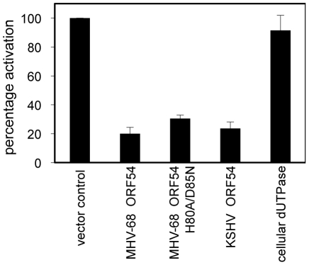
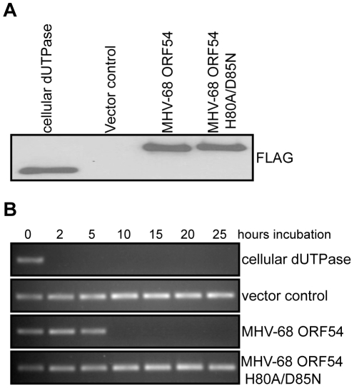
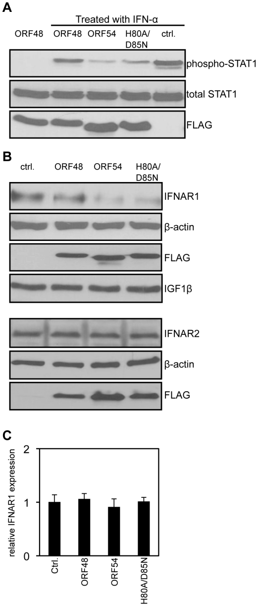
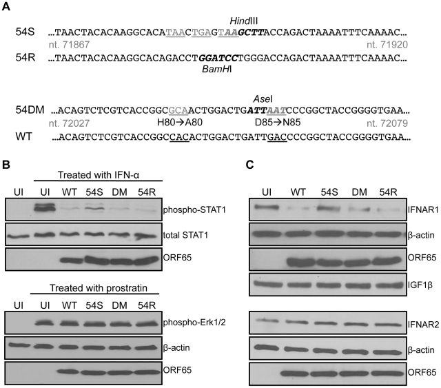
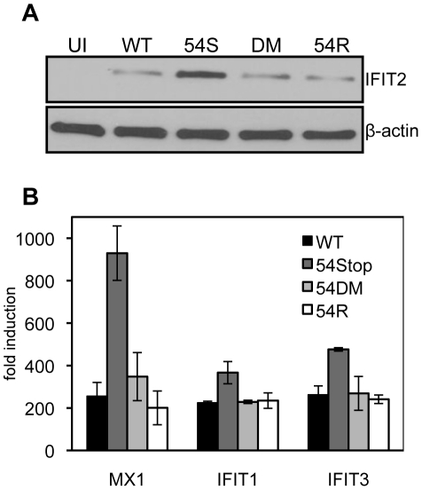
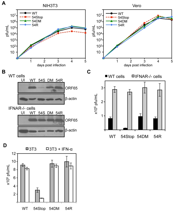
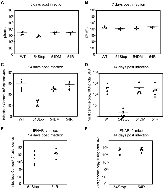
Gammaherpesviruses such as KSHV and EBV establish lifelong persistent infections through latency in lymphocytes. These viruses have evolved several strategies to counteract the various components of the innate and adaptive immune systems. We conducted an unbiased screen using the genetically and biologically related virus, MHV-68, to find viral ORFs involved in the inhibition of type I interferon signaling and identified a conserved viral dUTPase, ORF54. Here we define the contribution of ORF54 in type I interferon inhibition by ectopic expression and through the use of genetically modified MHV-68. ORF54 and an ORF54 lacking dUTPase enzymatic activity efficiently inhibit type I interferon signaling by inducing the degradation of the type I interferon receptor protein IFNAR1. Subsequently, we show in vitro that the lack of ORF54 causes a reduction in lytic replication in the presence of type I interferon signaling. Investigation of the physiological consequence of IFNAR1 degradation and importance of ORF54 during MHV-68 in vivo infection demonstrates that ORF54 has an even greater impact on persistent infection than on lytic replication. MHV-68 lacking ORF54 expression is unable to efficiently establish latent infection in lymphocytes, although it replicates relatively normally in lung tissues. However, infection of IFNAR-/- mice alleviates this phenotype, emphasizing the specific role of ORF54 in type I interferon inhibition. Infection of mice and cells by a recombinant MHV-68 virus harboring a site specific mutation in ORF54 rendering the dUTPase inactive demonstrates that dUTPase enzymatic activity is not required for anti-interferon function of ORF54. Moreover, we find that dUTPase activity is dispensable at all stages of MHV-68 infection analyzed. Overall, our data suggest that ORF54 has evolved anti-interferon activity in addition to its dUTPase enzymatic activity, and that it is actually the anti-interferon role that renders ORF54 critical for establishing an effective persistent infection of MHV-68.


Conflict of interest statement

The authors have declared that no competing interests exist.

Figures

Figure 1
Figure 1. MHV-68 and KSHV ORF54 diminish IFN-α induced activation of ISRE in a non-dUTPase related manner.
HEK 293T cells were transiently transfected with ISRE_firefly-luciferase, PGK_renilla-luciferase as an internal control, and with expression plasmids for each ORF, control plasmid, or murine cellular dUTPase, then were treated with 3×10⁴ U/mL of human IFN-α. Firefly luciferase values were normalized to renilla luciferase. Percentage of activation is calculated in comparison to control, an unrelated MHV-68 ORF or vector control. Error bars depict one standard error based on four independent trials.
Figure 2
Figure 2. Generation of a dUTPase-null MHV-68 ORF54.
A) Immunoblot against the FLAG epitope of immunoprecipitated ORFs or control demonstrates equal amounts of each protein was purified from transfected 293T cells and used in the dUTPase activity assay. B) A PCR reaction utilizing individual dNTPs, with dUTP replacing dTTP, was used to determine the presence of functional dUTP after incubation (between 0 and 25 hours) with purified murine cellular dUTPase, MHV-68 ORF 54, MHV-68 ORF54 H80A/D85N, and a vector control. The presence of a PCR product indicates intact dUTP, and thus a non-functional dUTPase.
Figure 3
Figure 3. ORF54 induces degradation of IFNAR1.
24 hours post transfection, 293T cells were treated with 1 µg/mL of puromycin to select for transfected cells. 48 hours post treatment, cells overexpressing vector control or FLAG-tagged MHV-68 ORFs were: A) treated with human IFN-α for 15 minutes. Immunoblot for phosphorylation of STAT1 on Tyr 701 was assayed first; the membrane was subsequently stripped twice to demonstrate equal levels of total STAT1 and expression of FLAG-ORFs as controls; B) Upper panels: Lysed to analyze level of IFNAR1. The membrane was subsequently stripped to demonstrate equal expression of the FLAG-ORFs and β-actin as controls, and IGF1β to demonstrate specificity of ORF54 to IFNAR1. Lower panels: The same lysates were also subjected to immunoblot against IFNAR2. The membrane was stripped twice to demonstrate equal expression of FLAG-ORFs and β-actin as controls. C) harvested for total RNA isolation. Human IFNAR1 transcript levels were quantified by RT-PCR, normalized first to actin, and are shown relative to vector control transfected cells. Ctrl. is the vector control, ORF48 is an unrelated MHV-68 ORF used as a negative control.
Figure 4
Figure 4. An ORF54-null virus has incomplete degradation of IFNAR1.
A) Schematic diagram depicting the construction of an MHV-68 ORF54 mutant with an insertion of a triple translational stop codon (54Stop), its revertant (54R), and a dUTPase-null double amino acid mutant with ORF54 histidine at position 80 mutated to alanine and aspartic acid at position 85 mutated to asparagine (54DM). Nucleotide coordinates are based on GenBank U97553. WT MHV-68 and 54R contain a BamHI restriction enzyme site that is altered to a HindIII site with the introduction of the triple stop codon. The base pair changes in 54DM allow the introduction of an AseI site that is absent in WT MHV-68. Base changes are depicted in gray and underlined; restriction enzyme recognition sites are bolded. B) Upper panels: NIH3T3 cells infected with each indicated virus at MOI 2 for 24 hours were treated with murine IFN-α for 15 minutes and then harvested for immunoblot against phosphorylation of Tyr701 of STAT1. Blots were stripped twice to demonstrate equal amounts of total STAT1 and comparable infection with expression of viral protein ORF65. Lower panels: NIH3T3 cells infected with each indicated virus at MOI 2 for 24 hours were treated with a final concentration of 160 µM of prostratin for 30 minutes and then harvested for immunoblot against phosphorylation of Thr202 and Tyr204 of Erk1/2. Blots were stripped twice to demonstrate equal amounts of β-actin and comparable infection with expression of viral protein ORF65. C) 293T cells infected with the indicated viruses at MOI 1 for 24 hours were lysed and analyzed for total amount of IFNAR1. Blots were stripped to demonstrate equal loading with β-actin, comparable infection with expression of viral protein ORF65, and specificity of ORF54 to IFNAR1 with IGF1β. The same lysates were also subjected to immunoblot against IFNAR2. The membrane was stripped twice to demonstrate equal loading with β-actin and comparable infection with expression of viral protein ORF65.UI = uninfected; 54S = 54Stop; DM = 54DM.
Figure 5
Figure 5. An ORF54-null virus infection promotes higher induction of interferon stimulated genes.
Bone marrow derived macrophages from wild type mice were infected at an MOI of 2. Equal infection was ensured by RT-PCR quantifying input viral genome copies from total infected cellular DNA 1 hour after virus was introduced to the culture. A) 24 hours post infection cells were harvested for immunoblot against murine IFIT2. The blot was stripped to demonstrate equal expression β-actin as a control. UI = uninfected; 54S = 54Stop; DM = 54DM. B) 24 hours post infection, RNA was harvested and reverse transcribed to cDNA. Transcripts for murine MX1, IFIT1, and IFIT3 were measured by real-time PCR. The fold induction was calculated by first normalizing each C(t) for the transcript to the actin C(t), and then comparing to uninfected samples.
Figure 6
Figure 6. ORF54-null virus has a moderate defect in the presence of type I IFN signaling.
A) In these multiple-step growth curves, NIH3T3 or Vero cells were infected at MOI 0.01 and harvested for 5 consecutive days post infection. Viral titers were calculated by plaque assay following three freeze and thaw cycles of the whole cell and supernatant lysate. This data is one representative from 3 independent trials. NIH3T3 curves are shown in the left panel, Vero curves in the right. B) Bone marrow derived macrophages isolated from IFNα/β receptor knock-out mice and wild-type controls were infected with each indicated virus at MOI 4. 60 hpi, cells were harvested and immunoblotted for viral late antigen ORF65. The blots were stripped and re-probed for β-actin. UI = uninfected; 54S = 54Stop; DM = 54DM. C) Infection was completed as described in B. At 60 hpi, cells were harvested and viral titers were calculated by plaque assay following three freeze and thaw cycles of the whole cell and supernatant lysate. The assay was done in triplicate; averages are shown here with error bars reflecting one standard error. For 54Stop compared to WT, 54DM, and 54R, p-values are 0.0004, 0.003, 0.0007, respectively. D) NIH3T3 cells were left alone or pretreated with 100 units/mL of IFN-α one day prior to infection by each indicated virus at MOI 0.01. Immediately following and 2 days post infection, IFN-α was supplemented into the media at 100 units/mL in the treated samples. On 4 days post infection, infected samples were harvested and subjected to three freeze and thaw cycles prior to titer by plaque assay. The assay was done in triplicate; averages are shown here with error bars reflecting one standard error. For 54Stop compared to WT, 54DM, and 54R, p-values are 0.0007, 0.003, 0.008, respectively. For 54Stop, the p-value comparing untreated and IFN-α treated cells is 0.022.
Figure 7
Figure 7. ORF54 non-dUTPase function is essential for establishment of latency in vivo, but not lytic infection.
Balb/C mice were infected with 500 pfu of each indicated virus. (A–B) Lung tissue was harvested for plaque assay to measure infectious titer. Infectious titer from lung lysates at A) 5 dpi and B) 7 dpi. P-values for 54S compared to WT, 54DM, and 54R in A are 0.047, 0.002, 0.018, respectively and in B are 0.048, 0.396, 0.022, respectively. (C–D) At 14 dpi, splenocytes were harvested to C) measure infectious centers which are derived from reactivating virus and D) to determine viral genome copies from 100 ng of total splenocyte DNA. P-values for 54Stop compared to WT, 54DM, and 54R in C are 0.043, 0.0003, 0.005, respectively, and in D are 0.007, 0.042, and 0.040, respectively. P-value of 54DM compared to WT in C is not significant, p = 0.159. E) IFNAR−/− mice were infected with 500 pfu of 54Stop and 54R viruses. At 14 dpi, splenocytes were harvested to measure infectious centers, the p-value between 54Stop and 54R is not significant, p = 0.248, and F) viral genome copies determined from 100 ng of total splenocyte DNA, the p-value between 54Stop and 54R is not significant, p = 0.387. In all Figures, each ▴ represents one animal, and a bar marks averages. For A,B n = 4; for C,D n = 6 except for WT virus where n = 5; for E and F, n = 5 for 54Stop and n = 6 for 54R.

Similar articles

Cited by

References

    1. Katze MG, He Y, Gale M., Jr Viruses and interferon: a fight for supremacy. Nat Rev Immunol. 2002;2:675–687. - PubMed
    1. Mossman KL, Ashkar AA. Herpesviruses and the innate immune response. Viral Immunol. 2005;18:267–281. - PubMed
    1. Alam R, Gorska M. 3. Lymphocytes. J Allergy Clin Immunol. 2003;111:S476–485. - PubMed
    1. Chaplin DD. 1. Overview of the immune response. J Allergy Clin Immunol. 2003;111:S442–459. - PubMed
    1. Takeuchi O, Akira S. Innate immunity to virus infection. Immunol Rev. 2009;227:75–86. - PMC - PubMed

Publication types

MeSH terms