. 2011;6(10):e24832.
doi: 10.1371/journal.pone.0024832. Epub 2011 Oct 6.

Progression of pathogenic events in cynomolgus macaques infected with variola virus

Affiliations

Progression of pathogenic events in cynomolgus macaques infected with variola virus

Victoria Wahl-Jensen et al. PLoS One. 2011.

Abstract

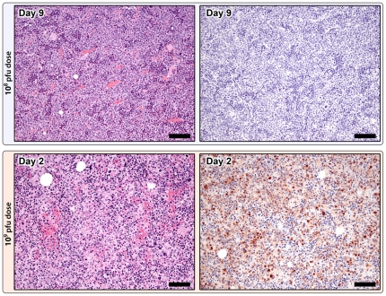
Smallpox, caused by variola virus (VARV), is a devastating human disease that affected millions worldwide until the virus was eradicated in the 1970 s. Subsequent cessation of vaccination has resulted in an immunologically naive human population that would be at risk should VARV be used as an agent of bioterrorism. The development of antivirals and improved vaccines to counter this threat would be facilitated by the development of animal models using authentic VARV. Towards this end, cynomolgus macaques were identified as adequate hosts for VARV, developing ordinary or hemorrhagic smallpox in a dose-dependent fashion. To further refine this model, we performed a serial sampling study on macaques exposed to doses of VARV strain Harper calibrated to induce ordinary or hemorrhagic disease. Several key differences were noted between these models. In the ordinary smallpox model, lymphoid and myeloid hyperplasias were consistently found whereas lymphocytolysis and hematopoietic necrosis developed in hemorrhagic smallpox. Viral antigen accumulation, as assessed immunohistochemically, was mild and transient in the ordinary smallpox model. In contrast, in the hemorrhagic model antigen distribution was widespread and included tissues and cells not involved in the ordinary model. Hemorrhagic smallpox developed only in the presence of secondary bacterial infections - an observation also commonly noted in historical reports of human smallpox. Together, our results support the macaque model as an excellent surrogate for human smallpox in terms of disease onset, acute disease course, and gross and histopathological lesions.


Conflict of interest statement

Competing Interests: The authors have declared that no competing interests exist.

Figures

Figure 1
Figure 1. Progression of cutaneous lesions in the 108 pfu group.
Day 3: Focal area of mild epidermal hyperplasia and keratinocyte swelling. Day 5: Marked epidermal hyperplasia with ballooning degeneration and early vesicle formation (*). Day 7: Intraepidermal pustule and early crust formation; dermal inflammation is primarily neutrophilic. Day 11: Resolution of epidermal hyperplasia and replacement by large serocellular crust; dermal inflammation is primarily lymphohistiocytic, H&E 10X; mag bars = 50 um.
Figure 2
Figure 2. Spleen. A–C: Poxviral immunohistochemistry in the 108 pfu group.
A: Day 1, Positive staining (brown) is localized to the marginal zone of primary follicles (*) and scattered individual cells within the sinusoids (arrows). Additional positive staining on Day 3 within the marginal zone of primary follicles (B) and within early germinal centers (C), anti-vaccinia IHC; 20X; mag bars = 50 um. D-F: Splenic changes in the 109 pfu premature death group. D: Diffuse red pulp hemorrhage and congestion, and marked white pulp lymphocytolysis and lymphoid depletion, H&E 20X; mag bar = 50 um. E: Widespread poxviral IHC staining (brown) is localized to the periarteriolar lymphoid sheaths (*), individual cells within the sinusoids (arrows), and follicular marginal zones (not shown), anti-vaccinia IHC; 20X; mag bar = 50 um. F: Ultrastructural appearance of mature virions, fibrin, and a degenerate cell, TEM; 40000X; mag bar = 500 nm.
Figure 3
Figure 3. Lymph node changes in the 108 pfu (top) and 109 pfu (bottom) dose groups.
108 pfu dose, Day 11: Marked lymphoid hyperplasia with large numbers of secondary follicles containing well developed germinal centers (*), H&E 5X; mag bar = 100 um. Poxviral immunohistochemistry (right) is negative, anti-vaccinia IHC; 5X; mag bar = 100 um. 109 pfu dose, premature death group, Day 2: Follicular depletion and lymphocytolysis with abundant apoptotic debris, H&E 20X; mag bar = 50 um. Poxviral IHC staining (brown) is localized to the mantle zone and scattered individual cells within the paracortex and sinuses, anti-vaccinia IHC; 20X; mag bar = 50 um.
Figure 4
Figure 4. Gonadal changes in males (top) and females (bottom) during smallpox.
Males (108 pfu dose), Day 5: Areas of testicular (seminiferous tubule) degeneration (*) adjacent to normal seminiferous tubules (arrows), H&E 10X; mag bar = 50 um. Poxviral IHC staining (brown) is localized to the interstitium surrounding degenerate tubules; normal areas are negative, anti-vaccinia IHC; 10X; mag bar = 50 um. Females (109 pfu), Day 2: H&E shows normal ovary with primordial, primary, secondary, and tertiary follicles, and remnant corpus luteum, H&E 5X; mag bar = 100 um. Poxviral IHC staining is primarily localized to the thecal cell layer of secondary and tertiary follicles, anti-vaccinia IHC; 5X; mag bar = 100 um.
Figure 5
Figure 5. Bone marrow changes in the 108 pfu (top) and 109 pfu (bottom) dose groups.
108 pfu dose, Day 9: Marked myeloid hyperplasia, H&E 20X; mag bar = 50 um. No poxviral IHC staining, anti-vaccinia IHC; 20X; mag bar = 50 um. 109 pfu premature death group, Day 2: Diffuse hematopoietic necrosis and multifocal hemorrhage, H&E 20X; mag bar = 50 um. Widespread poxviral staining (brown), anti-vaccinia IHC; 20X; mag bar = 50 um.
Figure 6
Figure 6. Cumulative poxviral immunohistochemistry scores by tissue type in the 108 pfu and 109 pfu dose groups.
IHC stains were subjectively scored on a 0–4 scale (0  =  none; 1 = minimal; 2 = mild; 3 = moderate; 4 = marked). For each tissue type, IHC scores for all 3 animals from the same dose and necropsy day group were summed. If, for an individual animal, more than one slide of the same tissue type was examined and scored, the average score for that tissue type was used in the summation.
Figure 7
Figure 7. Model of VARV pathogenesis.
Viremic blood is filtered through the spleen and liver following inoculation. Within 24 hours marginal zone macrophages and Kupffer cells begin expressing viral antigen indicating likely uptake and processing of the pathogen. When the immune response is effective these antigen presenting cells then traffic via the lymphatics to the lymph nodes where a hyperplastic response develops. Concurrently, trafficking to other preferred sites such as skin occurs, and lesions progress and regress as the ongoing adaptive immune response controls and clears the pathogen. In the face of an ineffective immune response, trafficking via macrophages also occurs, however, lymphoid necrosis rather than hyperplasia occurs, allowing unabated trafficking of the virus to other tissues. Infected cells undergo necrosis, pyroptosis, and/or apoptosis which, given the widespread state of infection, results in inflammation, hemorrhage and death.

Similar articles

Cited by

References

    1. Hopkins DR. Chicago: University of Chicago Press; 2002. The Greatest Killer: Smallpox in History.
    1. Fenner F. The eradication of smallpox. Prog Med Virol. 1977;23:1–21. - PubMed
    1. Fenner F. The global eradication of smallpox. Med J Aust. 1980;1:455–455. - PubMed
    1. Fenner F. A successful eradication campaign. Global eradication of smallpox. Rev Infect Dis. 1982;4:916–930. - PubMed
    1. Fenner F. Smallpox: emergence, global spread, and eradication. Hist Philos Life Sci. 1993;15:397–420. - PubMed

Publication types