. 2011;6(10):e25241.
doi: 10.1371/journal.pone.0025241. Epub 2011 Oct 5.

Dicer1 depletion in male germ cells leads to infertility due to cumulative meiotic and spermiogenic defects

Affiliations

Dicer1 depletion in male germ cells leads to infertility due to cumulative meiotic and spermiogenic defects

Yannick Romero et al. PLoS One. 2011.

Abstract

Background: Spermatogenesis is a complex biological process that requires a highly specialized control of gene expression. In the past decade, small non-coding RNAs have emerged as critical regulators of gene expression both at the transcriptional and post-transcriptional level. DICER1, an RNAse III endonuclease, is essential for the biogenesis of several classes of small RNAs, including microRNAs (miRNAs) and endogenous small interfering RNAs (endo-siRNAs), but is also critical for the degradation of toxic transposable elements. In this study, we investigated to which extent DICER1 is required for germ cell development and the progress of spermatogenesis in mice.

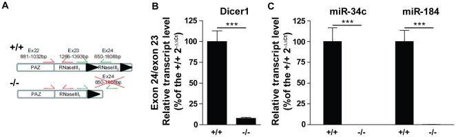
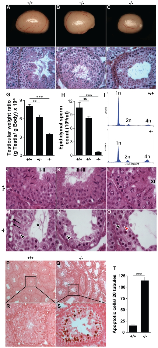
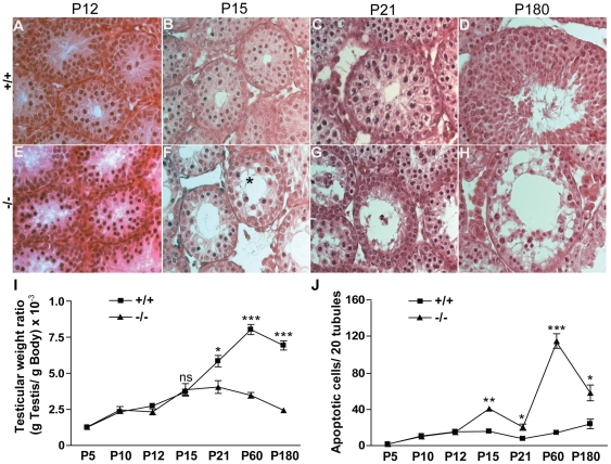
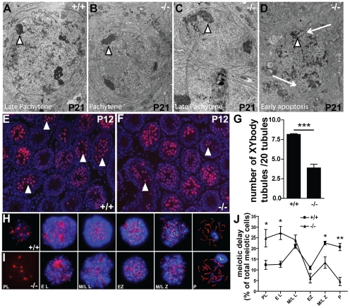
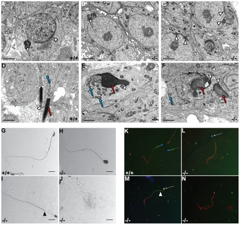
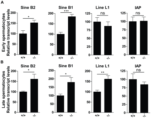
Principal findings: We show that the selective ablation of Dicer1 at the early onset of male germ cell development leads to infertility, due to multiple cumulative defects at the meiotic and post-meiotic stages culminating with the absence of functional spermatozoa. Alterations were observed in the first spermatogenic wave and include delayed progression of spermatocytes to prophase I and increased apoptosis, resulting in a reduced number of round spermatids. The transition from round to mature spermatozoa was also severely affected, since the few spermatozoa formed in mutant animals were immobile and misshapen, exhibiting morphological defects of the head and flagellum. We also found evidence that the expression of transposable elements of the SINE family is up-regulated in Dicer1-depleted spermatocytes.

Conclusions/significance: Our findings indicate that DICER1 is dispensable for spermatogonial stem cell renewal and mitotic proliferation, but is required for germ cell differentiation through the meiotic and haploid phases of spermatogenesis.


Conflict of interest statement

Competing Interests: The authors have declared that no competing interests exist.

Figures

Figure 1
Figure 1. Germ cell-specific deletion of Dicer1 and miRNA depletion in Ddx4-Cre;Dcr1fx/fx testes.
(A) Diagram of exons 22 to 24 of the Dcr1fx/fx (+/+) and Ddx4-Cre;Dcr1fx/fx (−/−) alleles; the protein domains encoded by each exon are also noted. Exon 24 is flanked with loxP sites (black triangles), and excision occurs upon Ddx4-Cre recombinase expression. The primer pairs used in real-time RT-PCR for quantifying the excision of exon 24 (green) compared to exon 23 (red) are shown. (B) Histogram showing the exon 24 to exon 23 ratio of Dicer1 mRNA in elutriated P60 spermatocytes isolated from +/+ (n = 3) and −/− (n = 3) testes; (C) Graph showing the expression of spermatocyte-specific miRNAs. Transcript abundance was quantified by real-time RT-PCR and normalized to 18s rRNA (B), U6 (C). Results are mean±SEM, ***p<0.0001 versus controls.
Figure 2
Figure 2. Reduction in testis size, apoptosis and near complete absence of mature spermatozoa in Ddx4-Cre;Dcr1fx/fx (−/−) testes.
At P60, testes from −/− (n = 16) mice (C) showed a 55% reduction (G) in weight compared to control Dcr1fx/fx (+/+; n = 11) (A) and Ddx4-Cre;Dcr1fx/wt (+/−; n = 6) (B) littermates. H&E staining of testes sections (D–F) revealed near complete absence of mature spermatozoa and elongated spermatids in −/− testes. (H) Epididymal sperm count analysis showed a ∼99% decreased in −/− epididymides. (I) DNA content histogram of P60 +/+ (upper panel) and −/− (lower panel) testes sorted by FACS using propidium iodide. (J–L) H&E staining of +/+ testes showing stage I–II (J), II–III (K) and XI (L) seminiferous tubules. (M–O) −/− tubules are devoid of elongated spermatids and show numerous seminiferous epithelium defects such as vacuolization (arrows), and Sertoli cell cytoplasmic lumen extensions (asterisk). Sparse (2–3) round spermatids (arrowhead in M), and zygotene spermatocytes (arrowhead in N) were found in tubules. (O) Germ cell association is disturbed in tubules as shown by the presence of leptotene spermatocyte (black arrowhead) and (2–3) few round spermatids (red arrowhead). Global TUNEL/eosin staining of P60 +/+ (P) and −/− (Q) testes revealed massive apoptosis (brown nuclei) in some specific −/− seminiferous tubules. Histological analysis indicated that meiotic cells are the most abundant apoptotic population found in −/− (S). (T) The overall number of TUNEL-positive cells is increased ∼8-fold in −/− compared to +/+ individuals. Results are mean ±SEM, *p<0.05, **p<0.01, ***p<0.001 versus controls. ns: not significant. Scale bar: 50 µm (A–F).
Figure 3
Figure 3. Tubular defects appear as early as P15 in Ddx4-Cre;Dcr1fx/fx mutant testes.
H&E staining of control (+/+) (A, B, C, D) and mutant (−/−) (E, F, G, H) testes at P12 (A&E), P15 (B&F) ,P21 (C&G) and P180 (D&H). The first anatomical defects including vacuolization (asterisk), apoptosis and germ cell disorganization appeared at P15 (F) and worsened by P21 (G). In aging individuals, at P180 (D&H), tubular histology is strongly affected, with numerous vacuoles and few germ cells remaining. Reduced testis weight ratio (I) correlated with an increase in apoptotic rate (J) during the first spermatogenic wave. For I and J, numbers of animals were n = 5 minimum for each genotype. Results are mean ±SEM, *p<0.05, **p<0.01, ***p<0.001 versus controls. ns: not significant.
Figure 4
Figure 4. Meiotic progression defects in Ddx4-Cre;Dcr1fx/fx (−/−) mutant testes.
Representative transmission electron micrographs from P21 control (A) and mutant (B–D) testes. Note the enlarged perinuclear heterochromatin areas (arrowheads) and the irregular and abnormal nucleus shape (arrows) in mutant pachytene spermatocytes typical of apoptotic cells. (E, F) Anti-γH2AX staining (red), present in the whole nucleus in early meiotic stages and restricted to the XY body from pachytene phase, revealed a reduction in the number of tubules containing XY body positive cells (arrowheads, punctual red staining) within −/− testis (F) compared to wild-type (E). DAPI (blue) was used for nuclear staining. (G) shows the quantification of XY body positive tubules per 20 tubules. Anti-SYCP3 (red), DMC1 (green) and γH2AX (blue) staining of chromosomal spread preparations from control (H), and mutant (I) P12 testes was used to quantify meiotic prophase I cells. (J) Note the higher number of early meiotic cells (i.e., Preleptotene and leptotene) and the reduced number of late meiotic cells (i.e. mid/Late zygotene and pachytene) in −/− cell preparations suggesting a delayed progression of germ cells into meiosis. Results are mean ±SEM, ns = not significant, *p<0.05, **p<0.01, ***p<0.001 versus controls. PL: Pre-leptotene, EL: Early leptotene, M/L L: mid/late leptotene, EZ: early zygotene, M/L Z: mid/late zygotene, P: Pachytene.
Figure 5
Figure 5. Spermiogenic and sperm alterations in Ddx4-Cre;Dcr1fx/fx (−/−) testes.
Representative transmission electron micrographs from P60 control (A&D) and mutant (B, C, E & F). In round spermatids (A–C), chromatoid body is normally shaped and located near the nucleus (red arrows), whereas acrosome is fragmented in mutant (arrowheads). In elongated spermatids (D–F), nuclear shape (red arrows), chromatin condensation, and acrosome (arrowheads) are abnormal in −/− (E and F) mice. Mitochondria showed hyperplasia of the intermembrane space or cristae (blue arrows, E). Scale bar: 2 µm. H&E staining of epididymal sperm spreads of +/+ (G) and −/− (H, I and J) adult mice. In contrast to +/+ animals (G), spermatozoa of −/− mice exhibited multiple defects such as head morphology, mid-piece defects (H and I) and reduced overall size (J). Black arrowhead shows the ectopic localization of the mid-piece in (I). Scale bars: 10 µm. Immunostaining of epididymal sperm spreads of control +/+ (K) and −/− (L, M and N) mice using DAPI staining to label nuclear DNA (blue), Mitotracker as a mitochondrial marker (green) and anti-α-tubulin to label the flagellum (red). Spermatozoa of −/− mice exhibited head morphology defects (L, M and N). White arrows show that some mutant spermatozoa display abnormal shaped nuclei and co-localized ectopic mitochondrial staining. White arrowhead in (M) indicates the ectopic localization of the mid-piece mitochondria compared to control individuals (K). Images were taken with a 40× objective.
Figure 6
Figure 6. Deregulation of transposable elements in Ddx4-Cre;Dcr1fx/fx (−/−) mutant spermatocytes.
Quantitative real-time PCR performed on elutriated germ cell fractions enriched either in leptotene/zygotene/early pachytene spermatocytes (A) or late pachytene/diplotene spermatocytes (B) originating from +/+ and −/− testes at P60. Results are mean ±SEM, ns = not significant, *p<0.05, **p<0.01, ***p<0.001 versus controls.

Similar articles

Cited by

References

    1. Eddy EM. Male germ cell gene expression. Recent Prog Horm Res. 2002;57:103–128. - PubMed
    1. Kim VN, Han J, Siomi MC. Biogenesis of small RNAs in animals. Nat Rev Mol Cell Biol. 2009;10:126–139. - PubMed
    1. He Z, Kokkinaki M, Pant D, Gallicano GI, Dym M. Small RNA molecules in the regulation of spermatogenesis. Reproduction. 2009;137:901–911. - PubMed
    1. Papaioannou MD, Nef S. microRNAs in the testis: building up male fertility. J Androl. 2010;31:26–33. - PubMed
    1. Buchold GM, Coarfa C, Kim J, Milosavljevic A, Gunaratne PH, et al. Analysis of microRNA expression in the prepubertal testis. PLoS One. 2010;5:e15317. - PMC - PubMed

Publication types

MeSH terms