. 2011;6(10):e25438.
doi: 10.1371/journal.pone.0025438. Epub 2011 Oct 5.

MMP-10/stromelysin-2 promotes invasion of head and neck cancer

Affiliations

MMP-10/stromelysin-2 promotes invasion of head and neck cancer

Elsayed Mohamed Deraz et al. PLoS One. 2011.

Erratum in

  • PLoS One. 2012;7(2). doi: 10.1371/annotation/ef18e199-e66d-43b9-97e0-96d1ab149193. Kiekhaee, Mohammad Reza [corrected to Keikhaee, Mohammad Reza]

Abstract

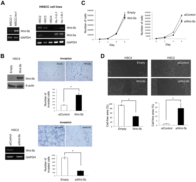
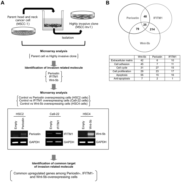
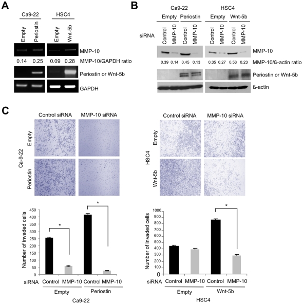
Background: Periostin, IFN-induced transmembrane protein 1 (IFITM1) and Wingless-type MMTV integration site family, member 5B (Wnt-5b) were previously identified as the invasion promoted genes of head and neck squamous cell carcinoma (HNSCC) by comparing the gene expression profiles between parent and a highly invasive clone. We have previously reported that Periostin and IFITM1 promoted the invasion of HNSCC cells. Here we demonstrated that Wnt-5b overexpression promoted the invasion of HNSCC cells. Moreover, stromelysin-2 (matrix metalloproteinase-10; MMP-10) was identified as a common up-regulated gene among Periostin, IFITM1 and Wnt-5b overexpressing HNSCC cells by using microarray data sets. In this study, we investigated the roles of MMP-10 in the invasion of HNSCC.

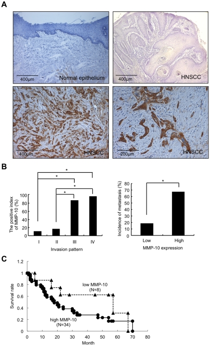
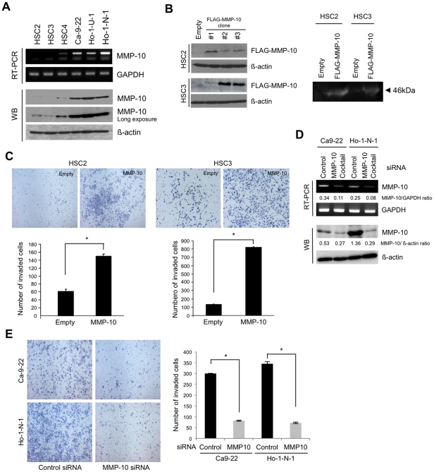
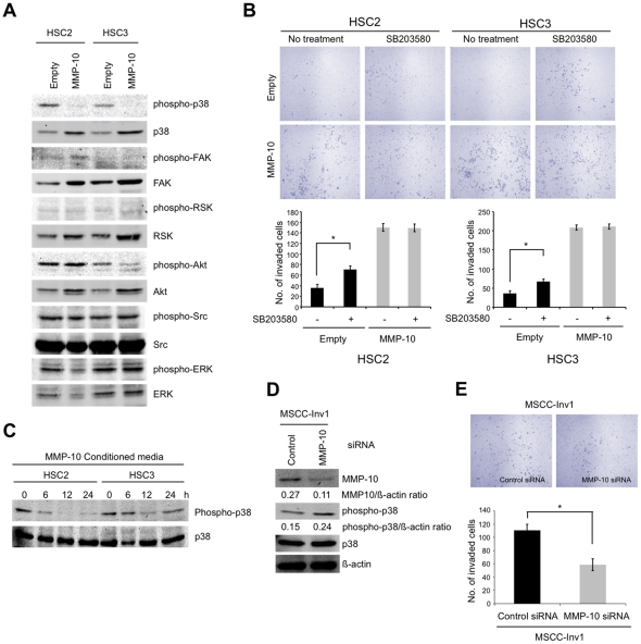
Methods and findings: We examined the expression of MMP-10 in HNSCC cases by immunohistochemistry. High expression of MMP-10 was frequently observed and was significantly correlated with the invasiveness and metastasis in HNSCC cases. Next, we examined the roles of MMP-10 in the invasion of HNSCC cells in vitro. Ectopic overexpression of MMP-10 promoted the invasion of HNSCC cells, and knockdown of MMP-10 suppressed the invasion of HNSCC cells. Moreover, MMP-10 knockdown suppressed Periostin and Wnt-5b-promoted invasion. Interestingly, MMP-10 overexpression induced the decreased p38 activity and MMP-10 knockdown induced the increased p38 activity. In addition, treatment with a p38 inhibitor SB203580 in HNSCC cells inhibited the invasion.

Conclusions: These results suggest that MMP-10 plays an important role in the invasion and metastasis of HNSCC, and that invasion driven by MMP-10 is partially associated with p38 MAPK inhibition. We suggest that MMP-10 can be used as a marker for prediction of metastasis in HNSCC.


Conflict of interest statement

Competing Interests: The authors have declared that no competing interests exist.

Figures

Figure 1
Figure 1. Wnt-5b promotes the invasion of HNSCC.
A, Expression of Wnt-5b in the highly invasive clone and HNSCC cell lines. Expression of Wnt-5b mRNA in MSCC-1, MSCC-Inv1 as well as six HNSCC cell lines; HSC2, HSC3, HSC4, Ca-9-22, Ho-1-N-1, and Ho-1-U-1 was examined by RT-PCR. Expression of Wnt-5a mRNA was also examined in 6 HNSCC cell lines. GAPDH was used as a loading control. B, Wnt-5b promoted the invasion of HNSCC cells. For generation of Wnt-5b-overexpressing cells, Wnt-5b expression vector was transfected into HSC4 cells without Wnt-5b expression. We obtained the stable clone and the expression of Wnt-5b was examined by Western blotting with anti-Wnt-5b polyclonal antibody (left upper panel). We used empty vector transfected cells (empty) as a control. β-actin expression was used as a loading control. The invasiveness of Wnt-5b-overexpressing cells (right upper panel) was examined by in vitro invasion assay. 1.5×10⁵ cells were placed on the upper compartment of the cell culture insert. After 12h of incubation, the penetrated cells onto the lower side of the membrane were fixed in formalin and stained with hematoxylin. HSC2 cells with Wnt-5b expression was transfected by Wnt-5b siRNA (siWnt-5b) or control siRNA (siControl). A scrambled sequence that does not show significant homology to rat, mouse or human gene sequences was used as a control. The effect of knockdown was evaluated by RT-PCR (left lower panel). GAPDH was used as a loading control. The invasiveness of Wnt-5b-knocked-down cells was examined by in vitro invasion assay (right lower panel). After 24 h of incubation, the penetrated cells onto the lower side of the membrane were fixed in formalin and stained with hematoxylin. The bars show the average values and SDs of three independent experiments. *Significantly different from empty vector transfected cells or control siRNA transfected cells at P<0.01. C, Cell proliferation of Wnt-5b-overexpressing cells and Wnt-5b-knocked-down cells. Cells were plated on 24 well plates, and trypsinized cells were counted by cell counter at 0, 2, 4 and 6 day. The bars show the average values and SDs of three independent experiments. D, Migration of Wnt-5b-overexpressing cells and Wnt-5b-knocked-down cells. Migration of the cells was determined by wound healing assay. At 24 h after scratching the cells, phase-contrast images (10× field) of the wound healing process were photographed digitally with an inverted microscope. The distance of the wound areas were measured on the images, set at 100% for 0 h, and the mean percentage of the total distances of the wound areas was calculated. The bars show the average values and SDs of three independent experiments. *Significantly different from empty vector transfected cells or control siRNA transfected cells at P<0.01.
Figure 2
Figure 2. MMP-10 and MMP13 are common upregulated genes by Periostin, IFITM1, and Wnt-5b overexpression.
A, Schema shows the strategy to identify the common target of invasion related molecules. Periostin, IFITM1, and Wnt-5b are identified as the invasion related molecules by comparing the gene expression profile between the parent (MSCC-1 cells) and a highly invasive clone (MSCC-Inv1 cells). To identify common targets of invasion related molecules, we compared the gene expression profiles of control vs. Periostin-overexpressing HSC2 cells, control vs. IFITM1-overexpressing Ca9-22 cells, and control vs. Wnt-5b-overexpressing HSC4 cells. Ectopic expression level of Periostin, IFITM1 and Wnt-5b in each cells are shown by RT-PCR. GAPDH expression was used as a loading control. B, Several upregulated genes were found between Periostin and IFITM1, Periostin and Wnt-5b, and Wnt-5b and IFITM1. By using Gene Ontology software, we identified the biological functions of these genes. The table shows these common upregulated genes comprising diverse families with variable biological functions.
Figure 3
Figure 3. High expression of MMP-10 is observed in HNSCC cases.
A, Immunohistochemical expression of MMP-10 in 116 HNSCC cases and 30 normal epithelia. Normal epithelium is completely negative for MMP-10 compared to HNSCC cases in which most tumor cells showed highly expression of MMP-10. Representative case of low MMP-10 expression in normal oral epithelium and HNSCC case (Jacobsson's classification Pattern I), and representative cases of high expression of MMP-10 (low and high magnification) in HNSCC cases (Jacobsson's classification Pattern IV) are shown. Scale bar is shown in each picture. B, Correlation between MMP-10 expression and invasion and metastasis in HNSCC cases. Left graph shows the relationship between MMP-10 expression and pattern of invasion. The Jacobsson's classification (Patterns I-IV) was used for evaluation of invasion pattern (Figure S1) . *Significantly different from pattern I or II at P<0.01. Right graph exhibits the relationship between MMP-10 expression and metastasis. *Significantly different from low expression of MMP-10 at P<0.01. C, MMP-10 expression and poor outcome. Forty-two Italian HNSCC cases were available for survival analysis. Kaplan-Meier curves show survival of HNSCC patients with high expression of MMP-10 (•, N = 34) or low expression of MMP-10 (▴, N = 8).
Figure 4
Figure 4. MMP-10 enhances the invasion of HNSCC cells.
A, Expression of MMP-10 mRNA and protein in six HNSCC cell lines. Expression of MMP-10 mRNA in HSC2, HSC3, HSC4, Ca-9-22, Ho-1-N-1, and Ho-1-U-1 was examined by RT-PCR. GAPDH was used as a loading control. MMP-10 protein expression level was evaluated in the six HNSCC cell lines by Western blot analysis. Images of short and long exposure of MMP-10 protein expression are shown. ß-actin was used as a loading control. B, Generation of MMP-10-overexpressing cells. We obtained several clones by transfection with pBICEP-FLAG-tagged MMP-10 in HSC2 and HSC3 cells. Ectopic expression of MMP-10 was determined by Western blotting with anti-FLAG antibody (left panel). β-actin was used as a loading control. Enzymatic activity of MMP-10 was detected by stromelysin zymography (right panel). Active form of MMP-10 (arrow head) was detected in conditioned media of MMP-10-overexpressing cells. C, Invasive activity of MMP-10-overexpressing HSC2 (left panel) and HSC3 (right panel) cells in comparison with empty vector transfected HSC2 and HSC3 cells (empty) by in vitro invasion assay. Cells were fixed after incubation of 12 h or 20 h in HSC2 cells or HSC3 cells, respectively. The figure shows the stained lower side of the membrane where the cells penetrated (upper panel). The graphs show the number of invaded cells in MMP-10-overexpressing and empty vector transfected cells (lower panel). The bars show the average values and SDs of three independent experiments. *Significantly different from empty vector transfected cells at P<0.01. D, MMP-10 siRNA suppressed the invasion of HNSCC cells. Cocktail of 3 different MMP-10 siRNAs was transiently transfected into Ca-9-22 and Ho-1-N-1 cells with MMP-10 expression. A scrambled sequence that does not show significant homology to rat, mouse or human gene sequences was used as a control. After 48 h of transfection, expression of MMP-10 mRNA and protein was examined by RT-PCR and Western blotting (WB), respectively. GAPDH mRNA and β-actin protein were used as a loading control. Densitometric analysis of MMP-10 expression was performed. MMP-10/GAPDH ratio is shown. E, Suppression of invasion by MMP-10 knockdown in Ca9-22 and Ho-1-N-1 cells. The invasiveness of MMP-10 knocked-down cells was examined by in vitro invasion assay in comparison with control siRNA transfected cells. A scrambled sequence that does not show significant homology to rat, mouse or human gene sequences was used as a control. After 24 h incubation, cells were fixed and the number of invaded cells was counted. The figure shows the stained lower side of the membrane where the cells penetrated (left panel). The graph shows the number of invaded cells (right panel). The bars show the average values and SDs of three independent experiments. *Significantly different from control siRNA transfected cells at P<0.01.
Figure 5
Figure 5. MMP-10 knockdown inhibits Periostin and Wnt-5b-promoted invasion.
A, The expression of MMP10 mRNA was examined by RT-PCR in control Ca9-22 cells, Periostin-overexpressing Ca9-22 cells, control HSC4 cells and Wnt-5b-overexpressing HSC4 cells. GAPDH expression was used as a loading control. B, MMP-10 knockdown into Periostin- and Wnt-5b-overexpressing cells. Cocktail of 3 different MMP-10 siRNAs was transiently transfected into Periostin-overexpressing Ca-9-22 cells and Wnt-5b-overexpressing HSC4 cells. A scrambled sequence that does not show significant homology to rat, mouse or human gene sequences was used as a control. After 48 h of transfection, MMP-10 protein level was examined by Western blot analysis with anti-MMP-10 antibody in MMP-10 siRNA transfected Periostin-overexpressing cells. ß-actin was used as a loading control. C, The invasiveness of MMP-10 siRNA transfected Periostin-overexpressing Ca-9-22 cells (left panel) and Wnt-5b-overexpressing HSC4 cells (right panel) was examined by in vitro invasion assay. After 18 h incubation of HSC4 cells and 24 h incubation of Ca9-22 cells, cells were fixed and the number of invaded cells was counted. The figure shows the stained lower side of the membrane where the cells penetrated (upper panel). Graphs show the number of invaded cells in knockdown and control cells (lower panel). The bars show the average values and SDs of three independent experiments. *Significantly different from control at P<0.01.
Figure 6
Figure 6. p38 inhibition is involved in promoting invasion by MMP-10.
A, p38 inhibition was noted in MMP-10 overexpressing cells. Levels of total and phosphorylated forms of p38, FAK, RSK, Akt, Src, and ERK in control and MMP-10 overexpresing cells by western blotting. B, Invasion of control and MMP-10 overexpressing cells after treatment by p38 inhibitor. The invasiveness of was examined by in vitro invasion assay in empty vector transfected cells and MMP-10 overexpressing cells with SB203580 treatment. Empty vector transfected cells were used as a control. After 12 h incubation, cells were fixed and the number of invaded cells was counted. The figure shows the stained lower side of the membrane where the cells penetrated (upper panel). Graphs show the number of invaded cells (lower panel). The bars show the average values and SDs of three independent experiments. *Significantly different from empty vector transfected cells without SB203580 treatment at P<0.01. C, p38 activity after treatment with conditioned media from MMP-10 overexpressing cells. Conditioned media from MMP-10 overexpressing HSC2 and HSC3 cells was collected after 48 h of plating. After adding conditioned media for 0, 6, 12 and 24 h, HSC2 or HSC3 cells were collected. Expression of phospho-p38 and p38 was examined by Western blot analysis. D, MMP-10 siRNA upregulates p38 activity in HNSCC cells. Cocktail of 3 different MMP-10 siRNAs was transiently transfected into MSCC-Inv1 cells. A scrambled sequence that does not show significant homology to rat, mouse or human gene sequences was used as a control. After 48 h of transfection, expression of MMP-10 protein was examined by Western blotting. Levels of total and phosphorylated forms of p38 were also examined by Western blotting. β-actin expression was used as a loading control. E, Suppression of invasion by MMP-10 knockdown in MSCC-Inv1 cells. The invasiveness was examined by in vitro invasion assay. After 6 h incubation, cells were fixed and the number of invaded cells was counted. The figure shows the stained lower side of the membrane where the cells penetrated (upper panel). The graph shows the number of invaded cells (lower panel). The bars show the average values and SDs of three independent experiments. *Significantly different from control at P<0.01.

Similar articles

Cited by

References

    1. Mao L, Hong WK, Papadimitrakopoulou VA. Focus on head and neck cancer. Cancer Cell. 2004;5:311–316. - PubMed
    1. Neville BW, Damm DD, Allen CM, Bouquot JE. Epithelial Pthology. Oral and Maxillofacial Pathology. Edited by Neville BW. Philadelphia, W B Saunders. 1995;295-302
    1. Mamelle G, Pampurik J, Luboinski B, Lancer R, Lusinchi A, et al. Lymph node prognostic factors in head and neck squamous cell carcinomas. Am J Surg. 1994;168:494–498. - PubMed
    1. Fidler IJ. Critical factors in the biology of human cancer metastasis: Twenty-eighth GHA Clowes Memorial Award Lecture. Cancer Res. 1990;50:6130–6138. - PubMed
    1. Howell GMS, Grandis JR. Molecular mediators of metastasis in head and neck squamous cell carcinoma. Head Neck. 2005;27:710–717. - PubMed

Publication types

MeSH terms