. 2011;6(10):e25480.
doi: 10.1371/journal.pone.0025480. Epub 2011 Oct 5.

Exogenous addition of a C-xylopyranoside derivative stimulates keratinocyte dermatan sulfate synthesis and promotes migration

Affiliations

Exogenous addition of a C-xylopyranoside derivative stimulates keratinocyte dermatan sulfate synthesis and promotes migration

Jun Muto et al. PLoS One. 2011.

Abstract

As C-Xyloside has been suggested to be an initiator of glycosaminoglycan (GAG) synthesis, and GAGs such as Dermatan sulfate (DS) are potent enhancers of fibroblast growth factor (FGF)--10 action, we investigated if a C-Xylopyranoside derivative, (C-β-D-xylopyranoside-2-hydroxy-propane, C-Xyloside), could promote DS production by cultured normal human keratinocytes, how this occurs and if C-Xyloside could also stimulate FGF-dependent cell migration and proliferation. C-Xyloside-treated keratinocytes greatly increased secretion of total sulfated GAGs. Majority of the induced GAG was chondroitin sulfate/dermatan sulfate (CS/DS) of which the major secreted GAG was DS. Cells lacking xylosyltransferase enzymatic activity demonstrated that C-Xyloside was able to stimulate GAG synthesis without addition to core proteins. Consistent with the observed increase in DS, keratinocytes treated with C-Xyloside showed enhanced migration in response to FGF-10 and secreted into their culture media GAGs that promoted FGF-10-dependent cellular proliferation. These results indicate that C-Xyloside may enhance epithelial repair by serving as an initiator of DS synthesis.


Conflict of interest statement

Competing Interests: This work was supported in part by an investigator initiated grant from L'Oreal. Nathalie Pineau and Lionel Breton are employees of L'Oréal Recherche. There are no patents, products in development or marketed products to declare. This does not alter the authors' adherence to all the PLoS ONE policies on sharing data and materials.

Figures

Figure 1
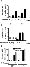
Figure 1. C-Xyloside induces sulfated GAGs from NHEK in culture media without the involvement of Xylosyltransferase.
Primary keratinocytes were stimulated with PBS (untreated cells) or C-Xyloside (0.3 mM or 1 mM). (A) Cultured media were collected after 24 and 48 hours. Total sulfated GAG was determined by Blyscan assay. (B), Increasing concentrations of C-Xyloside were added to keratinocytes. Culture media was collected after 24 hours and total sulfated GAG was determined by Blyscan assay. (C), CHO-K1 (black bars) and CHO-745 (white bars) cells were cultured in the presence or absence of C-Xyloside (1 mM) for 24 hours and the amount of sulfated GAG in the media was determined by Blyscan assay. Data are representative of three individual experiments, and presented as mean ± SEM (n = 3). N.D., not detected.
Figure 2
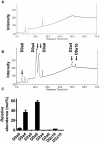
Figure 2. Monosaccharide composition analysis of GAGs release by NHEKs.
Media were harvested after 24 hours of culture (A) without or (B) with C-Xyloside (1 mM) stimulation and monosaccharide compositions were analyzed by HPAEC chromatograph as described under ‘materials and methods’. The peak at 17 min may represent a monosaccharide such as glucose in the media (Glc). Arrows indicate the retention time for galactosamine (GalNH2), glucuronic acid (GlcA), and iduronic acid (IdoA). PAD: pulsed amperometric detection; nC: nano-coulomb. Data are representative of three individual experiments.
Figure 3
Figure 3. Disaccharide analysis of synthesized GAGs.
Media were harvested after 24 hours of culture (A) without or (B) with C-Xyloside (1 mM) stimulation. GAG in culture media were digested with chondroitinase ABC and the disaccharides were resolved by anion-exhange HPLC with post-column derivitization and fluorescence detection (see materials and methods.) Values are expressed as molar percentage. Data are representative of three individual experiments. (C), the disaccharides were resolved by Glycan reductive isotope labeling/mass spectrometry (n = 2). Data are presented as mean ± range. D0a0, ΔUA-GalNAc; D0a4, ΔUA-GalNAc4S; D2a0, ΔUA2S-GalNAc; D0a6, ΔUA-GalNAc6S; D2a4, ΔUA2S-GalNAc4S; D2a6, ΔUA2S-GalNAc6S; D0a10, ΔUA-GalNAc4S6S; D2a10, ΔUA2S-GalNAc4S6S. ΔUA = 4, 5-unsaturated uronic acid.
Figure 4
Figure 4. C-Xyloside promotes FGF-10 dependent keratinocyte migration and cell proliferation.
(A), Primary keratinocytes were stimulated with DS alone, C-Xyloside (CX) alone, FGF-10 alone, C-Xyloside and FGF-10 combined, DS and FGF-10 combined or vehicle. Data are plotted as the mean ± SEM of three individual experiments with samples in quadruplicate. *, p<0.05 (B), Identical fields were photographed 0 and 24 hours after wounding. (C), Primary keratinocytes were treated with PBS or C-Xyloside in RPMI1640 media. All dilutions were done in this keratinocyte-conditioned medium (CM). Cells were incubated for 3 days at 37°C before performing a nonradioactive cell proliferation assay. Data are represented as the mean ± SEM of triplicate determinations. ***, p<0.001.

Similar articles

Cited by

References

    1. Penc SF, Pomahac B, Winkler T, Dorschner RA, Eriksson E, et al. Dermatan sulfate released after injury is a potent promoter of fibroblast growth factor-2 function. J Biol Chem. 1998;273:28116–28121. - PubMed
    1. Taylor KR, Rudisill JA, Gallo RL. Structural and sequence motifs in dermatan sulfate for promoting fibroblast growth factor-2 (FGF-2) and FGF-7 activity. J Biol Chem. 2005;280:5300–5306. - PubMed
    1. Ashikari S, Habuchi H, Kimata K. Characterization of heparan sulfate oligosaccharides that bind to hepatocyte growth factor. J Biol Chem. 1995;270:29586–29593. - PubMed
    1. Lyon M, Deakin JA, Rahmoune H, Fernig DG, Nakamura T, et al. Hepatocyte growth factor/scatter factor binds with high affinity to dermatan sulfate. J Biol Chem. 1998;273:271–278. - PubMed
    1. Maccarana M, Casu B, Lindahl U. Minimal sequence in heparin/heparan sulfate required for binding of basic fibroblast growth factor. J Biol Chem. 1993;268:23898–23905. - PubMed

Publication types

MeSH terms