. 2011;6(10):e25497.
doi: 10.1371/journal.pone.0025497. Epub 2011 Oct 5.

C/EBPβ-Thr217 phosphorylation signaling contributes to the development of lung injury and fibrosis in mice

Affiliations

C/EBPβ-Thr217 phosphorylation signaling contributes to the development of lung injury and fibrosis in mice

Martina Buck et al. PLoS One. 2011.

Abstract

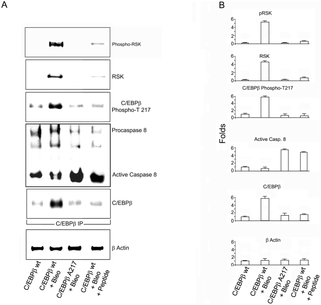
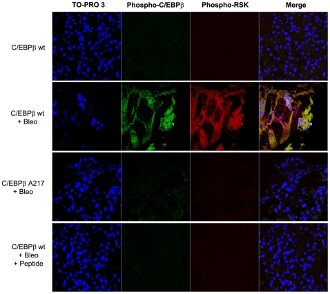
Background: Although C/EBPβ(ko) mice are refractory to Bleomycin-induced lung fibrosis the molecular mechanisms remain unknown. Here we show that blocking the ribosomal S-6 kinase (RSK) phosphorylation of the CCAAT/Enhancer Binding Protein (C/EBP)-β on Thr217 (a RSK phosphoacceptor) with either a single point mutation (Ala217), dominant negative transgene or a blocking peptide containing the mutated phosphoacceptor ameliorates the progression of lung injury and fibrosis induced by Bleomycin in mice.

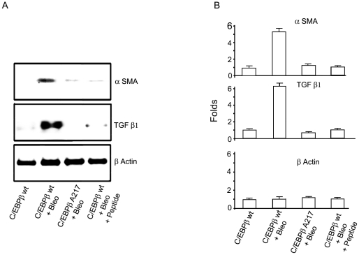
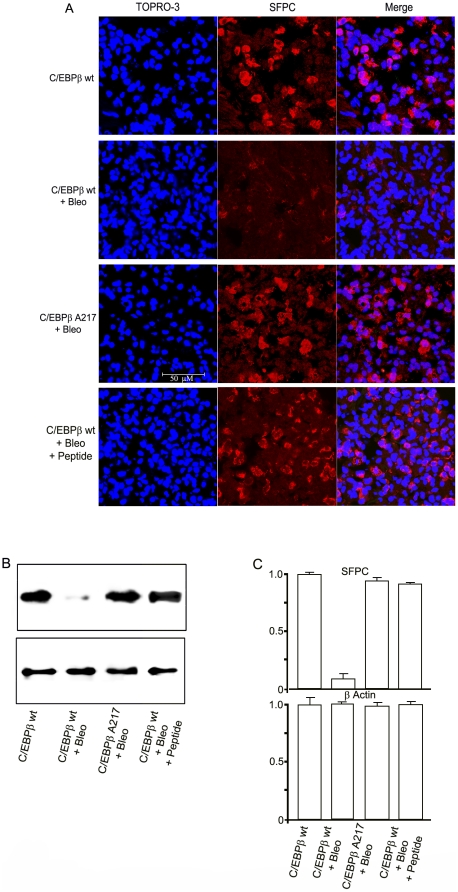
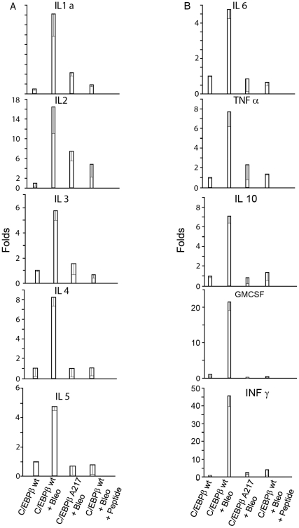
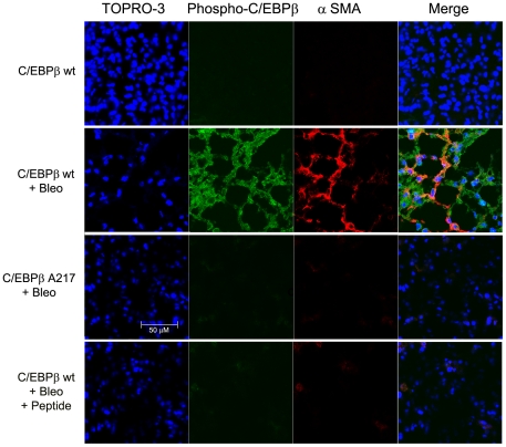
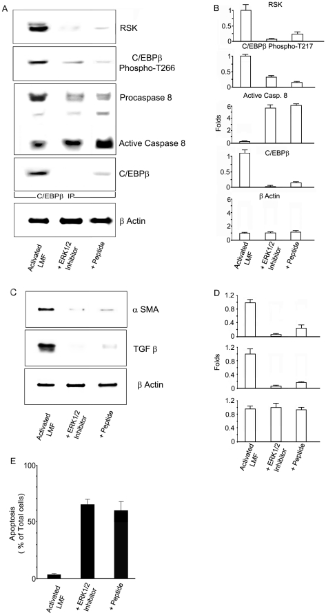
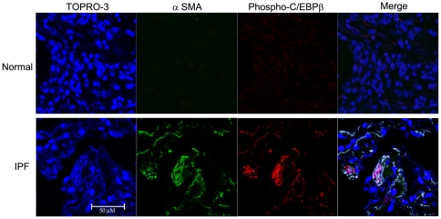
Methodology/principal findings: Mice expressing the non-phosphorylatable C/EBPβ-Ala217 transgene had a marked reduction in lung injury on day-13 after Bleomycin exposure, compared to C/EBPβ(wt) mice, judging by the decrease of CD68(+) activated monocytes/macrophages, bone marrow-derived CD45(+) cells and lung cytokines as well as by the normal surfactant protein-C expression by lung pneumocytes. On day-21 after Bleomycin treatment, C/EBPβ(wt) mice but not mice expressing the dominant negative C/EBPβ-Ala217 transgene developed severe lung fibrosis as determined by quantitative collagen assays. All mice were of identical genetic background and back-crossed to the parental wild-type inbreed FVB mice for at least ten generations. Treatment of C/EBPβ(wt) mice with a cell permeant, C/EBPβ peptide that inhibits phosphorylation of C/EBPβ on Thr217 (40 µg instilled intracheally on day-2 and day-6 after the single Bleomycin dose) also blocked the progression of lung injury and fibrosis induced by Bleomycin. Phosphorylation of human C/EBPβ on Thr266 (human homologue phosphoacceptor) was induced in collagen-activated human lung fibroblasts in culture as well as in activated lung fibroblasts in situ in lungs of patients with severe lung fibrosis but not in control lungs, suggesting that this signaling pathway may be also relevant in human lung injury and fibrosis.

Conclusions/significance: These data suggest that the RSK-C/EBPβ phosphorylation pathway may contribute to the development of lung injury and fibrosis.


Conflict of interest statement

Competing Interests: The authors have declared that no competing interests exist.

Figures

Figure 1
Figure 1. Blocking the C/EBPβ-Thr217 phosphorylation ameliorates the induction of lung fibrosis by Bleomycin.
FVB C/EBPβ wt mice received once intracheally either Bleomycin or control saline as described in Methods. Analysis was performed on day-21. The bars represent 50 µm. A. Representative Mallory's trichrome stain for lung fibrosis (in blue). We graded the coded lung samples with the standard clinical system (0–3; none, mild, moderate and severe). After Bleomycin treatment, C/EBPβwt mice had moderate to severe lung fibrosis compared to controls, while C/EBPβ-Ala217 mice of the same genetic background had mild or moderate lung fibrosis (mean scores : 2.8+/−0.4 vs. 1.5+/−0.5; n: 10 per group; P<0.001). Treatment of C/EBPβwt mice with the C/EBPβ peptide (on day-2 and day-6) after Bleomycin treatment ameliorated the lung fibrosis at day-21 (mean fibrosis score: 2.0+/−0.7; n: 5; P = 0.048). B. Representative Sirius red immunohistochemistry for collagen by light microscopy (in red) (L panels) and with a polarized light (R panels). Increase in lung collagen in a fibrotic pattern was observed in C/EBPβwt but not in C/EBPβ-Ala 217 mice, treated with Bleomycin. Treatment of C/EBPβwt mice with the C/EBPβ peptide (on day-2 and day-6) after Bleomycin treatment decreased the lung fibrosis at day-21. C, D and E. Quantitation lung fibrosis was performed by the Sirius red collagen–binding, collagen type 1 and hydroxyproline assays as described in Methods. The lung collagen content increased (fold from baseline) in C/EBPβwt mice (n: 7 per group; Sirius red collagen-binding: 2.8+/−0.3; collagen type: 3.6+/−0.5; and hydroxyproline: 8.7+/−0.9; P<0.0001 for all), while remaining closer to baseline in C/EBPβ-Ala217 mice treated with Bleomycin (n: 6 ; Sirius red collagen-binding: 1.1+/−0.2, P = 0.37 ; collagen type: 1.0+/−0.2, P = 0.46; and hydroxyproline: 3.1+/−1.0, P = 0.002). The specific fold changes in C/EBPβwt mice treated with Bleomycin and the C/EBPβ peptide included: Sirius red collagen-binding (0.94+/−0.2, P = 0.80); collagen type 1 (1.1+/−0.2, P = 0.934); and hydroxyproline (3.1+/−1.0, P = 0.002; n: 5 for all groups). Representative results from three independent studies for the transgenic mice and of two independent studies for the peptide.
Figure 2
Figure 2. Blocking the C/EBPβ-Thr217 phosphorylation decreases the Bleomycin-induced activation of LMF.
Mice were treated with Bleomycin or saline as described in Methods. Analysis was performed on day-21 A. Immunoblots for α-SMA, TGF-β and β-Actin were performed as described in Materials and methods. B. Pixel intensity was quantified by the Odyssey Western Infrared Detection Method and Odyssey Visualization protocol as described in Methods. The fold-increase in protein expression of lung fibrogenic indicators α-SMA and TGF-β1 were induced by Bleomycin in C/EBPβwt mice (n: 7 per group; α-SMA: 5.2+/−0.3, P<0.0001; TGF-β1: 5.9+/−0.3 P<0.0001) but not in C/EBPβ-Ala217 mice (n: 6 per group; α-SMA: 1.2+/−0.2, P = 0.15; TGF-β1 : 0.7+/−0.2 , P = 0.013) as measured by immunoblots Treatment of C/EBPβwt mice with the C/EBPβ peptide (on day-2 and day-6) after Bleomycin treatment also ameliorated the expression of α-SMA protein (1.0+/−0.18 , P = 0.428) and TGF-β1 protein (1.1+/−0.17, P = 0.793). β-Actin was used to correct for lung lysate input. Representative results from three independent studies.
Figure 3
Figure 3. Mice expressing the C/EBPβ-Ala217 transgene are refractory to the induction of lung injury.
C/EBPβ wt and C/EBPβ-Ala217 mice received once intracheally either Bleomycin or control saline as described in Methods. Analysis was performed on day-13. The bars represent 50 µm. A. Representative examples of the expression of Surfactant Protein C (SFPC), identified by confocal microscopy (red). It was decreased in the lungs of C/EBPβ wt mice treated with Bleomycin, but not in the lungs of C/EBPβ-Ala217 mice treated with Bleomycin or in the lungs of C/EBPβ wt treated with Bleomycin and the C/EBPβ peptide as described in Methods. Nuclei are identified with TO-PRO-3 (blue). Only background staining was observed when omitting the first antibody. B. Expression of SFPC as determined by immunoblots as described in Methods. C. Pixel intensity was quantified in a Kodak-4000 Imaging Station as described in Methods. C/EBPβ-Ala217 mice had much less lung injury than C/EBPβwt mice after Bleomycin treatment (n: 5 per group; 0.81+/−0.06 vs 0.04+/−0.25; P = 0.006). The expression of SFPC protein was decreased from baseline by Bleomycin treatment in both C/EBPβwt (P = 0.0003) and C/EBPβ-Ala217 mice (P = 0.0042). C/EBPβwt mice that received the C/EBPβ peptide had less lung injury than control C/EBPβwt mice after Bleomycin treatment, judging by the SFPC expression (n: 5; 0.78+/−0.05 vs 0.04+/−0.25; P = 0.008). The expression of SFPC protein was decreased from baseline by Bleomycin treatment in animals receiving the peptide (P = 0.0019). β-Actin was used to correct for lung lysate input. Representative results from two independent studies.
Figure 4
Figure 4. Mice expressing the C/EBPβ-Ala217 transgene are refractory to the induction of lung inflammation.
Animals received either a single intratracheal instillation of Bleomycin or saline as described in Methods. On day-13 , Bleomycin induced several fold the protein expression of cytokines IL-1-α, IL-2, IL-3, IL-4, IL-5, IL-6, IL-10, TNF-α, IFN-γ and GM-CSF in the lungs of C/EBPβwt animals (n:3 per group; P<0.01 for all cytokines). The protein cytokine induction was lower in the lungs from C/EBPβ-Ala217 mice than in the lungs of C/EBPβwt mice on day-13 after Bleomycin–induced lung injury (n: 3 per group; P<0.01 for all cytokines, except for IL-2; P = 0.56). C/EBPβwt mice that received the C/EBPβ peptide had less lung inflammation than control C/EBPβwt mice after Bleomycin treatment (n: 3 per group; P<0.05 for all cytokines).
Figure 5
Figure 5. Mice expressing the C/EBPβ-Ala217 transgene are refractory to lung myofibroblast activation.
Mice were treated with Bleomycin as described in Materials and methods. Analysis was performed on day-21 The bars represent 50 µm. A. Activated LMF, identified by confocal microscopy for α-smooth muscle actin (α-SMA; red), displayed C/EBPβ-PhosphoThr217 (green) in lungs of C/EBPβ wt treated with Bleomycin, but not in lungs of C/EBPβ-Ala217 mice treated with Bleomycin or in the lungs of C/EBPβ wt treated with Bleomycin and the C/EBPβ peptide. Co-localization of α-SMA and C/EBPβ-PhosphoThr217 is shown in yellow (merge). Nuclei are identified with TO-PRO-3 (blue). Only background staining was observed when omitting the first antibody.
Figure 6
Figure 6. Inhibition of the RSK signaling prevents C/EPBβ phosphorylation and stimulates the association of unphosphorylated C/EBPβ with active caspase 8 in Bleomycin-induced lung injury.
A. Immunoblots for phospho-RSK, RSK, C/EBPβ-phospho-Thr217, procaspase 8 and C/EBPβ were performed on C/EBPβ immunoprecipitates from lung protein lysates in an experiment conducted as described in (Fig. 1). Phospho-RSK, RSK and phosphorylated C/EBPβ were increased in C/EBPβ immunoprecipitates in lungs of C/EBPβwt treated with Bleomycin but not in C/EBPβ-Ala217 mice treated with Bleomycin or in the lungs of C/EBPβwt treated with Bleomycin and the C/EBPβ peptide. Active caspase 8 association with unphosphorylated C/EBPβ increased in C/EBPβ-Ala217 mice treated with Bleomycin and in C/EBPβ wt mice treated with Bleomycin and the C/EBPβ peptide. C/EBPβ was expressed as C/EBPβΔ154 also known as LIP. β-Actin was used to correct for lung lysate input. B. Pixel intensity was quantified by the Odyssey Western Infrared Detection Method and Odyssey Visualization protocol as described in Methods. After Bleomycin administration, C/EBPβ and phosphorylated C/EBPβ-Thr217 were induced several fold in the lungs of C/EBPβwt mice (n: 6 per group; C/EBPβ: 5.56+/−0.52, P<0.0001; phosphorylated C/EBPβ-Thr217: 5.48+/−0.37, P<0.0001), but not in the lungs of C/EBPβ-Ala217 mice (n: 6 per group; C/EBPβ: 1.20+/−0.11, P = 0.332; phosphorylated C/EBPβ-Thr217: 1.10+/−0.17, P = 0.510). There was an increased fold association between RSK and phosphorylated, activated RSK with phosphorylated C/EBPβ-Thr217 in lungs of C/EBPβwt mice treated with Bleomycin (n: 6 per group; RSK: 4.85+/−0.31, P<0.0001; phosphorylated, activated RSK: 5.07+/−0.37, P<0.0001) but not C/EBPβ-Ala217 mice treated with Bleomycin (n: 6 per group; RSK: 0.87+/−0.12, P = 0.242; phosphorylated, activated RSK: 1.01+/−0.21, P = 0.633). The fold association between C/EBPβ with active caspase 8 was greater in the lungs from C/EBPβ-Ala217 mice after Bleomycin administration (n: 6 per group; active caspase 8: 5.65+/−0.40, P<0.0001) than in the lungs from C/EBPβwt mice after Bleomycin administration (n: 6 per group; active caspase 8: 0.99+/−0.12, P = 0.362). After Bleomycin administration, mice that received the C/EBPβ peptide had a lower fold induction than control C/EBPβwt mice after Bleomycin treatment in the expression of the following lung protein: (n: 7 , controls; n:6, Bleomycin and n:5, peptide ; C/EBPβ: 1.23+/−0.39, P = 0.479 vs controls and P = 0.0001 vs. Bleomycin; phosphorylated C/EBPβ-Thr217: 1.03+/−0.18 , P = 0.258 vs. controls and P = 0.0001 vs. Bleomycin). There was an increased fold association between RSK and phosphorylated, activated RSK with phosphorylated C/EBPβ-Thr217 in lungs of C/EBPβwt mice treated with Bleomycin and the peptide (n: 7, controls; n:6, Bleomycin and n:5, peptide; RSK: 2.17+/−0.22, P<0.0001 vs. control and P<0.0001 vs Bleomycin ; phosphorylated, activated RSK: 1.44+/−0.22, P<0.170 vs. controls and P<0.0001 vs Bleomycin). The fold association between C/EBPβ with active caspase 8 was greater in the lungs from C/EBPβwt mice treated with the Ac-KAla217VD-CHO peptide after Bleomycin administration (n: 5; active caspase 8: 5.65+/−0.40, P<0.0001) that in the lungs from C/EBPβwt mice after Bleomycin administration.
Figure 7
Figure 7. Induction and co-localization of active RSK and C/EBPβ-PhosphoThr217 in Bleomycin-induced lung fibrosis.
Mice were treated with Bleomycin as described in (Fig. 1) and confocal microscopy was performed as described in Methods. Activated RSK-PhosphoSer380 (red) and C/EBPβ-PhosphoThr217 (green) were induced and co-localized at day-21 in the lungs of C/EBPβ wt treated with Bleomycin, but not in C/EBPβ-Ala217 mice treated with Bleomycin or in the lungs of C/EBPβ wt treated with Bleomycin and the C/EBPβ peptide. Nuclei are identified with TO-PRO-3 (blue). Only background staining was observed when omitting the first antibody. The bar represents 50 µm.
Figure 8
Figure 8. Inhibition of the RSK signaling prevents C/EPBβ phosphorylation and stimulates apoptosis in activated lung myofibroblasts.
A. Immunoblots for RSK, C/EBPβ-phospho-Thr217, procaspase 8 and C/EBPβ were performed on C/EBPβ immunoprecipitates from activated primary human LMF lysates as described in Methods. RSK and phosphorylated C/EBPβ were induced in activated LMF but decreased in activated LMF treated with the ERK1/2 inhibitor (10 µg for 24 hr) or with the C/EBPβ peptide (200 µg for 24 hr). Inactive procaspase 8 was associated with phosphorylated C/EBPβ in untreated, activated LMF, while active caspase 8 was associated with unphosphorylated C/EBPβ in activated LMF treated with the ERK1/2 inhibitor or with the C/EBPβ peptide. Human LMF expressed full-length C/EBPβ from the second AUG . β-Actin was used to correct for lung lysate input. We performed single analysis of the samples. Results from triplicate samples of two independent experiments are shown. B. The ERK1/2 inhibitor decreased the fold association of RSK with C/EBPβ (n: 6 per group; 0.07+/−0.01, P<0.0001), and the fold expression of C/EBPβ (n: 6 per group; 0.05+/−0.006, P<0.0001) and C/EBPβ-PhosphoThr266 (n: 6 per group; 0.33+/−0.078, P<0.0001) in activated, human lung fibroblasts. There was also an increased fold association between unphosphorylated human C/EBPβ and active caspase 8 (n: 6 per group; active caspase 8; 5.70+/−0.59, P<0.0001). The cell permeant Ac-KAla217VD-CHO peptide also inhibited C/EBPβ expression (n: 6 per group; 0.12+/−0.01, P<0.0001), the phosphorylation of C/EBPβ-Thr266 (n: 6 per group; 0.17+/−0.03, P<0.0001), the association of RSK with C/EBPβ (n: 6 per group; 0.24+/−0.08, P<0.0001) and increased the association of RSK with C/EBPβ (n: 6 per group; 0.24+/−0.08, P<0.0001). We performed single analysis of the samples. C. α-SMA and TGF-β were induced in activated LMF but the expression of these fibrogenic genes was inhibited by treatment with the ERK1/2 inhibitor (10 µg for 24 hr) or with the peptide (200 µg for 24 hr). β-Actin was used to correct for lung lysate input. Representative results from two independent studies. D. The ERK1/2 inhibitor decreased the fold expression of α-SMA (n: 6 per group; 0.24+/−0.11, P<0.0001) and TGF-β1 (n: 6 per group; 0.14+/−0.02, P<0.0001). The cell permeant Ac-KAla217VD-CHO peptide also inhibited the fold expression of α-SMA (n: 6 per group; 0.24+/−0.11, P<0.0001) and TGF-β1 (n: 6 per group; 0.14+/−0.02, P<0.0001). We performed single analysis of the samples. E. Annexin-V-PE binding in vivo in activated LMF was increased after treatment with the ERK1/2 inhibitor (20 µg for 8 hr) or with the peptide (200 µg for 24 hr). Values are the percentage of cells expressing annexin-V-PE binding as described in Methods. Activated, human lung fibroblasts treated with the ERK1/2 inhibitor (n: 6; 66.33+/−5.68%, P<0.0001) or with the Ac-KAla217VD-CHO peptide (n: 6; 61.00+/−9.27%, P<0.0001) displayed increased percent annexin-V binding compared to control (n: 6; 4.15+/−0.94%). We performed single analysis of the samples. Results from triplicate samples of three independent experiments are shown.
Figure 9
Figure 9. Increased expression of C/EBPβ-PhosphoThr266 in activated lung myofibroblasts of human lung fibrosis.
Representative confocal microscopy of 2 IPF patients with severe lung fibrosis and 2 matched control subjects. C/EBPβ-PhosphoThr266 (in red) and α-SMA (in green) were present in activated LMF only in lungs of patients with lung fibrosis (lower panel). Co-localization of C/EBPβ-PhosphoThr217 and α-SMA is shown in white/yellow (merge). Nuclei are identified with TO-PRO-3 (blue). The bar represents 50 µm.

Similar articles

Cited by

References

    1. Crystal RG, Bitterman PB, Mossman B, Schwarz MI, Sheppard D, et al. Future Research Directions in Idiopathic Pulmonary Fibrosis: Summary of a National Heart, Lung, and Blood Institute Working Group. Am J Respir Crit Care Med. 2002;166:236–246. - PubMed
    1. Orens JB, Estenne M, Arcasoy S, Conte JV, Corris P, et al. International guidelines for the selection of lung transplant candidates: 2006 update–a consensus report from the Pulmonary Scientific Council of the International Society for Heart and Lung Transplantation. J Heart Lung Transplant. 2006;25:745–755. - PubMed
    1. Raghu G, Weycker D, Edelsberg J, Bradford WZ, Oster G. Incidence and prevalence of idiopathic pulmonary fibrosis. Am J Res Critoical Care Med. 2006;174:810–816. - PubMed
    1. Olson AL, Swigris JJ, Lezotte DC, Norris JM, Wilson CG, et al. Mortality from pulmonary fibrosis increased in the United States from 1992 to 2003. Am J Respir Crit Care Med. 2007;176:277–284. - PubMed
    1. Meneghin A, Hogaboam CM. Infectious disease, the innate immunity response, and fibrosis. J Clin Invest. 2007;117:530–538. - PMC - PubMed

Publication types

MeSH terms