. 2011;6(10):e25529.
doi: 10.1371/journal.pone.0025529. Epub 2011 Oct 6.

Functional reconstitution of a voltage-gated potassium channel in giant unilamellar vesicles

Affiliations

Functional reconstitution of a voltage-gated potassium channel in giant unilamellar vesicles

Sophie Aimon et al. PLoS One. 2011.

Abstract

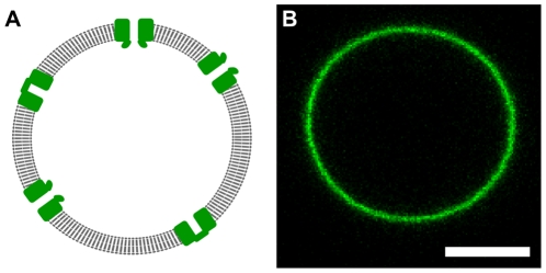
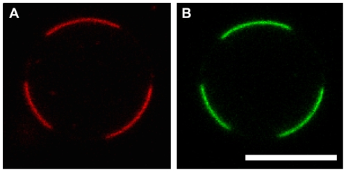
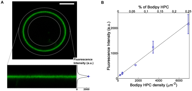
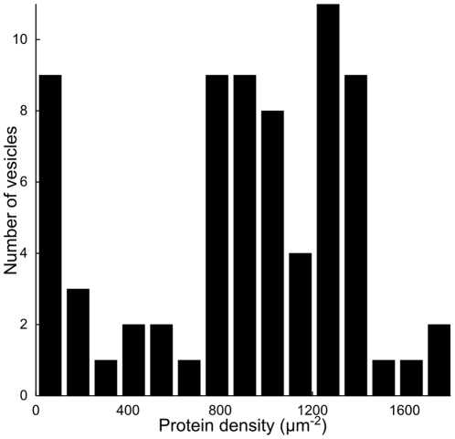
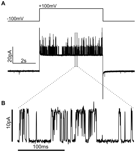
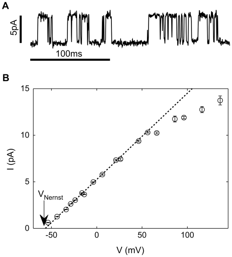
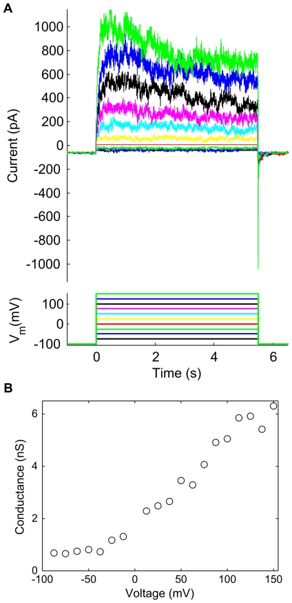
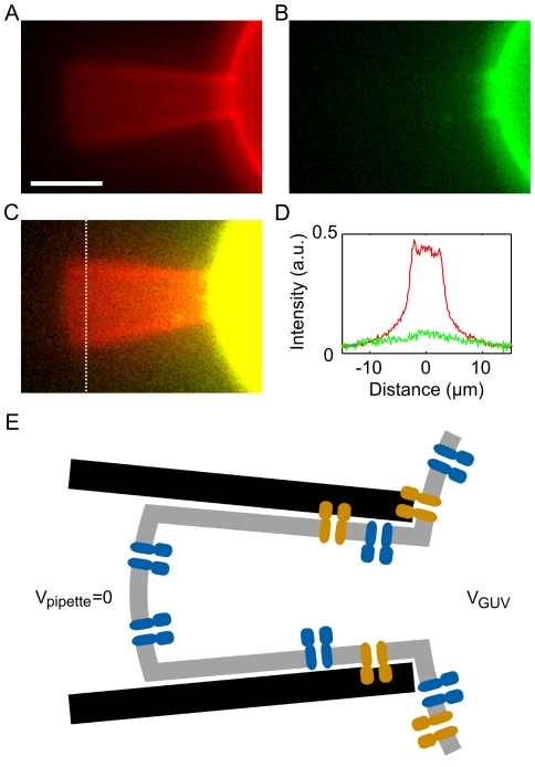
Voltage-gated ion channels are key players in cellular excitability. Recent studies suggest that their behavior can depend strongly on the membrane lipid composition and physical state. In vivo studies of membrane/channel and channel/channel interactions are challenging as membrane properties are actively regulated in living cells, and are difficult to control in experimental settings. We developed a method to reconstitute functional voltage-gated ion channels into cell-sized Giant Unilamellar Vesicles (GUVs) in which membrane composition, tension and geometry can be controlled. First, a voltage-gated potassium channel, KvAP, was purified, fluorescently labeled and reconstituted into small proteoliposomes. Small proteoliposomes were then converted into GUVs via electroformation. GUVs could be formed using different lipid compositions and buffers containing low (5 mM) or near-physiological (100 mM) salt concentrations. Protein incorporation into GUVs was characterized with quantitative confocal microscopy, and the protein density of GUVs was comparable to the small proteoliposomes from which they were formed. Furthermore, patch-clamp measurements confirmed that the reconstituted channels retained potassium selectivity and voltage-gated activation. GUVs containing functional voltage-gated ion channels will allow the study of channel activity, distribution and diffusion while controlling membrane state, and should prove a powerful tool for understanding how the membrane modulates cellular excitability.


Conflict of interest statement

Competing Interests: The authors have declared that no competing interests exist.

Figures

Figure 1
Figure 1. Giant unilamellar vesicle containing reconstituted KvAP.
A) Cartoon of ion channels (green) inserted into the single lipid bilayer (gray; thickness ∼5 nm) of a GUV (diameter ∼10 µm; not at scale). Many techniques used to study cell membranes can be applied to GUVs because they are unilamellar and of comparable size. B) Confocal image of a representative GUV containing KvAP labeled with Alexa488. Scale bar: 5 µm.
Figure 2
Figure 2. Partitioning of KvAP between co-existing liquid domains in GUVs.
A) Confocal fluorescence image of TR-DHPE, a red fluorescent lipid that preferentially segregates into the liquid disordered phase. B) KvAP (green) is enriched in the same membrane domains. The GUV was formed from a lipid mixture of DPhPC, DPPC, and Chol (6∶2∶2 by mole with 0.25% of TR-DHPE) and KvAP was included at a protein to lipid ratio of 1∶20 (by mass). The gain has been adjusted to show the membrane more clearly. Scale bar: 5 µm.
Figure 3
Figure 3. Calibration of GUV confocal fluorescence.
A) Confocal images of the equatorial plane of GUVs containing Bodipy-HPC (top) were transformed into a coordinate system centered on the membrane contour (bottom) in order to calculate the membrane intensity profile (right). Membrane fluorescence intensity was characterized by the profile maximum (blue cross). Scale bar: 5 µm. B) Membrane fluorescence intensity as a function of Bodipy-HPC density. Each point represents ∼10 vesicles (except for the third point (density 1800 µm−2) which represents a single vesicle) and error bars show the sample standard deviation. The slope of this calibration curve, Mref, relates membrane fluorescence intensity to fluorophore density.
Figure 4
Figure 4. Histogram of protein density in GUVs.
GUVs were formed using a low salt buffer (5 mM KCl, 1 mM HEPES, 400 mM sucrose) and protein density measured via quantitative confocal fluorescence (described in Text S4). The average protein density of the GUVs (1000±700 proteins/µm2) is comparable to the protein density of the (EPC∶EPA) SUVs from which they were formed (1600±400 proteins/µm2).
Figure 5
Figure 5. Activity of GUV Membrane Patches.
A) GUV membrane patch current in response to an applied voltage step. Patch and bath solutions were both 100 mM KCl, 4 mM HEPES, pH 7.2, and the patch was formed from a DPhPC GUV containing fluorescent KvAP. B) Section of the trace showing distinct jumps in conductance (∼100 pS) that are consistent with the opening and closing of individual channels.
Figure 6
Figure 6. Ionic selectivity.
A) Single channel current driven by ionic diffusion ([K+]bath = 100 mM, [K+]pipette = 10 mM, VNernst = −58 mV) overcome a small negative applied membrane potential (V = −4 mV). B) Dependence of single channel current, I, on membrane voltage, V. A linear fit for negative voltages (dotted line) gives a conductance of 93±2 pS and reversal voltage of −57±3 mV.
Figure 7
Figure 7. Voltage-dependent gating.
A) Response of patch membrane current to a transient step in voltage. Pipette and bath solutions both contained 100 mM KCl, and the membrane was held for 30 seconds at −100 mV between successive voltage steps. Membrane current was filtered at 500 Hz. B) Peak conductivity as a function of voltage.
Figure 8
Figure 8. Protein concentration in the patch membrane.
A–B) Epi-fluorescence image of the patch pipette and GUV showing the lipid (A, red) and KvAP (B, green). Red and green channels are scaled to be equal on the GUV body. C) Merged image with contrast increased to show the absence of protein (green) fluorescence in the patch. Scale Bar: 5 µm. D) Fluorescence intensity across the patch membrane (dotted line in C) of lipid (red) and KvAP (green). E) Schematic illustrating how adhesion between the membrane and pipette could favor the “correct” (blue; intracellular domain facing into the GUV interior) insertion over the “reverse” insertion (orange). The larger intracellular domain of the “reverse” insertion may stick more easily to the patch pipette walls, thereby excluding it from the patch membrane.

Similar articles

Cited by

References

    1. Hille B. Ion Channels of Excitable Membranes (3rd Edition) Sunderland: Sinauer Associates; 2001.
    1. Marban E. Cardiac channelopathies. Nature. 2002;415:213–8. - PubMed
    1. Ryan DP, Placek LJ. Episodic neurological channelopathies. Neuron. 2010;68:282–292. - PubMed
    1. Kullmann DM. Neurological channelopathies. Annu Rev Neurosci. 2010;33:151–172. - PubMed
    1. Lai HC, Jan LY. The distribution and targeting of neuronal voltage-gated ion channels. Nat Rev Neurosci. 2006;7:548–562. - PubMed

Publication types

MeSH terms

Substances