. 2011;6(10):e25625.
doi: 10.1371/journal.pone.0025625. Epub 2011 Oct 5.

The B-Raf status of tumor cells may be a significant determinant of both antitumor and anti-angiogenic effects of pazopanib in xenograft tumor models

Affiliations

The B-Raf status of tumor cells may be a significant determinant of both antitumor and anti-angiogenic effects of pazopanib in xenograft tumor models

Brunilde Gril et al. PLoS One. 2011.

Abstract

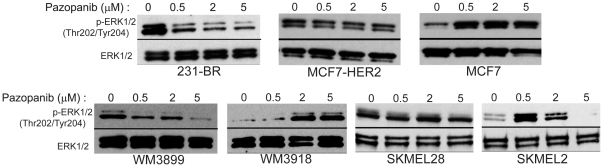
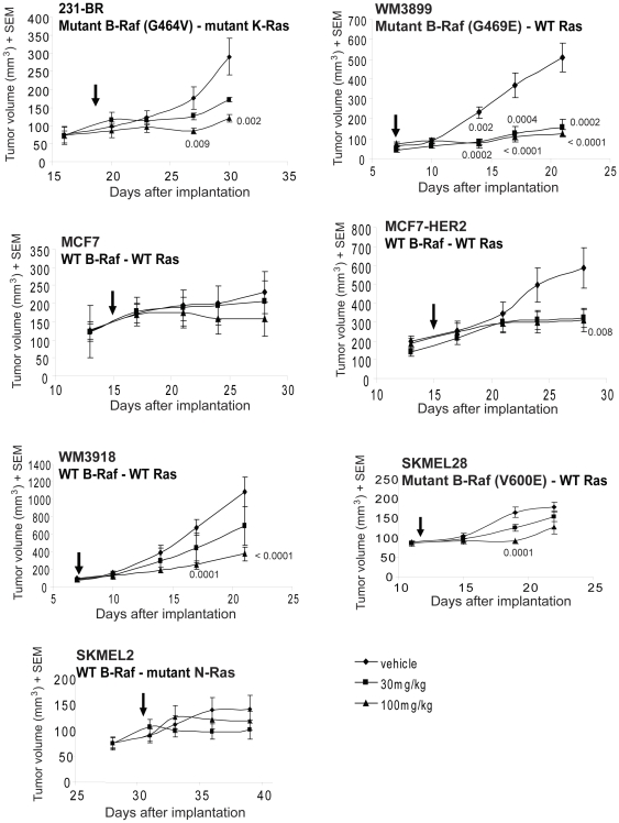
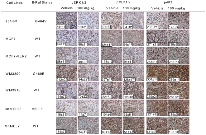
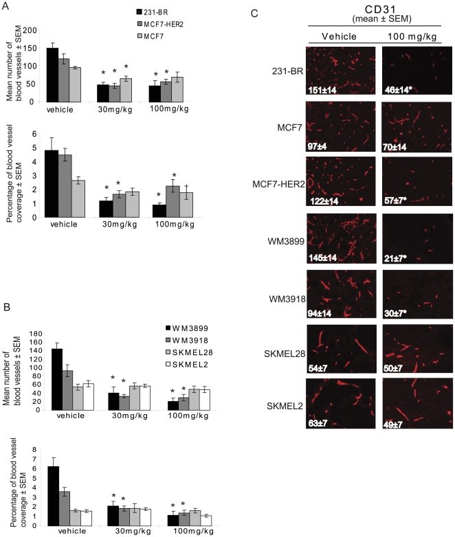
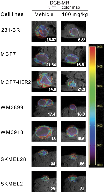
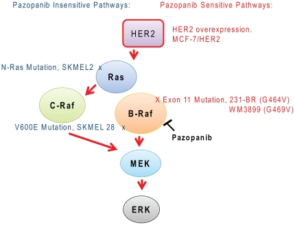
Pazopanib is an FDA approved Vascular Endothelial Growth Factor Receptor inhibitor. We previously reported that it also inhibits tumor cell B-Raf activity in an experimental brain metastatic setting. Here, we determine the effects of different B-Raf genotypes on pazopanib efficacy, in terms of primary tumor growth and anti-angiogenesis. A panel of seven human breast cancer and melanoma cell lines harboring different mutations in the Ras-Raf pathway was implanted orthotopically in mice, and tumor growth, ERK1/2, MEK1/2 and AKT activation, and blood vessel density and permeability were analyzed. Pazopanib was significantly inhibitory to xenografts expressing either exon 11 mutations of B-Raf, or HER2 activated wild type B-Raf; no significant inhibition of a xenograft expressing the common V600E B-Raf mutation was observed. Decreased pMEK staining in the responsive tumors confirmed that B-Raf was targeted by pazopanib. Interestingly, pazopanib inhibition of tumor cell B-Raf also correlated with its anti-angiogenic activity, as quantified by vessel density and area. In conclusion, using pazopanib, tumor B-Raf status was identified as a significant determinant of both tumor growth and angiogenesis.


Conflict of interest statement

Competing Interests: The authors declare a collaborative research agreement with GlaxoSmithKline. This does not alter the authors' adherence to all the PLoS ONE policies on sharing data and materials.

Figures

Figure 1
Figure 1. Effect of pazopanib on ERK activation.
Tumor cells were serum starved overnight and subsequently treated with pazopanib or DMSO for 24 hours. After treatment, cells were stimulated with 10 ng/mL VEGF for 10 minutes, and cell lysates were analyzed by immunoblot for pERK1/2. The data shown are representative of two conducted experiments.
Figure 2
Figure 2. Effect of pazopanib on primary tumor growth of breast cancer and melanoma cell lines.
Breast cancer lines were implanted in the mammary fat pad, while melanoma cells were implanted subcutaneously. Mice were randomly chosen to receive vehicle or 30 or 100 mg/kg pazopanib, twice daily for 10–14 days. Tumor size was measured twice weekly. Arrows indicate when treatment started. P values are shown for the tumors in which the decrease in tumor size achieved significance (p<0.01) at a given dose of pazopanib. The results shown for 231-BR, MCF7 and MCF7-HER2 cell lines are representative of two experiments. Raw data means and SEM are presented; analysis was performed on cubed root transformed data.
Figure 3
Figure 3. Pazopanib inhibition of primary tumor growth correlates with B-Raf inhibition.
Tumor cell lines and the B-Raf genotypes are listed. Representative photographs of pERK1/2, pMEK1/2 and pAKT immunostaining are shown for the vehicle and the 100 mg/kg pazopanib treated groups. The mean number of positive cells ± SEM is shown on each representative image. Asterisks (*) indicate a statistically significant difference between the pazopanib and vehicle treated group (p<0.01). (See Figure S3 for the 30 mg/kg dose data).
Figure 4
Figure 4. Anti-angiogenic effect of pazopanib on primary tumor xenografts correlates with B-Raf status.
Panel A: Breast cancer xenografts. Panel B- Melanoma xenografts. In each panel the top graph represents the mean number of blood vessels per tumor section (n = 5 mice) ± SEM and the bottom graph represents the percentage of area covered by blood vessels (n = 5 mice) ± SEM for the vehicle treated group, the 30 mg/kg group and the 100 mg/kg group. Panel C: Representative photographs of CD31 staining for vehicle and 100 mg/kg treatment groups. Asterisks (*) indicate a statistically significant difference between the pazopanib and vehicle treated groups (p<0.01). (See Figure S4 for representative photographs of CD31 staining).
Figure 5
Figure 5. Effect of pazopanib on blood vessel permeability of breast and melanoma cell xenografts.
Dynamic Contrast- Enhanced Magnetic Resonance Imaging (DCE-MRI) was performed on 5 mice per group at the end point. Representative photographs and Ktrans calculations are presented.
Figure 6
Figure 6. Schematic of pazopanib signaling pathways.
The standard receptor tyrosine kinase activation of the ERK pathway is shown. Pazopanib sensitive alterations, including HER2 overexpression and B-Raf exon 11 mutations, are shown on the right in red. Insensitive alterations are shown on the left in blue. Tumor cell lines featuring each alteration are named.

Similar articles

Cited by

References

    1. Kumar R, Knick VB, Rudolph SK, Johnson JH, Crosby RM, et al. Pharmacokinetic-pharmacodynamic correlation from mouse to human with pazopanib, a multikinase angiogenesis inhibitor with potent antitumor and antiangiogenic activity. Mol Cancer Ther. 2007;6:2012–2021. - PubMed
    1. Gril B, Palmieri D, Qian Y, Smart D, Ileva L, et al. Pazopanib reveals a role for tumor cell B-Raf in the prevention of HER2+ breast cancer brain metastasis. Clin Cancer Res. 2010;17:142–153. - PMC - PubMed
    1. Carbonell WS, Ansorge O, Sibson N, Muschel R. The vascular basement membrane as “soil” in brain metastasis. PLoS One. 2009;4:e5857. - PMC - PubMed
    1. Kusters B, Leenders WP, Wesseling P, Smits D, Verrijp K, et al. Vascular endothelial growth factor-A(165) induces progression of melanoma brain metastases without induction of sprouting angiogenesis. Cancer Res. 2002;62:341–345. - PubMed
    1. Leenders W, Kusters B, Verrijp K, Maass C, Wesseling P, et al. Antiangiogenic Therapy of Cerebral Melanoma Metastases Results in Sustained Tumor Progression via Vessel Co-Option. Clin Cancer Res. 2004;10:6222–6230. - PubMed

Publication types

MeSH terms