. 2011;6(10):e25813.
doi: 10.1371/journal.pone.0025813. Epub 2011 Oct 5.

3D Mapping of the SPRY2 domain of ryanodine receptor 1 by single-particle cryo-EM

Affiliations

3D Mapping of the SPRY2 domain of ryanodine receptor 1 by single-particle cryo-EM

Alex Perálvarez-Marín et al. PLoS One. 2011.

Abstract

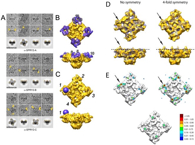
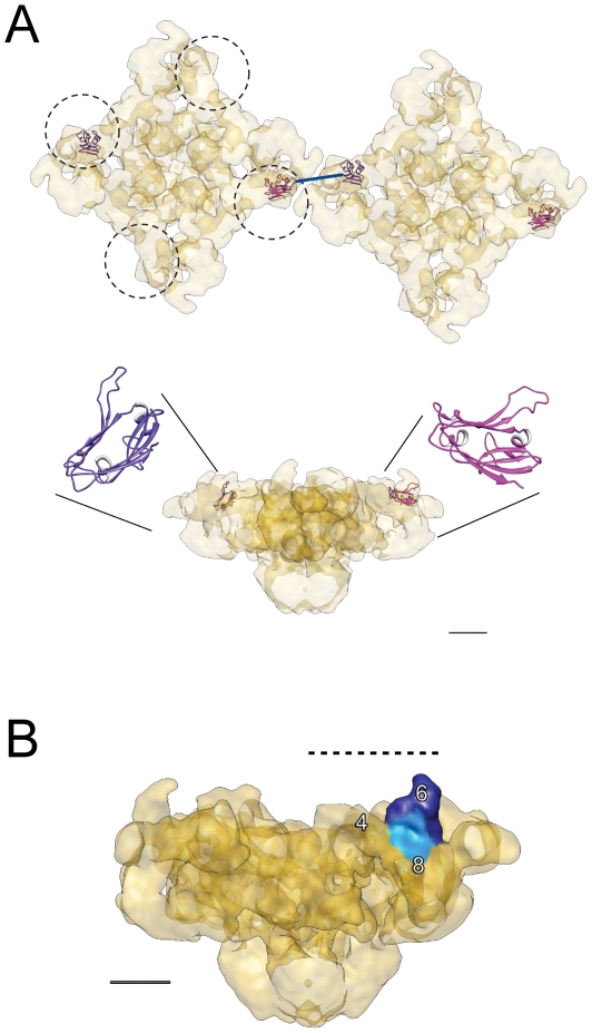
The type 1 skeletal muscle ryanodine receptor (RyR1) is principally responsible for Ca(2+) release from the sarcoplasmic reticulum and for the subsequent muscle contraction. The RyR1 contains three SPRY domains. SPRY domains are generally known to mediate protein-protein interactions, however the location of the three SPRY domains in the 3D structure of the RyR1 is not known. Combining immunolabeling and single-particle cryo-electron microscopy we have mapped the SPRY2 domain (S1085-V1208) in the 3D structure of RyR1 using three different antibodies against the SPRY2 domain. Two obstacles for the image processing procedure; limited amount of data and signal dilution introduced by the multiple orientations of the antibody bound in the tetrameric RyR1, were overcome by modifying the 3D reconstruction scheme. This approach enabled us to ascertain that the three antibodies bind to the same region, to obtain a 3D reconstruction of RyR1 with the antibody bound, and to map SPRY2 to the periphery of the cytoplasmic domain of RyR1. We report here the first 3D localization of a SPRY2 domain in any known RyR isoform.


Conflict of interest statement

Competing Interests: The authors have declared that no competing interests exist.

Figures

Figure 1
Figure 1. Immunoblotting of RyR1.
Immunodetection of the SPRY2 domain in purified RyR1 samples after SDS-PAGE (*) and native conditions (**) and in rabbit muscle vesicles (RyR1 in native conditions) (***). The antibodies used are the following: anti-SPRY2-A, anti-SPRY2-B, anti-SPRY2-C and anti-RyR-34C.
Figure 2
Figure 2. Cryo-EM fields of ice-embedded particles of RyR1 incubated with anti-SPRY2 antibodies.
A. Anti-SPRY2-A antibody. B. Anti-SPRY2-B antibody. C. Anti-SPRY2-C antibody. Scale bar, 100 nm.
Figure 3
Figure 3. 3D reconstruction of the RyR1 anti-SPRY2 binding site.
A. Unprocessed RyR1 particles incubated with the specified anti-SPRY2 antibody (raw images in upper row, antibody position indicated in yellow in middle row) compared to a calculated 2D projection (bottom row) for RyR1 containing four 30 Å radius spheres at the preliminary proposed binding region for the anti-SPRY2 antibody. For easier visualization the projection of the spheres has been highlighted in semitransparent yellow. B. 3D volumes of RyR1 displayed in two different orientations illustrating the preliminary location for the SPRY2 domain (purple shadowing). C. Preliminary assigned location for the anti-SPRY2 antibody (purple sphere) in the RyR1 3D reconstruction, at the vicinity of the domains 5 and 6. A set of four such spheres in the first, second, third and fourth repeats of the RyR1 (indicated by numerals) originated the 2D projections shown in the bottom rows of panel A. D. Low-resolution 3D reconstructions of RyR1 with anti-SPRY2 antibody (golden surface) in two orthogonal positions, without (left) and with (right) the use of fourfold symmetry. The black arrows point at the main difference between the 3D maps corresponding to the RyR1-antibody and the RyR1 control (purple mesh). E. 3D reconstruction with superimposed contour maps indicating the density level in the selected one-pixel slice (dashed line in panel D) for the non symmetrized and the fourfold symmetrized 3D reconstructions. The corresponding contour map for the control 3D volume (panel D, purple mesh) is displayed as a reference (bottom). The gradient scale indicates the density level in arbitrary units.
Figure 4
Figure 4. Docking of a homology model for the SPRY2 domain in the context of the RyR1-DHPR and inter-RyR1 interactions found in the triad junction.
A. Docking of a homology model for the SPRY2 domain in the anti-SPRY2 binding site of RyR1. The two RyR1s are in the typical lattice arrangement and the dashed-line circles indicate the region of overlap with the DHPR . The two SPRY2 orientations within the RyR1 originate from two slightly different starting locations for the antibody-binding site: that obtained from the unsymmetrized 3D volume (purple) and that obtained from the fourfold symmetrized 3D volume (magenta). The RyR1 with docked SPRY2 is shown in two orthogonal orientations. The distance between two proximal SPRY2 domains of two neighboring RyR1s measures 60 Å (blue line). B. Domains of RyR1 relevant for the interaction with the DHPR: DR2 region (domains 6–8, light blue [39]), and region of overlap with DHPR delimited by dashed line. The SPRY2 domain (domain 6) is shown in dark blue. The side view of RyR1 is rotated 45° around the fourfold axis with respect to the side views shown in panel A and Fig. 3, panels B–D. Scale bars, 5 nm.

Similar articles

Cited by

References

    1. Sencer S, Papineni RV, Halling DB, Pate P, Krol J, et al. Coupling of RYR1 and L-type calcium channels via calmodulin binding domains. J Biol Chem. 2001;276:38237–38241. - PubMed
    1. Cornea RL, Nitu F, Gruber S, Kohler K, Satzer M, et al. FRET-based mapping of calmodulin bound to the RyR1 Ca2+ release channel. Proc Natl Acad Sci U S A. 2009;106:6128–6133. - PMC - PubMed
    1. Samso M, Wagenknecht T. Apocalmodulin and Ca2+-calmodulin bind to neighboring locations on the ryanodine receptor. J Biol Chem. 2002;277:1349–1353. - PubMed
    1. Samso M, Shen X, Allen PD. Structural characterization of the RyR1-FKBP12 interaction. J Mol Biol. 2006;356:917–927. - PubMed
    1. Wagenknecht T, Radermacher M, Grassucci R, Berkowitz J, Xin HB, et al. Locations of calmodulin and FK506-binding protein on the three-dimensional architecture of the skeletal muscle ryanodine receptor. J Biol Chem. 1997;272:32463–32471. - PubMed

Publication types

MeSH terms

Substances