. 2011;6(10):e25938.
doi: 10.1371/journal.pone.0025938. Epub 2011 Oct 5.

A human multi-epitope recombinant vaccinia virus as a universal T cell vaccine candidate against influenza virus

Affiliations

A human multi-epitope recombinant vaccinia virus as a universal T cell vaccine candidate against influenza virus

Alan G Goodman et al. PLoS One. 2011.

Abstract

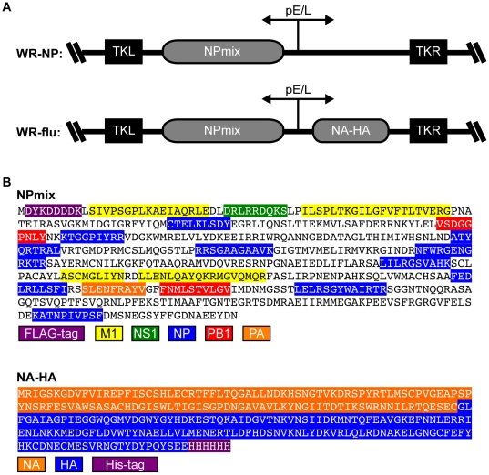
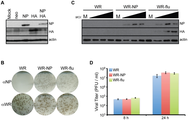
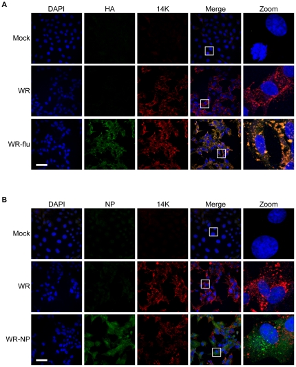
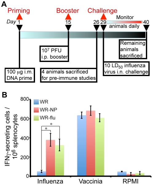
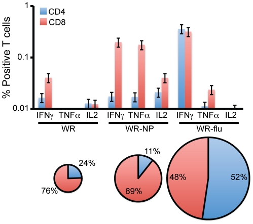
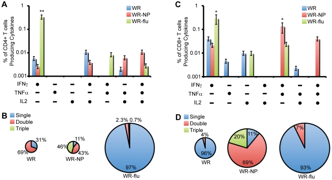
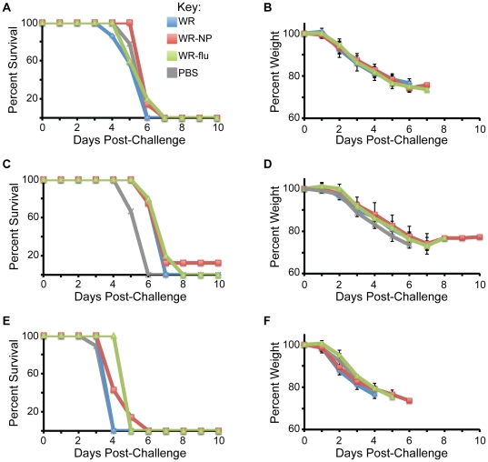
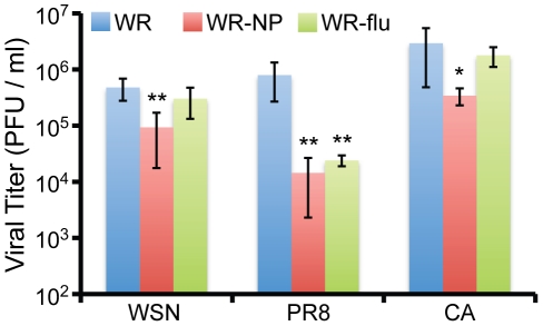
There is a need to develop a universal vaccine against influenza virus infection to avoid developing new formulations of a seasonal vaccine each year. Many of the vaccine strategies for a universal vaccine target strain-conserved influenza virus proteins, such as the matrix, polymerase, and nucleoproteins, rather than the surface hemagglutinin and neuraminidase proteins. In addition, non-disease-causing viral vectors are a popular choice as a delivery system for the influenza virus antigens. As a proof-of-concept, we have designed a novel influenza virus immunogen based on the NP backbone containing human T cell epitopes for M1, NS1, NP, PB1 and PA proteins (referred as NPmix) as well as a construct containing the conserved regions of influenza virus neuraminidase (N-terminal) and hemagglutinin (C-terminal) (referred as NA-HA). DNA vectors and vaccinia virus recombinants expressing NPmix (WR-NP) or both NPmix plus NA-HA (WR-flu) in the cytosol were tested in a heterologous DNA-prime/vaccinia virus-boost vaccine regimen in mice. We observed an increase in the number of influenza virus-specific IFNγ-secreting splenocytes, composed of populations marked by CD4(+) and CD8(+) T cells producing IFNγ or TNFα. Upon challenge with influenza virus, the vaccinated mice exhibited decreased viral load in the lungs and a delay in mortality. These findings suggest that DNA prime/poxvirus boost with human multi-epitope recombinant influenza virus proteins is a valid approach for a general T-cell vaccine to protect against influenza virus infection.


Conflict of interest statement

Competing Interests: The authors have declared that no competing interests exist.

Figures

Figure 1
Figure 1. Design and characterization of vaccine constructs.
(A) Recombinant influenza virus gene constructs (NPmix or NA-HA) were inserted at the TK locus of the Western Reserve (WR) of vaccinia virus and are driven by the synthetic early/late promoter (pE/L). The cloning vectors (shown) were introduced into the wild-type WR virus by homologous recombination and iterative plaque purification. (B) Amino acid sequences of NPmix and NA-HA recombinant influenza virus protein constructs. The backbone for the NPmix construct is influenza virus NP into which was inserted other influenza virus protein human T cell epitopes. Human T cell epitopes for influenza virus M1, NS1, NP, PB1, and PA proteins are indicated. The NA-HA construct consists of the conserved regions of H5N1 influenza virus neuraminidase (N-terminal amino acids 108–231) and hemagglutinin (C-terminal amino acids 347–511).
Figure 2
Figure 2. Recombinant viruses synthesize NP and HA proteins and replicate to similar levels.
(A) 10 µg of pCIneo (neo) vectors containing the NPmix (NP) or NA-HA (HA) inserts were transfected into BSC40 cells. For double transfections, 5 µg of each vector was used. 48 h post-transfection, cells were lysed and levels of influenza virus proteins were determined using antibodies for NP and HA. (B–D) BSC40 cells were infected with WR, WR-NP, or WR-flu at an MOI of 0.01 (B, D), 0.1, 1, or 10 (C) PFU/cell. (B) At 24 h p.i., cells were fixed and plaques were stained with NP antibody. (C) At 24 h p.i., the levels of NP and HA in the lysates were determined by immunoblot analysis. (D) At 8 or 24 h p.i., infectious virus present in the cells was measured in triplicate standard plaque assay on BSC40 cells.
Figure 3
Figure 3. Recombinant influenza virus proteins are present in the cytosol of infected cells.
BSC40 cells were infected with WR, WR-flu (A), or WR-NP (B) at an MOI of 1 PFU/cell. At 24 h p.i., cells were fixed with 2% paraformaldehyde, permeabilized, and stained with DAPI or antibodies recognizing influenza virus HA (A), NP (B), or vaccinia virus 14 K (A27 gene). Bar = 25 µm.
Figure 4
Figure 4. Immunogenicity of WR-NP and WR-flu in mice.
(A) Immunization schedule. BALB/c mice were primed with 100 µg of DNA (either 100 µg pCIneo-NPmix or empty vector, or 50 µg pCIneo-NPmix+50 µg pCIneo-HANA) intramuscularly (i.m.) at the start of the vaccination protocol. Two weeks later, the mice were boosted by intraperitoneal (i.p.) infection with 107 PFU of WR, WR-NP, or WR-flu. Eleven days post-boost, four mice were sacrificed to analyze the adaptive immune response. The remaining mice were challenged with influenza virus A/WSN/33, A/PR/8/34, or A/California/07/09. (B) Vaccine-elicited T cell responses of splenocytes 25 d after the start of the immunization protocol were measured in triplicate for each immunization group by fresh IFNγ ELISPOT assay following stimulation with influenza virus NP peptide TYQRTRALV, vaccinia virus E3 peptide VGPSNSPTF, or RPMI media alone. The results represent the mean number of IFNγ-secreting cells per 106 splenocytes from three biological replicates ± standard deviations. P values from a two-tailed t test assuming nonequal variance are indicated (*, P<0.05).
Figure 5
Figure 5. Phenotypic analysis of vaccine-induced CD4+ and CD8+ T cell responses.
The same groups of splenocytes as described in Figure 4 were stimulated with the influenza virus NP-specific peptide and analyzed using polychromatic flow cytometry. The results represent the mean number of CD4+ and CD8+ T cells secreting IFNγ, TNFα, or IL2 in each immunization group using three biological replicates ± standard error. The background from unstimulated controls was subtracted in all cases. The pie charts represent the magnitude and percentage of CD4+ and CD8+ T cells secreting cytokines in each immunization group.
Figure 6
Figure 6. Polyfunctionality of influenza virus-specific CD4+ and CD8+ T cells.
(A, C) Functional composition of CD4+ (A) or CD8+ (C) T cells responses against influenza virus NP peptide based on the secretion of IFNγ, IL-2, or TNFα. All the possible combinations of the responses are shown on the x-axis, whereas the percentages of the functionally distinct cell populations are shown on the y-axis. Bars correspond to the fraction of different functionally distinct T-cell populations within total CD4+ or CD8+ populations. Responses are grouped and color-coded on the basis of the number functions. (*, P<10−5; **, P<10−25) (B, D) The pie chart summarizes the data and each slice of the pie correspond to the fraction of CD4+ T cells with a given number of functions within the total CD4+ (B) or CD8+ (D) T cell populations. The size of the pie chart represents the magnitude of the specific influenza virus immune response induced.
Figure 7
Figure 7. Vaccination delays mortality in influenza virus-challenged mice.
Two weeks following the end of the vaccination protocol, 5–8 mice from each vaccination group and 9 mice from control PBS-inoculated animals were infected with 10×LD50 of the A/WSN/33 (A, B), A/PR/8/34 (C, D), or A/California/07/09 (E, F) strains of influenza virus. Mice were sacrificed when body weight reached 75% of starting weight.
Figure 8
Figure 8. Vaccination reduces the levels of infectious virus in the lungs of influenza virus-challenged mice.
Two weeks following the end of the vaccination protocol, mice from each vaccination group were infected with 10×LD50 of the A/WSN/33 (WSN), A/PR/8/34 (PR8), or A/California/07/09 (CA) strains of influenza virus. Mice were sacrificed at 5 d p.i. (except for some mice challenged with CA that died at 3–4 d p.i), and diaphragmatic lung lobes were isolated and homogenized. Levels of infectious virus were determined in triplicate by plaque assay on MDCK cells. The results represent the mean activity of 5–8 independent samples per group ± standard deviation. P values from a two-tailed t test assuming nonequal variance are indicated (*, P<0.05; **, P<0.01).

Similar articles

Cited by

References

    1. Regoes RR, Bonhoeffer S. Emergence of Drug-Resistant Influenza Virus: Population Dynamical Considerations. Science. 2006;312:389–391. - PubMed
    1. Gibbs MJ, Armstrong JS, Gibbs AJ. Recombination in the Hemagglutinin Gene of the 1918 “Spanish Flu”. Science. 2001;293:1842–1845. - PubMed
    1. Taubenberger JK, Reid AH, Janczewski TA, Fanning TG. Integrating historical, clinical and molecular genetic data in order to explain the origin and virulence of the 1918 Spanish influenza virus. Philos Trans R Soc Lond B Biol Sci. 2001;356:1829–1839. - PMC - PubMed
    1. Li KS, Guan Y, Wang J, Smith GJD, Xu KM, et al. Genesis of a highly pathogenic and potentially pandemic H5N1 influenza virus in eastern Asia. Nature. 2004;430:209–213. - PubMed
    1. Thompson WW, Shay DK, Weintraub E, Brammer L, Bridges CB, et al. Influenza-associated hospitalizations in the United States. JAMA. 2004;292:1333–1340. - PubMed

Publication types

MeSH terms