. 2011;6(10):e25945.
doi: 10.1371/journal.pone.0025945. Epub 2011 Oct 6.

Generating a knockdown transgene against Drosophila heterochromatic Tim17b gene encoding mitochondrial translocase subunit

Affiliations

Generating a knockdown transgene against Drosophila heterochromatic Tim17b gene encoding mitochondrial translocase subunit

Mikael Garabedian et al. PLoS One. 2011.

Abstract

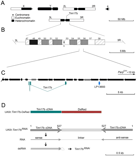
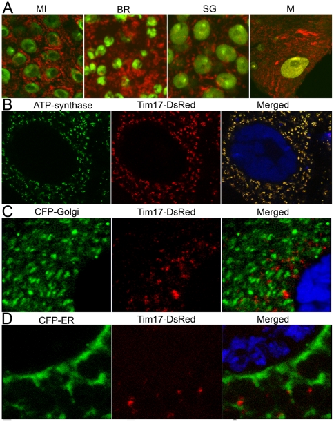
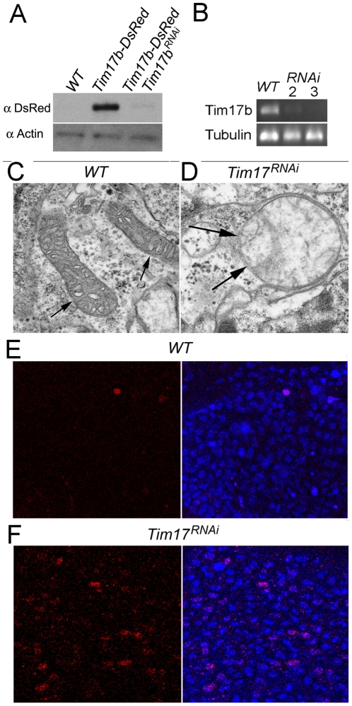
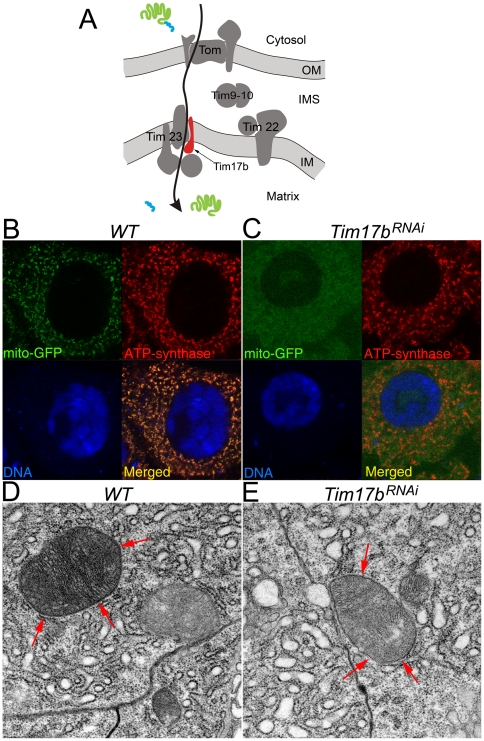
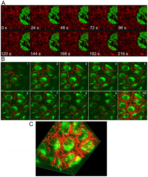
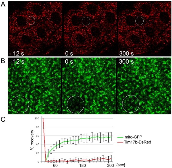
Heterochromatic regions of eukaryotic genomes contain multiple functional elements involved in chromosomal dynamics, as well as multiple housekeeping genes. Cytological and molecular peculiarities of heterochromatic loci complicate genetic studies based on standard approaches developed using euchromatic genes. Here, we report the development of an RNAi-based knockdown transgenic construct and red fluorescent reporter transgene for a small gene, Tim17b, which localizes in constitutive heterochromatin of Drosophila melanogaster third chromosome and encodes a mitochondrial translocase subunit. We demonstrate that Tim17b protein is required strictly for protein delivery to mitochondrial matrix. Knockdown of Tim17b completely disrupts functions of the mitochondrial translocase complex. Using fluorescent recovery after photobleaching assay, we show that Tim17b protein has a very stable localization in the membranes of the mitochondrial network and that its exchange rate is close to zero when compared with soluble proteins of mitochondrial matrix. These results confirm that we have developed comprehensive tools to study functions of heterochromatic Tim17b gene.


Conflict of interest statement

Competing Interests: The authors have declared that no competing interests exist.

Figures

Figure 1
Figure 1. Tim17b gene is located within constitutive heterochromatin of third chromosome.
A. Schematic illustration of Drosophila melanogaster chromosomes. Heterochromatic regions are shown in black. B. An ideogram of chromosome 3 heterochromatin. C. A scheme summarizing the sequence organization around Tim17b locus. Retrotransposable and transposable repeated DNA are shown by black arrows (arrowhead – 3′ end). Genes identified by homology to known cDNAs are shown below (blue boxes correspond to exons). Distance to closest characterized locus (Parp) is indicated. D. Generation of UAS-Tim17b-DsRed and UAS-Tim17bRNAi transgenes. Structure of transgenic constructs is shown. Nucleotide positions of Tim17b cDNA are indicated.
Figure 2
Figure 2. Tim17b-DsRed recombinant protein is localized to mitochondria.
A. Tim17b-DsRed (red) recombinant protein labels mitochondria in all Drosophila tissues. MI – mid-intestine; BR – brain; SG – salivary glands; M – body wall muscles. DNA is stained with OliGreen dye (green). B–D. Tim17b-DsRed recombinant protein is co-localized with mitochondrial protein ATP-synthase (B), but not with Golgi (C) or Endoplasmic Reticulum (ER) (D) markers. The dissected larval salivary glands expressing Tim17b-DsRed (red) were stained with anti-ATP-synthase antibody (green) and DNA binding dye Draq5 (blue) (B). The dissected larval salivary glands co-expressing Tim17b-DsRed (red) and CFP-Golgi (green) (C) or CFP-ER (green) proteins (D) were stained with DNA binding dye Draq5 (blue).
Figure 3
Figure 3. Expression of Tim17bRNAi transgene disrupts Tim17b protein production.
A. Co-expression of Tim17b-DsRed recombinant protein with Tim17bRNAi eliminates Tim17b-DsRed protein production. Western blot hybridization was used to compare the amount of Tim17b-DsRed protein in Gal4 69B; UAS-Tim17b-DsRed and Gal4 69B; UAS-Tim17b-DsRed; UAS-Tim17bRNAi larvae. Anti-Actin antibody was used as a loading control. B. Expression of Tim17bRNAi disrupts intrinsic Tim17b mRNA production. RT-PCR using intrinsic Tim17b-specific primers demonstrates that accumulation of Tim17b mRNA is abolished in Tim17bRNAi-expressing animals. The numbers 2 and 3 indicate age of animals collected for analysis after egg laying. Primers specific to Tubulin mRNA were used as a loading control. CD. Expression of Tim17bRNAi disrupts mitochondria. The structure of wild-type mitochondria detected by TEM (C) is affected in Tim17bRNAi-expressing animals (D). Arrows indicate mitochondria. EF. Expression of Tim17bRNAi increases apoptosis in larval brain. WT – wild- type first-instar larvae (E). Tim17bRNAi-expressing first-instar larvae (F). Dissected larval brains were stained using ApopTag (red), which detects apoptotic cells. DNA visualized using OliGreen dye (blue).
Figure 4
Figure 4. Elimination of Tim17b function by RNAi expression disrupts protein delivery to mitochondrial matrix.
A. Model of organization and function of mitochondrial translocase complexes TOM, TIM9-10, TIM23 and TIM22. Arrow shows pathway of protein with cleavable N-terminal signal translocation through TOM-TIM23 into matrix. Position of Tim17b protein (red) in TIM23 complex is shown. OM – outer membrane; IMS – intermembrane space; IM – inner membrane. Green – protein of mitochondrial matrix. Blue – cleavable N-terminal signal peptide. B–C. Tim17 is required for mito-GFP delivery to mitochondria. The dissected larval mid-intestine expressing mito-GFP (green) (B) or co-expressing mito-GFP (green) with Tim17bRNAi (C) were stained with anti-ATP-synthase antibody (red) and TOTO3 DNA binding dye (blue). Displacement of mito-GFP from mitochondria is clearly seen in Tim17bRNAi–expressing tissues. D E. TEM analysis of tissues shown in B C panels demonstrates that mitochondrial structure has still not been compromised by the time mito-GPF delivery to mitochondria is disrupted. Red arrows indicate mitochondria.
Figure 5
Figure 5. Use of Tim17b-DsRed fluorescent reporter for time-lapse microscopy of mitochondria, 3D reconstruction of mitochondrial network and mitochondrial proteins dynamics assay.
A. Time-lapse microscopy of live Drosophila tissue. Tim17b-DsRed is shown in red color. DNA is detected using Draq5 dye (green). B–C. 3D-mitochondrial network within mid-intestinal cells. 1–9 represent individual confocal sections. PR – X-Y projection. C. 3D reconstruction of the mitochondrial network is presented. Tim17b-DsRed is shown in red color. DNA is detected using TOTO3 dye (green).
Figure 6
Figure 6. Fluorescence Recovery After Photobleaching (FRAP) assay for Tim17b-DsRed and mito-GFP proteins.
A. Tim17b-DsRed (red). B. mito-GFP (green). White circle shows position of photobleached area (A B). In contrast to soluble GFP protein of mitochondrial matrix, assay demonstrates that Tim17b, which is a transmembrane protein, has a very slow replacement/dynamic rate (C).

Similar articles

Cited by

References

    1. Heitz E. Das geterochromatinder moose. J Jh Wiss. 1933;69:762–818.
    1. Blower MD, Sullivan B, Karpen GH. Conserved Organization of Centromeric Chromatin in Flies and Humans. Dev Cell. 2002;2:319–330. - PMC - PubMed
    1. Adams MD, Celniker SE, Holt RA, Evans CA, Gocayne JD et al. The Genome Sequence of Drosophila melanogaster. Science. 2000;287:2185–2195. - PubMed
    1. Gatti M, Pimpinelli S. Cytological and genetical analysis of the Y chromosome of Drosophila melanogaster. . Chromosoma. 1983;88:593–617.
    1. Lohe AR, Hilliker AJ, Roberts PA. Mapping Simple Repeated DNA Sequences in Heterochromatin of Drosophila melanogaster. Genetics. 1993;134:1149–1174. - PMC - PubMed

Publication types

MeSH terms

LinkOut - more resources