. 2011;6(10):e25984.
doi: 10.1371/journal.pone.0025984. Epub 2011 Oct 6.

Membrane-bound steel factor maintains a high local concentration for mouse primordial germ cell motility, and defines the region of their migration

Affiliations

Membrane-bound steel factor maintains a high local concentration for mouse primordial germ cell motility, and defines the region of their migration

Ying Gu et al. PLoS One. 2011.

Abstract

Steel factor, the protein product of the Steel locus in the mouse, is a multifunctional signal for the primordial germ cell population. We have shown previously that its expression accompanies the germ cells during migration to the gonads, forming a "travelling niche" that controls their survival, motility, and proliferation. Here we show that these functions are distributed between the alternatively spliced membrane-bound and soluble forms of Steel factor. The germ cells normally migrate as individuals from E7.5 to E11.5, when they aggregate together in the embryonic gonads. Movie analysis of Steel-dickie mutant embryos, which make only the soluble form, at E7.5, showed that the germ cells fail to migrate normally, and undergo "premature aggregation" in the base of the allantois. Survival and directionality of movement is not affected. Addition of excess soluble Steel factor to Steel-dickie embryos rescued germ cell motility, and addition of Steel factor to germ cells in vitro showed that a fourfold higher dose was required to increase motility, compared to survival. These data show that soluble Steel factor is sufficient for germ cell survival, and suggest that the membrane-bound form provides a higher local concentration of Steel factor that controls the balance between germ cell motility and aggregation. This hypothesis was tested by addition of excess soluble Steel factor to slice cultures of E11.5 embryos, when migration usually ceases, and the germ cells aggregate. This reversed the aggregation process, and caused increased motility of the germ cells. We conclude that the two forms of Steel factor control different aspects of germ cell behavior, and that membrane-bound Steel factor controls germ cell motility within a "motility niche" that moves through the embryo with the germ cells. Escape from this niche causes cessation of motility and death by apoptosis of the ectopic germ cells.


Conflict of interest statement

Competing Interests: The authors have declared that no competing interests exist.

Figures

Figure 1
Figure 1. Expression of Steel factor in mouse embryos.
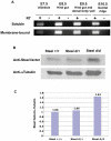
(A) RT-PCR analysis of membrane-bound and soluble Steel factor. cDNA was prepared from dissected E7.5 allantois, E8.5 hind gut, E9.5 hind gut and dorsal body wall, and E10.5 genital ridge. (B) Expression levels of soluble Steel factor protein in embryos of different Steel-dickie genotypes measured by western blot. (C) Densitometric analysis of western blots in (B). α-tubulin antibody was used as a loading control.
Figure 2
Figure 2. PGC number in Steeld/d embryos and in vitro culture.
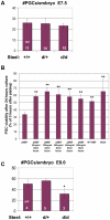
(A) There was no significant change in PGC numbers at E7.5 in Steeld/d embryos compared to their littermates. “n” indicates the number of embryos used for quantitation. (B) PGC number after 24 hours in vitro culture in medium with or without soluble recombinant Steel factor on different feeder layer cells. Y axis represents the ratio of PGC number 24 hours after plating versus 3 hours after plating. ΔMEF: primary MEF from Steel-null embryos. M220: stromal cell line express only membrane-bound Steel factor. ** = p<0.01. (C) PGC number reduced significantly in E8.0 Steeld/d embryos compared to their littermates. “n” indicates the number of embryos used for quantitation. * = p<0.05.
Figure 3
Figure 3. PGC migration in Steeld/d embryos at E7.5.
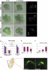
(Column A, B) Frames at t = 0 and t = 6 hours respectively from movies of E7.5 embryos with different Steel-dickie genotypes. (Column C) Tracks were made from PGCs in the allantois (white boxes) that remained in the plane of the confocal image throughout the movies. The white line in C indicates the boundary between the extraembryonic tissues (EEM), and the posterior end of the embryo (PEM). Scale bars in (A–C): 100 µm. (D) The maximum velocity, average velocity, and displacement of E7.5 PGCs with different Steel-dickie genotypes. PGCs in Steeld/d embryos showed significantly decreased velocities and displacement compared to wild type littermates. “n” indicates the number of PGCs used for quantitation. Units on the “Y” axis vary based upon parameter, and are indicated below the bar charts. ** = p<0.01. (E) The percentage of PGCs which enter the posterior of the embryo is significantly reduced in Steeld/d embryos. (F) The percentage of PGCs which form clusters is dramatically increased in Steeld/d embryos. “n” indicates the number of embryos used for quantitation for (E) and (F). ** = p<0.01. (G) PGCs in E8.0 wild type and Steeld/d embryos. The left diagram shows an E8.0 embryo with PGCs migrating along the hind gut (yellow). Arrow shows the direction of PGC migration. Red box indicates the area shown in the image on the right.
Figure 4
Figure 4. Effects of soluble recombinant Steel factor on PGC motility in Steeld/d embryos at E7.5 and in vitro culture.
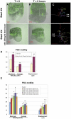
(Column A, B) Frames at t = 0 and t = 6 hours respectively from movies of E7.5 Steeld/d embryos with or without addition of 200 ng/ml soluble recombinant Steel factor. (Column C) Tracks were made from PGCs in the allantois (white boxes) that remained in the plane of the confocal image throughout the movies. The white line indicates the boundary between the extraembryonic tissues (EEM), and the posterior end of the embryo (PEM). Scale bars in (A–C): 100 µm. (D) The maximum velocity, average velocity, and displacement of PGCs in E7.5 Steeld/d embryos were significantly increased by adding of 200 ng/ml soluble recombinant Steel factor into culture medium for 6 hours. “n” indicates the number of PGCs used for quantitation. Units on the “Y” axis vary based upon parameter, and are indicated below the bar charts. ** = p<0.01. (E) The maximum velocity, average velocity, and displacement of PGCs after 24 hours in vitro culture with increasing concentration of soluble recombinant Steel factor on Steel-null MEFs (ΔMEF). Units on the “Y” axis vary based upon parameter, and are indicated below the bar charts. * = p<0.05.
Figure 5
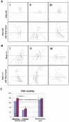
Figure 5. Effects of soluble recombinant Steel factor on PGC directions.
Directions of individual PGC migration in Steeld/d embryos (A) or wild type embryos (B) with or without addition of 200 ng/ml soluble recombinant Steel factor. The boundary between the extraembryonic tissues (EEM), and the posterior end of the embryo (PEM), is marked by a line. Column I, II, and III are representative images from 3 different embryos of the same genotype labeled on the left. (C) The maximum velocity, average velocity, and displacement of PGCs in E7.5 wild type embryos with or without addition of 200 ng/ml soluble recombinant Steel factor into culture medium for 6 hours. “n” indicates the number of PGCs used for quantitation. Units on the “Y” axis vary based upon parameter, and are indicated below the bar charts. ** = p<0.01.
Figure 6
Figure 6. Effects of soluble recombinant Steel factor on PGC motility in wild type embryos at E11.0.
(Column A-C) Frames at t = 0, t = 6 and t = 12 hours respectively from movies of E11.0 wild type embryo slices with or without addition of 200 ng/ml soluble recombinant Steel factor. (D) The relative expression level of membrane-bound and soluble Steel factor mRNA in E10.5 genital ridges, E11.5 genital ridges and E11.5 midline mysenchyme as determined by real-time RT-PCR. *p<0.05, **p<0.01. (E and F) E-cadherin expression in PGCs without (E) or with (F) addition of Steel factor. Upper panels show E-cadherin (red) staining. The lower panels show the merged images with PGC marker Stella-GFP (green). No difference in expression of E-cadherin was observed between groups.

Similar articles

Cited by

References

    1. Hayashi K, de Sousa Lopes SM, Surani MA. Germ cell specification in mice. Science. 2007;316:394–396. - PubMed
    1. Ohinata Y, Payer B, O'Carroll D, Ancelin K, Ono Y, et al. Blimp1 is a critical determinant of the germ cell lineage in mice. Nature. 2005;436:207–213. - PubMed
    1. Anderson R, Copeland TK, Scholer H, Heasman J, Wylie C. The onset of germ cell migration in the mouse embryo. Mech Dev. 2000;91:61–68. - PubMed
    1. Gu Y, Runyan C, Shoemaker A, Surani A, Wylie C. Steel factor controls primordial germ cell survival and motility from the time of their specification in the allantois, and provides a continuous niche throughout their migration. Development. 2009;136:1295–1303. - PubMed
    1. Molyneaux KA, Stallock J, Schaible K, Wylie C. Time-lapse analysis of living mouse germ cell migration. Dev Biol. 2001;240:488–498. - PubMed

Publication types

MeSH terms

LinkOut - more resources