. 2011;6(10):e25998.
doi: 10.1371/journal.pone.0025998. Epub 2011 Oct 5.

TLR1/TLR2 heterodimers play an important role in the recognition of Borrelia spirochetes

Affiliations

TLR1/TLR2 heterodimers play an important role in the recognition of Borrelia spirochetes

Marije Oosting et al. PLoS One. 2011.

Abstract

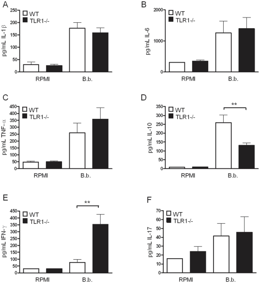
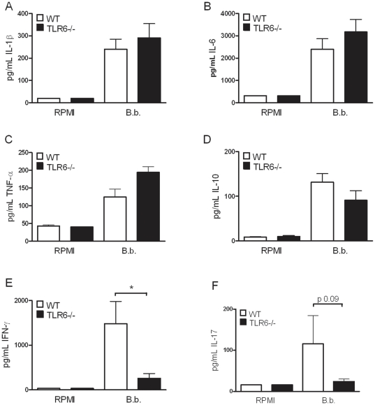
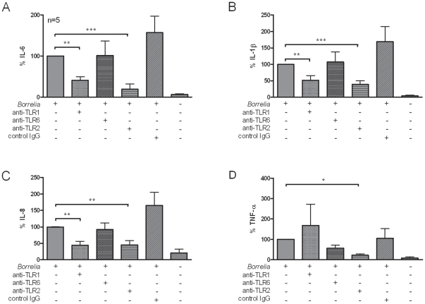
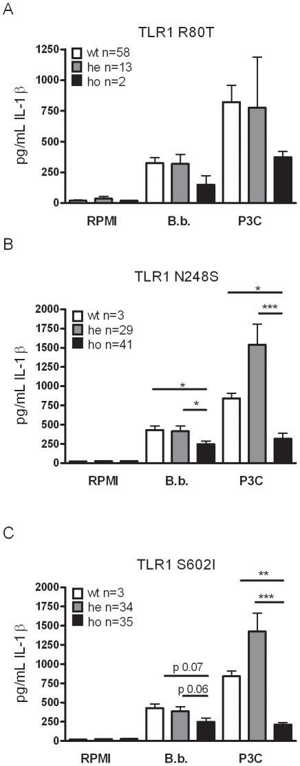
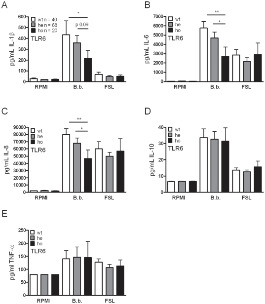
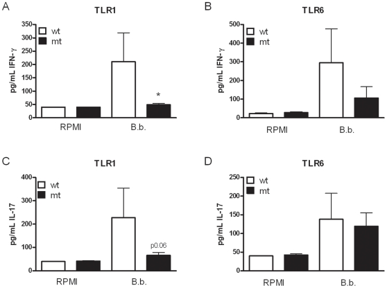
After infection with Borrelia species, the risk for developing Lyme disease varies significantly between individuals. Recognition of Borrelia by the immune system is mediated by pattern recognition receptors (PRRs), such as TLRs. While TLR2 is the main recognition receptor for Borrelia spp., little is known about the role of TLR1 and TLR6, which both can form functionally active heterodimers with TLR2. Here we investigated the recognition of Borrelia by both murine and human TLR1 and TLR6. Peritoneal macrophages from TLR1- and TLR6- gene deficient mice were isolated and exposed to Borrelia. Human PBMCs were stimulated with Borrelia with or without specific TLR1 and TLR6 blocking using specific antibodies. Finally, the functional consequences of TLR polymorphisms on Borrelia-induced cytokine production were assessed. Splenocytes isolated from both TLR1-/- and TLR6-/- mice displayed a distorted Th1/Th2 cytokine balance after stimulation with B.burgdorferi, while no differences in pro-inflammatory cytokine production were observed. In contrast, blockade of TLR1 with specific neutralizing antibodies led to decreased cytokine production by human PBMCs after exposure to B.burgdorferi. Blockade of human TLR6 did not lead to suppression of cytokine production. When PBMCs from healthy individuals bearing polymorphisms in TLR1 were exposed to B.burgdorferi, a remarkably decreased in vitro cytokine production was observed in comparison to wild-type controls. TLR6 polymorphisms lead to a minor modified cytokine production. This study indicates a dominant role for TLR1/TLR2 heterodimers in the induction of the early inflammatory response by Borrelia spirochetes in humans.


Conflict of interest statement

Competing Interests: The authors have declared that no competing interests exist.

Figures

Figure 1
Figure 1. In vitro cytokine production by TLR1−/− cells after stimulation with Borrelia.
1×10⁵ peritoneal macrophages from five C57Bl/6 mice were stimulated separately for 24 hours with 1×10⁶ live B.burgdorferi per mL. Levels of IL-1β (A), IL-6 (B), and TNF-α (C) were measured in the supernatants and compared to the cytokine production induced by cells deficient in expressing TLR1 (black bars, represent TLR1−/− cytokine responses). Spleen cells (5×10⁶/well) of both wild-type (black bars) and TLR1−/− (white bars) mice were stimulated for 5 days with 1×10⁶ live Borrelia per mL and levels of IL-10, IFN-γ, and IL-17 were measured in the supernatant using ELISA (D–F, respectively). Bars represent the mean ± SEM of 5 animals per group. **p<0.01 (for comparisons between wild-type and knock-out mice), Mann-Whitney U-test, experiments were performed in duplicates.
Figure 2
Figure 2. Recognition of Borrelia species by immune cells of TLR6−/− mice.
Peritoneal macrophages (1×10⁵/well) of C57Bl/6 wild-type or TLR6 gene deficient mice (n = 5 per group) were stimulated for 24 hours with 1×10⁶ live Borrelia per mL. Using ELISA or RIA, IL-1β (A), IL-6 (B), or TNF-α (C) levels were measured in pg/mL. 5×10⁶ spleen cells/well were stimulated with 1×10⁶ live Borrelia per mL. IL-10, IFN-γ, and IL-17 levels were determined in the supernatant of 5-days spleen cell culture (D–F, respectively). White bars represent cytokine induction after stimulation of wild-type cells, black bars the TLR6 knockout cells. An asterisk indicates that the P-value is <0.05 (for comparisons between wild-type and knock-out mice), 5 animals per group, Mann-Whitney U-test. Bars represent the mean ± SEM, experiments were performed in duplicates.
Figure 3
Figure 3. Borrelia is recognized by human TLR1 and TLR2.
(A) Peripheral blood mononuclear cells (PBMCs, 5×10⁵/well) from 6 healthy volunteers were stimulated for 24 h with 1×10⁶ B.burgdorferi per mL (grey bars). IL-6 production in the supernatant was measured using ELISA and showed in % where Borrelia induced cytokine production is set as 100% cytokine induction. Bars represent the means ± SEM. **p<0.01; ***p<0.001 (Mann-Whitney). Borrelia IL-6 production 100% was 30271±8607 pg/mL. Anti-TLR1, 10 µg/mL specific antibody; anti-TLR6, 10 µg/mL specific antibody; anti-TLR2, 10 µg/mL specific antibody; control IgG, mouse IgG1<$>\scale 80%\raster="rg1"<$> isotype control 10 µg/mL. (B) IL-1β production measured in supernatant after 24 hours culture of PBMCs stimulated with or without 1×10⁵ B.burgdorferi per mL or in the presence or absence of 10 µg/mL antibody. Borrelia IL-1β production 100% was 576±295 pg/mL. (C) IL-8 production. Borrelia IL-8 production 100% was 121±33 ng/mL (D) TNF-α production after 24 hours of stimulation. Borrelia TNF-α production 100% was 6528±2716 pg/mL Bars represent the means ± standard error of the means; *p<0.05; **p<0.01; ***p<0.001, Mann-Whitney U-test. The data shown are from three independent experiments each performed in duplicate.
Figure 4
Figure 4. Functional consequences of human TLR1 SNPs in cytokine production.
Peripheral blood mononuclear cells (PBMCs) from healthy volunteers were stimulated for 24 h with different stimuli, including Borrelia burgdorferi (1×10⁶ spirochetes/mL), and 10 µg/mL Pam3Cys. After stimulation, supernatants were collected, and cytokine levels were measured by enzyme-linked immunosorbent assay. The TLR1 status of these individuals was determined before PBMC stimulation, and they were separated into 3 groups—one group did not displayed the R80T SNP (A), N248S SNP (B), or S602I SNP (C) in TLR1 (white bars; wt; wild-type), one group had a heterozygous mutation (grey bars), and one group had a homozygous mutation (black bars). Data are means ± standard errors. IL-1β, interleukin 1β; RPMI, Roswell Park Memorial Institute 1640 medium; *p<0.05; **p<0.01; ***p<0.001, Mann-Whitney U-test. The data shown are from three independent experiments each performed in duplicate.
Figure 5
Figure 5. Less important role for human TLR6 in the induction of cytokines after Borrelia stimulation.
Peripheral blood mononuclear cells (PBMCs) from 128 healthy volunteers carrying the S249N SNP in TLR6 were stimulated with either medium, 1×10⁶ per mL Borrelia spirochetes, or 1 µg/mL FSL-1 for 24 h and cytokines were measured using ELISA; (interleukin 1β [IL-1β],(A); IL-6,(B); IL-8,(C); IL-10, (D); and tumor necrosis factor α [TNF-α],(E)). Bars represent individuals carrying no SNP (wild-type, wt, white bars), heterozygous SNP carriers (he, grey bars), or homozygous variation (ho, black bars). Data represent the mean ± SEM, *p<0.05; **p<0.01; Mann-Whitney U-test. The data shown are from three independent experiments each performed in duplicate.
Figure 6
Figure 6. Human TLR1 is involved in Borrelia-induced IFN-γ.
Peripheral blood mononuclear cells (PMBCs, 5×10⁵/ well) from individuals bearing a SNP in TLR1 (N248S) or TLR6 (S249P) were stimulated with medium, 1×10⁶ Borrelia per mL, or Pam3Cys (10 µg/mL), in the presence of 10% human pool serum. After 7 days of incubation, IFN-γ (A, B) and IL-17 (C, D) levels were determined in the supernatant using ELISA. Bars represent individuals carrying no SNP (wild-type, wt, white bars, n≥3 for TLR1 SNPs and n≥5 for TLR6 SNPs), or individuals bearing the SNP in one allele or in both alleles (mutant, mt, black bars, n≥6 for TLR1 SNPs and n≥7 for TLR6 SNPs). Data represent the means ± the standard error of the means, Mann-Whitney U test, *p<0.05. The data shown are from three independent experiments each performed in duplicate.

Similar articles

Cited by

References

    1. Burgdorfer W, Barbour AG, Hayes SF, Benach JL, Grunwaldt E, et al. Lyme disease-a tick-borne spirochetosis? Science. 1982;216:1317. - PubMed
    1. Balmelli T, Piffaretti JC. Association between different clinical manifestations of Lyme disease and different species of Borrelia burgdorferi sensu lato. . Res Microbiol. 1995;146:329. - PubMed
    1. Glickstein L, Moore B, Bledsoe T, Damle N, Sikand V, et al. Inflammatory cytokine production predominates in early Lyme disease in patients with erythema migrans. Infect Immun. 2003;71:6051. - PMC - PubMed
    1. Cruz AR, Moore MW, La Vake CJ, Eggers CH, Salazar JC, et al. Phagocytosis of Borrelia burgdorferi, the Lyme disease spirochete, potentiates innate immune activation and induces apoptosis in human monocytes. Infect Immun. 2008;76:56. - PMC - PubMed
    1. Hoshino K, Takeuchi O, Kawai T, Sanjo H, Ogawa T, et al. Cutting edge: Toll-like receptor 4 (TLR4)-deficient mice are hyporesponsive to lipopolysaccharide: evidence for TLR4 as the Lps gene product. J Immunol. 1999;162:3749. - PubMed

Publication types

MeSH terms