. 2011;6(10):e26032.
doi: 10.1371/journal.pone.0026032. Epub 2011 Oct 6.

Apolipoprotein J/Clusterin is a novel structural component of human erythrocytes and a biomarker of cellular stress and senescence

Affiliations

Apolipoprotein J/Clusterin is a novel structural component of human erythrocytes and a biomarker of cellular stress and senescence

Marianna H Antonelou et al. PLoS One. 2011.

Abstract

Background: Secretory Apolipoprotein J/Clusterin (sCLU) is a ubiquitously expressed chaperone that has been functionally implicated in several pathological conditions of increased oxidative injury, including aging. Nevertheless, the biological role of sCLU in red blood cells (RBCs) remained largely unknown. In the current study we identified sCLU as a component of human RBCs and we undertook a detailed analysis of its cellular topology. Moreover, we studied the erythrocytic membrane sCLU content during organismal aging, in conditions of increased organismal stress and accelerated RBCs senescence, as well as during physiological in vivo cellular senescence.

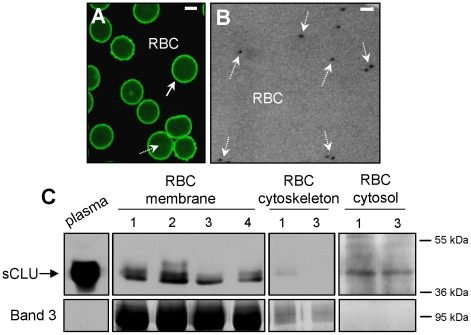
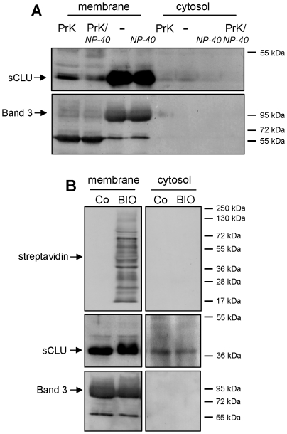
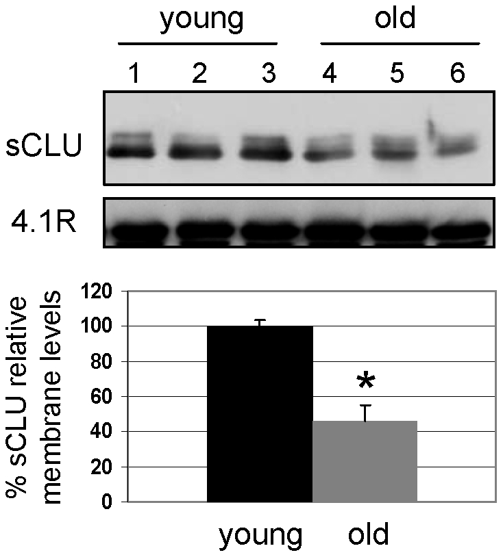
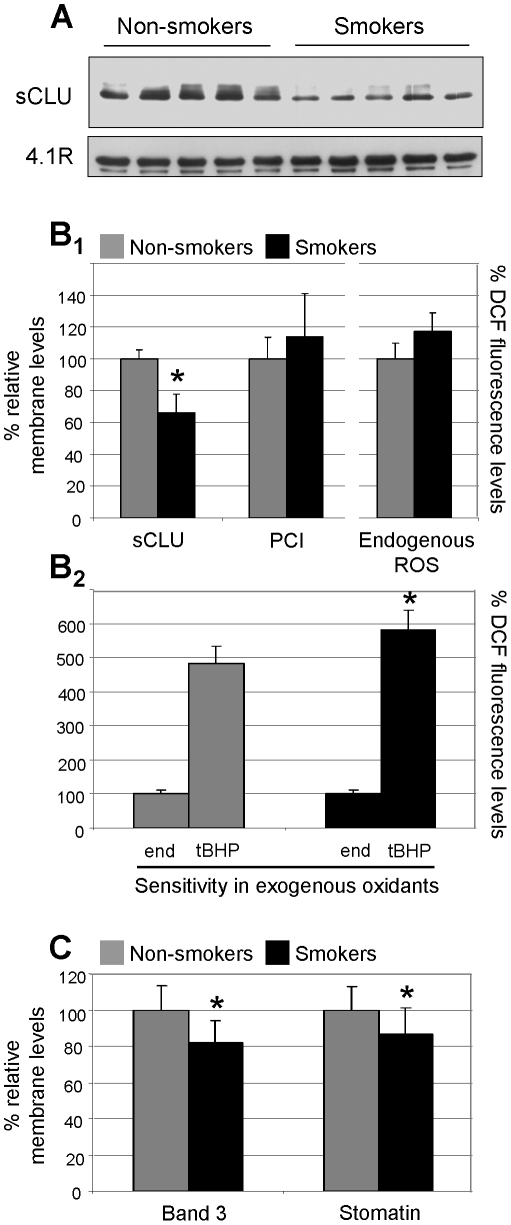
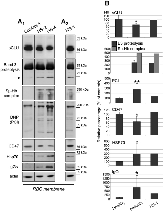
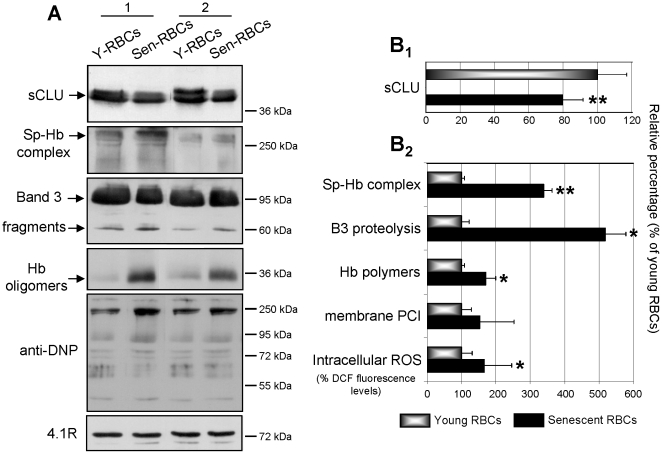
Methodology/principal findings: By using a combination of molecular, biochemical and high resolution microscopical methods we found that sCLU is a novel structural component of RBCs extra- and intracellular plasma membrane and cytosol. We observed that the RBCs membrane-associated sCLU decreases during organismal aging or exposure to acute stress (e.g. smoking), in patients with congenital hemolytic anemia, as well as during RBCs in vivo senescence. In all cases, sCLU reduction paralleled the expression of typical cellular senescence, redox imbalance and erythrophagocytosis markers which are also indicative of the senescence- and oxidative stress-mediated RBCs membrane vesiculation.

Conclusions/significance: We propose that sCLU at the mature RBCs is not a silent remnant of the erythroid precursors, but an active component being functionally implicated in the signalling mechanisms of cellular senescence and oxidative stress-responses in both healthy and diseased organism. The reduced sCLU protein levels in the RBCs membrane following cell exposure to various endogenous or exogenous stressors closely correlates to the levels of cellular senescence and redox imbalance markers, suggesting the usefulness of sCLU as a sensitive biomarker of senescence and cellular stress.


Conflict of interest statement

Competing Interests: The authors have declared that no competing interests exist.

Figures

Figure 1
Figure 1. sCLU is a structural component of human RBCs plasma membrane and cytosol.
(A) CLSM immunofluorescence or (B) TEM immunogold localization of sCLU in RBCs derived from healthy donors (representative of preparations in 3 or 2 subjects, respectively). Solid (A) or dashed (A and B) arrows indicate sCLU localization at the periphery of RBCs or intracellularly, respectively. Bars, CLSM, 3 µm; TEM, 100 nm. (C) Representative immunoblot analysis of isolated plasma (5 µg of total protein per lane), membrane (20 µg), cytoskeleton (20 µg) and cytosol (200 µg) purified RBCs fractions probed with an anti-sCLU antibody. Band 3 probing was used to demonstrate fraction purity and equal protein loading; numbers 1 to 4 denote different subjects (out of the 18 tested). Molecular weight markers are shown to the right of the blots.
Figure 2
Figure 2. RBCs sCLU localizes at both extra- and intracellular sides of the plasma membrane.
(A) Representative immunoblot analysis of purified membrane and cytosol fractions of untreated RBCs (-); RBCs treated with NP-40 (NP-40) or RBCs digested with proteinase K in the absence (PrK) or presence of NP-40 (PrK/NP-40). Samples (N = 3) were probed with specific anti-sCLU and anti-Band 3 antibodies. (B) Immunobloting of streptavidin, sCLU and Band 3 in purified membrane and cytosolic fractions of control (Co) or biotinylated (BIO) RBCs (N = 3). Molecular weight markers are shown to the right of the blots.
Figure 3
Figure 3. RBCs sCLU membrane levels are decreased during in vivo organismal aging.
Representative immunoblot analysis and collective densitometry of sCLU relative levels at the membrane of RBCs derived from young (N = 13) or old (N = 10) non-smoking healthy subjects. Probing with anti-4.1R protein was used as reference for equal protein loading. Densitometric data were normalized against the sCLU values of the young donors. Error bars, ±s.d; asterisk, significance at p<0.05.
Figure 4
Figure 4. Decreased sCLU membrane levels in the RBCs of (otherwise healthy) smokers.
(A) Representative immunoblot analysis of sCLU membrane levels in RBCs derived from healthy middle-aged cigarette smoking (N = 8) or non-smoking (N = 8) subjects (upper panel). 4.1R protein probing (lower panel) was used as reference for equal protein loading. (B1) Collective densitometric analysis of sCLU relative membrane content; PCI (data from not shown immunoblots) and endogenous cellular ROS from the smoking and non-smoking subjects. (B2) ROS measurement in RBCs from smokers and non-smokers challenged with exogenous tBHP (100 µM). (C) Densitometric analyses of Band 3 and stomatin relative membrane levels (data from not shown immunoblots). Presented data for sCLU, Band 3 and stomatin are mean values of each protein relative proportion against a reference membrane protein (4.1R) followed by normalization to samples derived from controls (non-smokers). ROS values represent the mean ± s.d. of dichlorofluorescein (DCF) fluorescence levels of two independent experiments (done in triplicates) following normalization to a standard protein quantity. Values represent the relative percentage to either the non-smokers (control) (Fig. B1) or to the endogenous ROS (Fig. B2) measurements. Error bars, ±s.d.; asterisks, significance at p<0.05.
Figure 5
Figure 5. Reduced sCLU membrane levels in hemolytic anemia correlate to markers of RBCs senescence, redox imbalance and erythrophagocytosis.
(A1) Representative immunoblots of RBCs plasma membrane preparations from a healthy subject (Control-1) and two patients with hereditary spherocytosis (HS-2 and HS-4) exhibiting decreased sCLU membrane levels. (A2) Representative immunoblot analysis of a RBCs membrane preparation from the splenectomized patient HS-1; sCLU membrane levels are similar to controls. Immunoblots were also probed with antibodies against Band 3, Hb, dinitrophenylhydrazone (DNP) residues (oxyblot analysis), CD47, Hsp70, IgGs and actin (used as loading control); in some cases compositions of different blots is shown. Molecular weight markers are indicated at the right of each blot. (B) Densitometric analyses of sCLU and cellular senescence or oxidative stress markers in respective immunoblots. Shown data are the mean values of the proteins' relative proportion against a reference membrane protein followed by normalization to the controls (N = 10, non-smoking, age-matched subjects). Error bars, ±s.d.; single or double asterisks, difference of patients vs. controls at significance level of p<0.05 or p<0.01, respectively.
Figure 6
Figure 6. Loss of membrane sCLU during in vivo RBCs senescence.
Analysis of sCLU membrane content, as well as of cellular aging and oxidative stress markers in young and senescent RBCs. (A) Representative immunoblots of membrane preparations from young (Y) and senescent (Sen) RBCs fractionated from the peripheral blood of two subjects (1 and 2); blots were probed with antibodies against sCLU, spectrin, Band 3, Hb, dinitrophenylhydrazone (DNP) moiety and 4.1R protein (used as loading reference). Molecular weight markers are shown to the right of the blots. (B) Densitometric analyses of sCLU (B1) and aging or oxidative stress markers presentation (B2) in respective immunoblots from middle-aged non-smoking volunteers (N = 6). Presented data are mean values of each protein relative proportion against a reference membrane protein (4.1R) followed by normalization to the young RBCs (set to 100% of RBCs membrane sCLU content). ROS values represent the mean ± s.d. of dichlorofluorescein (DCF) fluorescence levels of six independent experiments (done in triplicates) following normalization to a standard protein quantity. Error bars, ±s.d.; single or double asterisks indicate difference of young vs. senescent RBCs at p<0.05 or p<0.01, respectively.

Similar articles

Cited by

References

    1. Olearczyk JJ, Stephenson AH, Lonigro AJ, Sprague RS. NO inhibits signal transduction pathway for ATP release from erythrocytes via its action on heterotrimeric G protein Gi. Am J Physiol Heart Circ Physiol. 2004;287:H748–754. - PubMed
    1. Bosman GJ, Werre JM, Willekens FL, Novotny VM. Erythrocyte aging in vivo and in vitro: structural aspects and implications for transfusion. Transfus Med. 2008;18:335–347. - PubMed
    1. Antonelou MH, Kriebardis AG, Papassideri IS. Aging and death signalling in mature red cells: from basic science to transfusion practice. Blood Transfus. 2010;8(Suppl 3):s39–s47. - PMC - PubMed
    1. Mohandas N, Groner W. Cell membrane and volume changes during red cell development and aging. Ann N Y Acad Sci. 1989;554:217–224. - PubMed
    1. Kay M. Immunoregulation of cellular life span. Ann N Y Acad Sci. 2005;1057:85–111. - PubMed

Publication types

MeSH terms