. 2010 Fall;7(4):185-95.
Epub 2010 Sep 30.

In vitro Growth and Characterization of Stem Cells from Human Dental Pulp of Deciduous Versus Permanent Teeth

Affiliations

In vitro Growth and Characterization of Stem Cells from Human Dental Pulp of Deciduous Versus Permanent Teeth

Mr Baghaban Eslaminejad et al. J Dent (Tehran). 2010 Fall.

Abstract

Objective: By date investigations have indicated the presence of stem cells within the pulp tissue of both temporary and permanent human teeth. In the present study, these stem cells were compared in terms of their growth kinetics and culture requirements.

Materials and methods: Stem cells within the pulp of the human third molar (permanent tooth) and the deciduous incisor (temporary tooth) were isolated, culture-expanded and characterized. Then the proliferation potential of the cells was compared using multiple cell growth indices as PDT (Population doubling time), colonogenic activity and growth curve. Furthermore, the cultures of both cells were optimized for maximal proliferation.

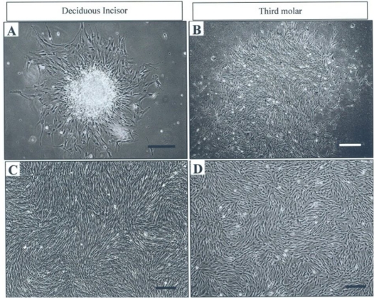
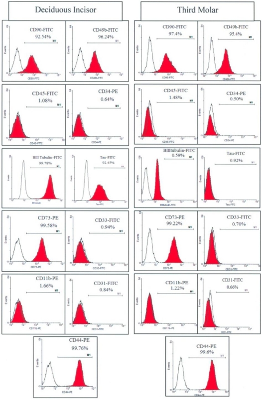
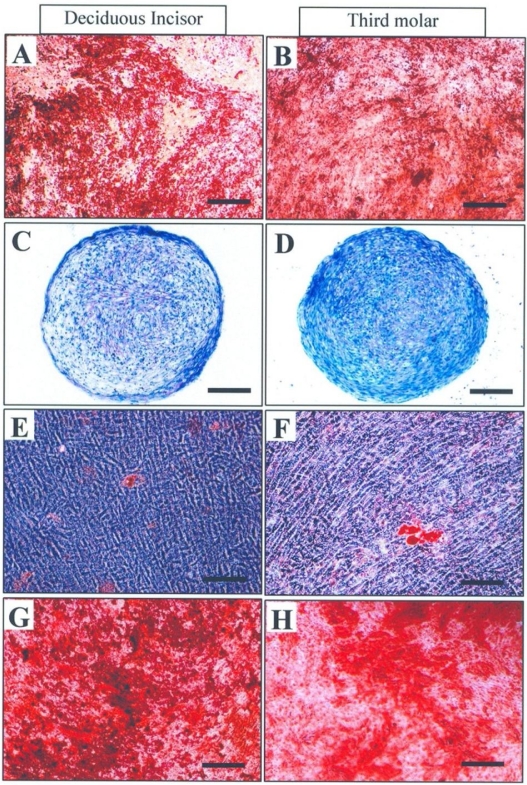
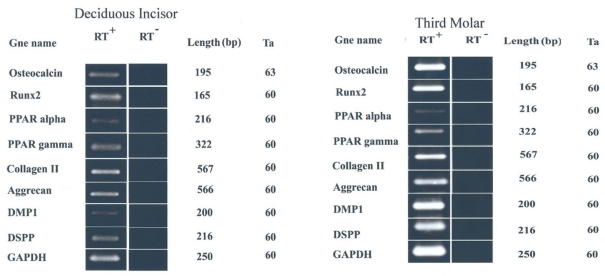
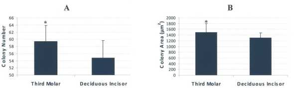
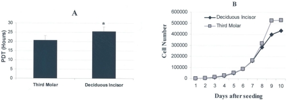
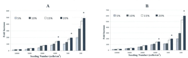
Results: Stem cells of either pulp tissue appeared as fibroblastic cells capable of differentiating into osteoblastic, odontoblastic, adipocytic and chondrocytic cell lineages. In contrast to molar stem cells, those from the incisor tooth expressed neurogenic markers of ßIII Tubulin and Tau. Based on in vitro growth data, the cells from third molar tended to have a lower PDT value (20.79, SD=2.8 versus 25.55, SD=2.9 hours), higher colonogenic activity and better growth curve than those from the deciduous incisor (P<0.05). Both cells exhibited high expansion rate when being plated in a medium with 20% phosphate buffer solution at a density of 100 cells/cm(2).

Conclusion: Given the high proliferation capacity, the stem cells from the human third molar would be an appropriate candidate for use in experimental, preclinical and even clinical setups.

Keywords: Cell Differentiation; Cell Proliferation; Molar, Third; Tooth, Deciduous.


Figures

Fig 1
Fig 1
Pulp cell cultures from human deciduous incisors and third molar teeth. Primary cultures (A and B), The cells in confluent culture at passage 3 (C and D). Bar=100 μm
Fig 2
Fig 2
Flow cytometric analysis of surface antigens on stem cells derived from human deciduous incisors and third molar teeth.
Fig 3
Fig 3
Differentiation of human dental pulp stem cells: either cell was succeeded to differentiate along odontoblastic (A, B), chondrogenic (C, D), adipocytic (E, F) and osteoblastic (G, H) cell lineages. Bar=100 μm
Fig 4
Fig 4
RT-PCR analysis of gene expression in the differentiated cells isolated from human deciduous incisors and third molar teeth. Both cells were successfully expressed odontoblastic-, chondrogenic-, adipogenic- and osteogenic-related genes.
Fig 5
Fig 5
Colonogenic assays performed for stem cells derived from human deciduous incisors and third molar teeth. A) The cells from the third molar appeared to create significantly more colonies than their counterpart from deciduous teeth. B) Likewise, the colonies of third molar cells were larger than those of deciduous incisors. * indicates the significant difference of P<0.05.
Fig 6
Fig 6
Population doubling time (PDT) and growth curve determined for stem cells derived from human deciduous incisors and third molar teeth. (A) Third molar cells tended to have a shorter PDT value compared to deciduous incisors (P<0.05). * indicates the significant difference of P<0.05. (B) According to the growth curve, third molar cells reached plateau sooner than deciduous incisor cells. Both cells exhibited a lag time of a few days in culture.
Fig 7
Fig 7
Culture optimization for better growth of the stem cells isolated from human deciduous incisors (A) and molar teeth (B). The cells underwent considerable fold increase when are plated at low density in a medium with 20% FBS.

Similar articles

Cited by

References

    1. Gronthos S, Mankani M, Brahim J, Robey PG, Shi S. Postnatal human dental pulp stem cells (DPSCs) in vitro and in vivo. Proc Natl Acad Sci USA. 2000 Dec 5;97(25):13625–30. - PMC - PubMed
    1. Miura M, Gronthos S, Zhao M, Lu B, Fisher LW, Robey PG, et al. SHED: stem cells from human exfoliated deciduous teeth. Proc Natl Acad Sci USA. 2003 May 13;100(10):5807–12. - PMC - PubMed
    1. Seo BM, Miura M, Gronthos S, Bartold PM, Batouli S, Brahim J, et al. Investigation of multipotent postnatal stem cells from human periodontal ligament. Lancet. 2004 Jul 10–16;364(9429):149–55. - PubMed
    1. Sonoyama W, Liu Y, Yamaza T, Tuan RS, Wang S, Shi S, et al. Characterization of the apical papilla and its residing stem cells from human immature permanent teeth: a pilot study. J Endod. 2008 Feb;34(2):166–71. - PMC - PubMed
    1. Morsczeck C, Moehl C, Götz W, Heredia A, Schäffer TE, Eckstein N, et al. In vitro differentiation of human dental follicle cells with dexamethasone and insulin. Cell Biol Int. 2005 Jul;29(7):567–75. - PubMed

LinkOut - more resources