. 2012 Feb;14(2):226-38.
doi: 10.1111/j.1462-5822.2011.01714.x. Epub 2011 Nov 3.

The cooperative action of bacterial fibronectin-binding proteins and secreted proteins promote maximal Campylobacter jejuni invasion of host cells by stimulating membrane ruffling

Affiliations

The cooperative action of bacterial fibronectin-binding proteins and secreted proteins promote maximal Campylobacter jejuni invasion of host cells by stimulating membrane ruffling

Tyson P Eucker et al. Cell Microbiol. 2012 Feb.

Abstract

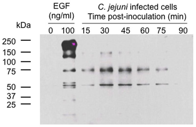
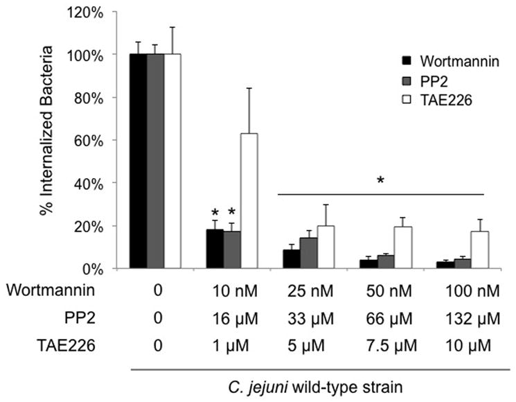
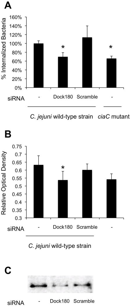
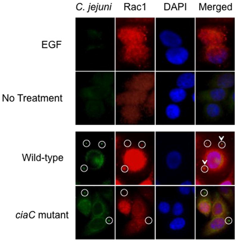
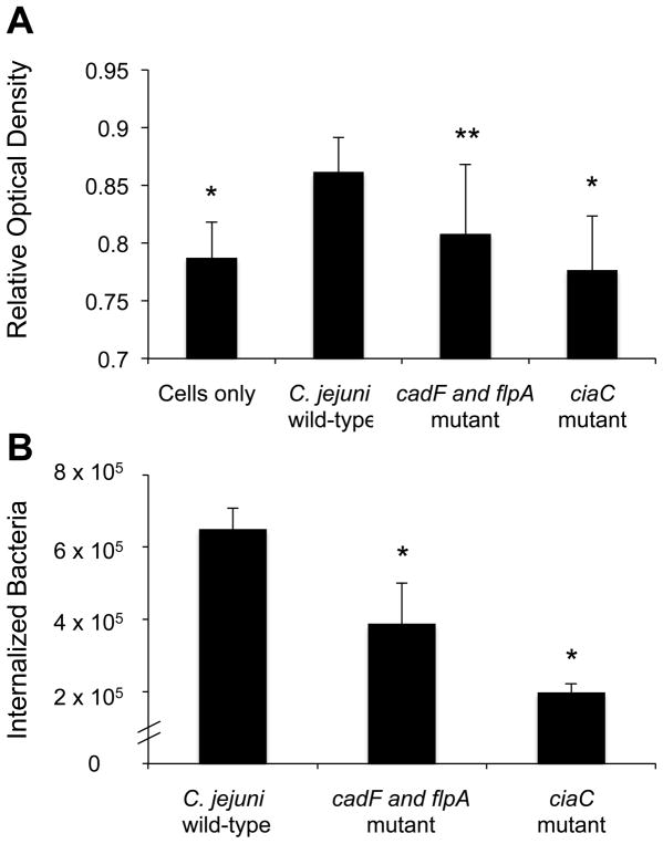
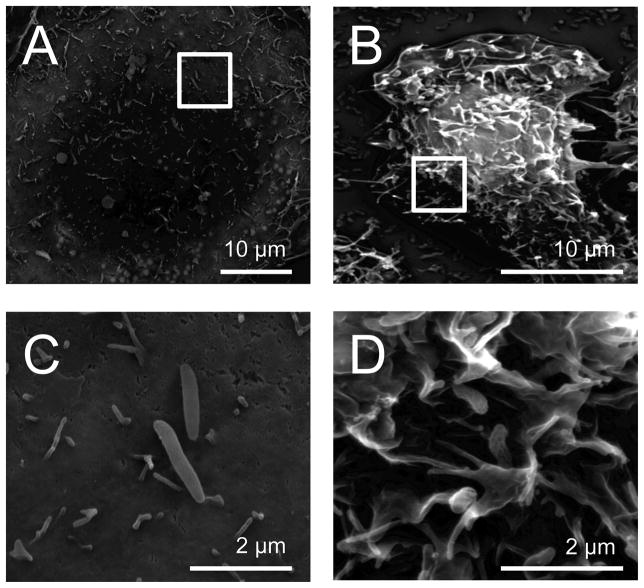
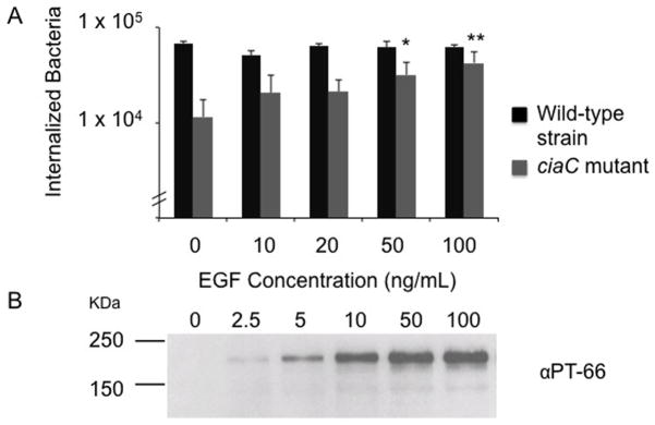
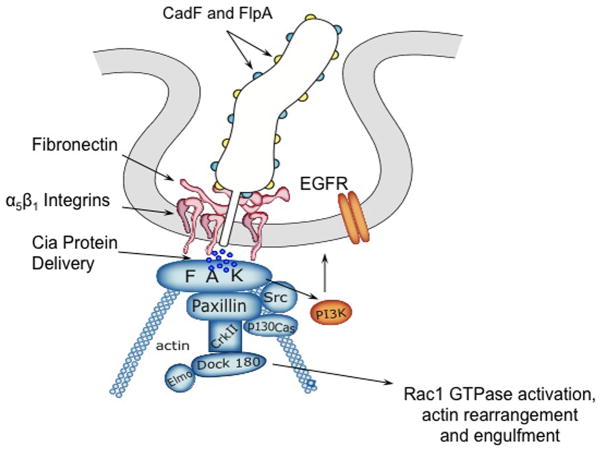
This study was performed to elucidate the host cell scaffolding and signalling molecules that Campylobacter jejuni utilizes to invade epithelial cells. We hypothesized that the C. jejuni fibronectin-binding proteins and secreted proteins are required for cell signalling and maximal invasion of host cells. C. jejuni binding to host cells via the CadF and FlpA fibronectin-binding proteins activated the epidermal growth factor (EGF) pathway, as evidenced by inhibitor studies and immunoprecipitation coupled with immunoblot analysis using antibodies reactive against total and active EGF receptor. Inhibitor studies revealed maximal C. jejuni host cell invasion was dependent upon PI3-Kinase, c-Src and focal adhesion kinase (FAK), all of which are known to participate in cytoskeletal rearrangements. Knockdown of endogenous Dock180, which is a Rac1-specific guanine nucleotide exchange factor, using siRNA revealed that C. jejuni invasion was significantly reduced compared with cells treated with scrambled siRNA. We further demonstrated that the C. jejuni Cia proteins are, in part, responsible for Rho GTPase Rac1 recruitment and activation, as judged by immunofluorescence microscopy and Rac1 activation. Based on these data, we present a model that illustrates that C. jejuni utilizes a coordinated mechanism involving both adhesins and secreted proteins to promote membrane ruffling and host cell invasion.


Figures

Fig. 1
Fig. 1
Infection of INT 407 cells with C. jejuni results in an increase in host cell tyrosine phosphorylated proteins. INT 407 cells were infected with C. jejuni for 15 to 90 min. Triton X-100 soluble host cell proteins were extracted, and subjected to SDS-PAGE coupled with immunoblot analysis using the PT-66 α-phosphotyrosine antibody. INT 407 cells treated with 100 ng/ml of EGF served as the positive control, whereas uninoculated INT 407 cells were used as a negative control. Equal amounts of total protein were added to each well of the gel.
Fig. 2
Fig. 2
Effect of host cell PI3-Kinase (wortmannin), c-Src (PP2), and FAK (TAE226) inhibitors on C. jejuni invasion. Each inhibitor was tested on a different day. The invasion assay was performed as indicated in the “Experimental Procedures,” whereby the bacteria were incubated with the INT 407 cells for 3 hr prior to gentamicin treatment. Values represent percent of internalized (gentamicin-protected) bacteria ± standard deviation compared to untreated cells. The asterisks indicate significance between untreated and treated samples (P < 0.01), as determined using Student’s t test.
Fig. 3
Fig. 3
Dock180 participates in C. jejuni invasion of epithelial cells. HeLa cells were transfected with Dock180 or scrambled (Scr) siRNA. Panels: A) Invasion of the C. jejuni wild-type strain in untreated, Dock180 siRNA-treated, and Scr siRNA-treated cells. Also shown is the number of bacteria internalized for the C. jejuni ciaC mutant. The values represent the mean number of internalized bacteria ± standard deviation. B) Rac1 activation in host cells infected with C. jejuni wild-type strain. Whole cell lysates were processed after 15 minutes of incubation and analyzed for activated Rac1 by G-LISA. The mean ± standard deviation of total active Rac1 is indicated in Relative Optical Density. The data shown represent at least 10 samples of each condition analyzed in duplicate. The asterisks indicate significance (P < 0.01) in the invasiveness or Rac1 activation of the C. jejuni wild-type strain in untreated versus Dock180-treated cells, as determined using the Student’s t test. C) Whole cell lysates of untreated, Dock180 siRNA-treated, and Scr siRNA-treated INT 407 cells (from left to right) analyzed by immunoblotting with an α-Dock180 antibody.
Fig. 4
Fig. 4
Rac1 is recruited to sites of C. jejuni attachment. The localization of Rac1 in C. jejuni-infected cells was examined by immunofluorescence microscopy as outlined in “Experimental Procedures.” EGF-treated cells served as a positive control and uninoculated (non-EGF treated) INT 407 cells were used as a negative control. EGF treatment of INT 407 cells resulted in Rac1 enriched sites, whereas Rac1 was dispersed in uninoculated INT 407 cells. The cell-associated bacteria are indicated by the circles, whereas the C. jejuni adjacent to sites of accumulated Rac1 are indicated by the circles with the arrowhead. Approximately 33% of the C. jejuni wild-type bacteria were co-localized with Rac1, whereas only 13% of the C. jejuni ciaC mutant bacteria were co-localized with Rac1.
Fig. 5
Fig. 5
Rac1 is activated in INT 407 cells in response to C. jejuni infection. INT 407 cells were inoculated with the C. jejuni wild-type strain, cadF flpA mutant, and ciaC mutant. A) Whole cell lysates were processed after 15 minutes of incubation and analyzed for activated Rac1 by G-LISA. The mean ± standard deviation of total active Rac1 is indicated in Relative Optical Density. The data shown represent at least 10 samples of each condition analyzed in duplicate. B) C. jejuni invasion of host cells as determined by gentamicin protection assay. Values represent mean ± standard deviation of internalized bacteria/well of a 24-well tissue culture tray. The asterisks indicate a significant difference (P < 0.01) in internalization as compared to the wild-type strain. The asterisks indicate a significance difference (*P < 0.01, **P < 0.05) in Rac1 activity for the wild-type strain versus uninoculated cells and cells inoculated with the C. jejuni ciaC and cadF flpA mutants.
Fig. 6
Fig. 6
Inoculation of INT 407 cells with a C. jejuni wild-type strain induces membrane ruffling. Scanning electron microscopy of INT 407 cells infected with C. jejuni. A) Cell infected with a ciaC mutant; B) Cell infected with a C. jejuni wild-type strain; C) Increased magnification of the image (inset box) shown in Panel A; and D) Increased magnification of the image (inset box) shown in Panel B.
Fig. 7
Fig. 7
Stimulation of the EGF signaling pathway rescues the invasiveness of a C. jejuni ciaC mutant. Panels: A) INT 407 cells were incubated with the concentrations of EGF indicated for 30 minutes prior to inoculation with either the C. jejuni wild-type strain or the ciaC mutant. Values represent mean ± standard deviation of internalized bacteria/well of a 24-well tissue culture tray. The asterisks (* P < 0.05, ** P < 0.01) indicate a significant increase in the number of internalized bacteria for cells treated with EGF and inoculated with the ciaC mutant versus untreated cells inoculated with the ciaC mutant. B) Immunoblot probed with the PT-66 α-phosphotyrosine antibody. INT 407 cells were treated with EGF and cell lysates prepared after a 30 min incubation period. The blot was exposed for a short period of time in order to show that the EGFR (Mr = 180 kDa) is activated in a dose-dependent manner in response to increasing concentrations of EGF.
Fig. 8
Fig. 8
Model of the host cell EGF signaling pathway and proteins downstream of the EGF receptor that are activated in response to C. jejuni invasion. Indicated in blue are components of the focal complex, (FC) and indicated in orange are other components that likely participate in C. jejuni internalization. C. jejuni binding to Fn results in integrin occupancy and clustering, which in turn, leads to integrin activation. The activation of EGFR and FAK then occurs through from the association with the tails of clustered β1 integrins. Activated FAK autophosphorylates Tyr397, resulting in association of c-Src. c-Src phosphorylates additional residues within FAK as well as other members of the FC, including paxillin and p130Cas. This activation results in the recruitment of CrkII and Dock180, the latter of which associates with ELMO to expose its catalytic domain. The Dock180/ELMO complex activates the Rho family monomeric G protein Rac1, leading to a local restructuring of the actin cytoskeleton.

References

    1. Bain J, Plater L, Elliott M, Shpiro N, Hastie CJ, McLauchlan H, et al. The selectivity of protein kinase inhibitors: a further update. Biochem J. 2007;408:297–315. - PMC - PubMed
    1. Balasubramanian N, Scott DW, Castle JD, Casanova JE, Schwartz MA. Arf6 and microtubules in adhesion-dependent trafficking of lipid rafts. Nat Cell Biol. 2007;9:1381–1391. - PMC - PubMed
    1. Beardsley A, Fang K, Mertz H, Castranova V, Friend S, Liu J. Loss of caveolin-1 polarity impedes endothelial cell polarization and directional movement. J Biol Chem. 2005;280:3541–3547. - PubMed
    1. Buchsbaum RJ, Connolly BA, Feig LA. Interaction of Rac exchange factors Tiam1 and Ras-GRF1 with a scaffold for the p38 mitogen-activated protein kinase cascade. Mol Cell Biol. 2002;22:4073–4085. - PMC - PubMed
    1. Buelow DR, Christensen JE, Neal-McKinney JM, Konkel ME. Campylobacter jejuni survival within human epithelial cells is enhanced by the secreted protein Cial. Mol Microbiol. 2011 In press. - PMC - PubMed

Publication types

MeSH terms

Substances