

Functional promiscuity of the COG0720 family
- PMID: 21999246
- PMCID: PMC3262898
- DOI: 10.1021/cb200329f
Functional promiscuity of the COG0720 family
Abstract
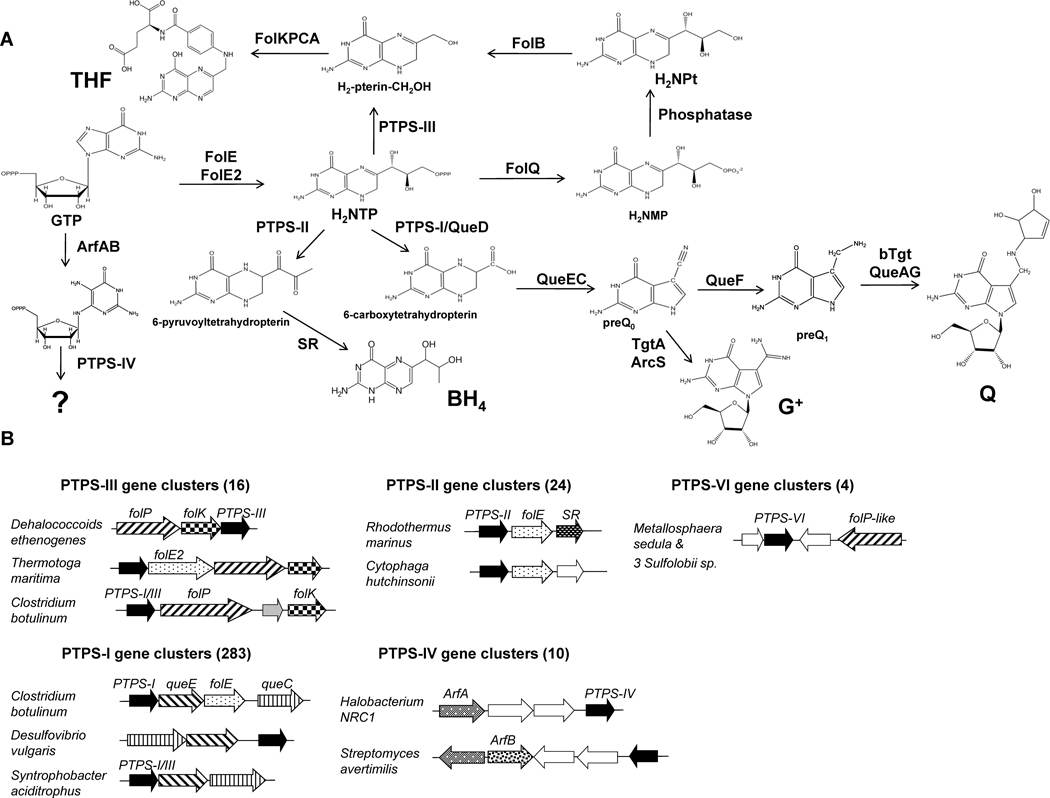
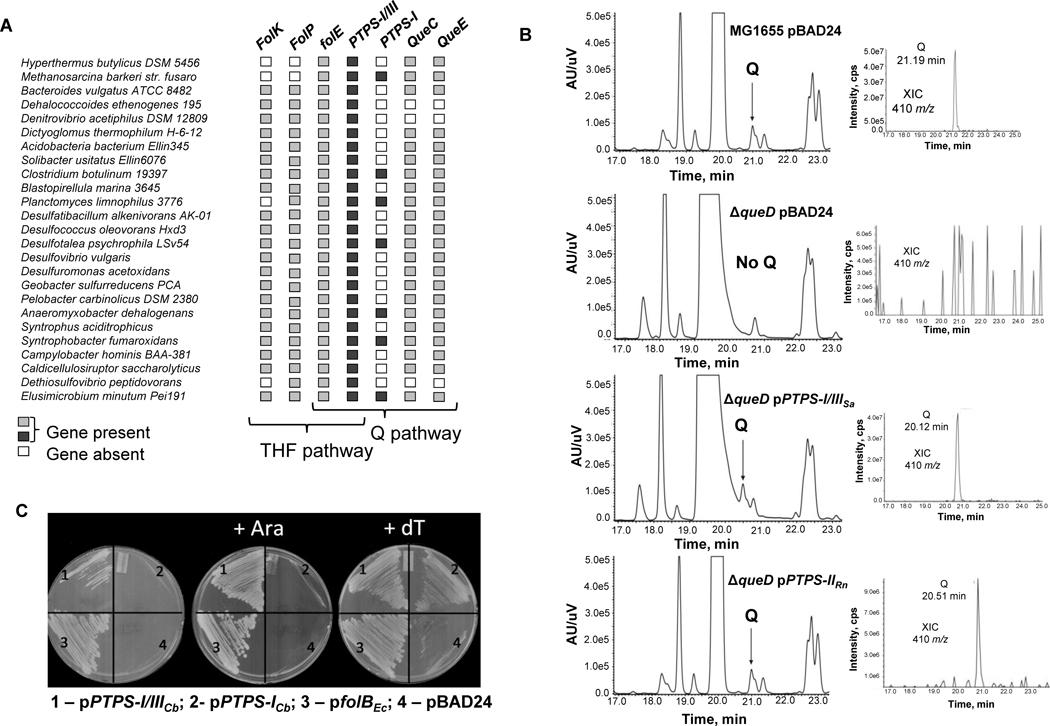
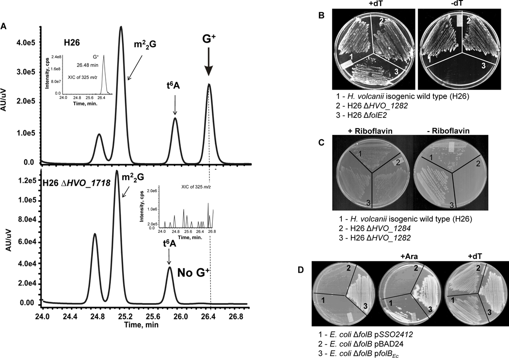
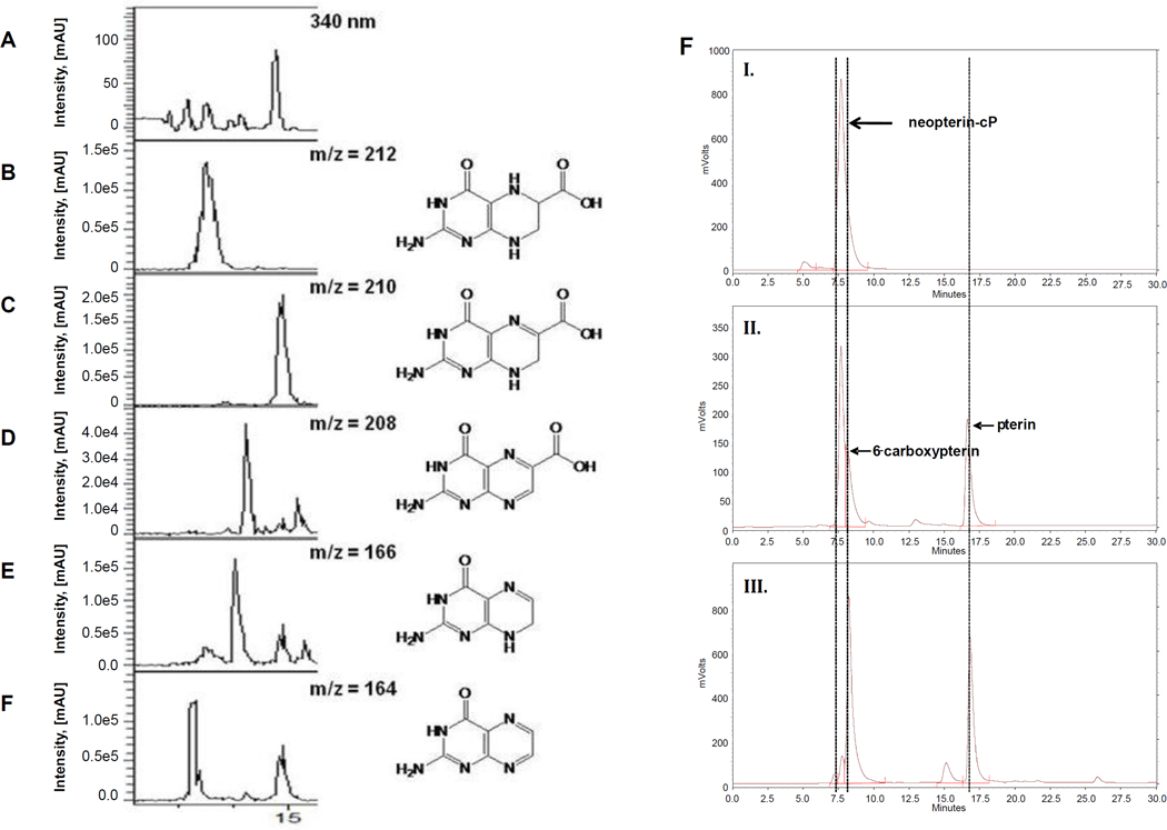
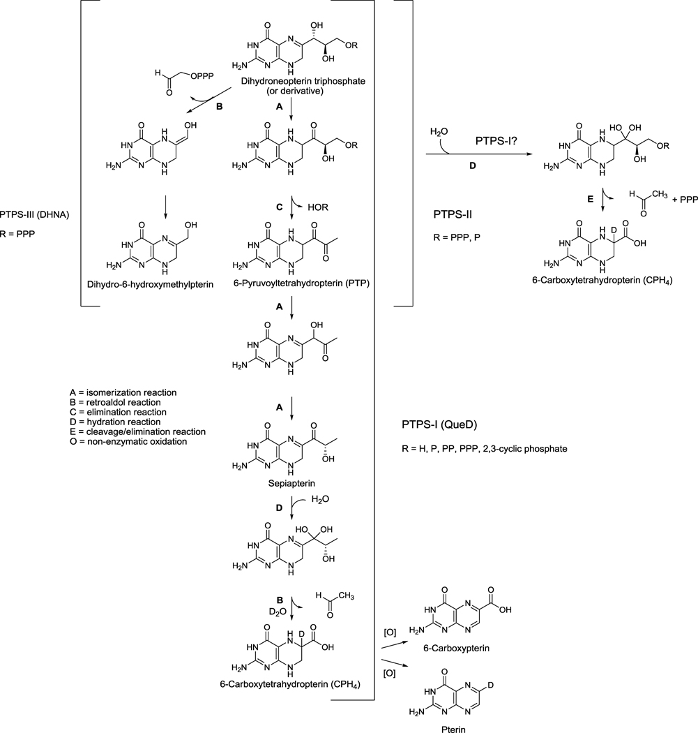
The biosynthesis of GTP derived metabolites such as tetrahydrofolate (THF), biopterin (BH(4)), and the modified tRNA nucleosides queuosine (Q) and archaeosine (G(+)) relies on several enzymes of the Tunnel-fold superfamily. A subset of these proteins includes the 6-pyruvoyltetrahydropterin (PTPS-II), PTPS-III, and PTPS-I homologues, all members of the COG0720 family that have been previously shown to transform 7,8-dihydroneopterin triphosphate (H(2)NTP) into different products. PTPS-II catalyzes the formation of 6-pyruvoyltetrahydropterin in the BH(4) pathway, PTPS-III catalyzes the formation of 6-hydroxylmethyl-7,8-dihydropterin in the THF pathway, and PTPS-I catalyzes the formation of 6-carboxy-5,6,7,8-tetrahydropterin in the Q pathway. Genes of these three enzyme families are often misannotated as they are difficult to differentiate by sequence similarity alone. Using a combination of physical clustering, signature motif, phylogenetic codistribution analyses, in vivo complementation studies, and in vitro enzymatic assays, a complete reannotation of the COG0720 family was performed in prokaryotes. Notably, this work identified and experimentally validated dual function PTPS-I/III enzymes involved in both THF and Q biosynthesis. Both in vivo and in vitro analyses showed that the PTPS-I family could tolerate a translation of the active site cysteine and was inherently promiscuous, catalyzing different reactions on the same substrate or the same reaction on different substrates. Finally, the analysis and experimental validation of several archaeal COG0720 members confirmed the role of PTPS-I in archaeosine biosynthesis and resulted in the identification of PTPS-III enzymes with variant signature sequences in Sulfolobus species. This study reveals an expanded versatility of the COG0720 family members and illustrates that for certain protein families extensive comparative genomic analysis beyond homology is required to correctly predict function.
Figures
Similar articles
-
6-pyruvoyltetrahydropterin synthase paralogs replace the folate synthesis enzyme dihydroneopterin aldolase in diverse bacteria.J Bacteriol. 2009 Jul;191(13):4158-65. doi: 10.1128/JB.00416-09. Epub 2009 Apr 24. J Bacteriol. 2009. PMID: 19395485 Free PMC article.
-
Structural basis of a novel activity of bacterial 6-pyruvoyltetrahydropterin synthase homologues distinct from mammalian 6-pyruvoyltetrahydropterin synthase activity.Acta Crystallogr D Biol Crystallogr. 2014 May;70(Pt 5):1212-23. doi: 10.1107/S1399004714002016. Epub 2014 Apr 26. Acta Crystallogr D Biol Crystallogr. 2014. PMID: 24816091
-
Comparative genomics guided discovery of two missing archaeal enzyme families involved in the biosynthesis of the pterin moiety of tetrahydromethanopterin and tetrahydrofolate.ACS Chem Biol. 2012 Nov 16;7(11):1807-16. doi: 10.1021/cb300342u. Epub 2012 Sep 7. ACS Chem Biol. 2012. PMID: 22931285 Free PMC article.
-
The pathway from GTP to tetrahydrobiopterin: three-dimensional structures of GTP cyclohydrolase I and 6-pyruvoyl tetrahydropterin synthase.Biol Chem. 1997 Mar-Apr;378(3-4):185-92. Biol Chem. 1997. PMID: 9165069 Review.
-
Biosynthesis of the 7-deazaguanosine hypermodified nucleosides of transfer RNA.Bioorg Chem. 2003 Feb;31(1):24-43. doi: 10.1016/s0045-2068(02)00513-8. Bioorg Chem. 2003. PMID: 12697167 Review.
Cited by
-
AGeNNT: annotation of enzyme families by means of refined neighborhood networks.BMC Bioinformatics. 2017 May 25;18(1):274. doi: 10.1186/s12859-017-1689-6. BMC Bioinformatics. 2017. PMID: 28545394 Free PMC article.
-
Biosynthesis and function of 7-deazaguanine derivatives in bacteria and phages.Microbiol Mol Biol Rev. 2024 Mar 27;88(1):e0019923. doi: 10.1128/mmbr.00199-23. Epub 2024 Feb 29. Microbiol Mol Biol Rev. 2024. PMID: 38421302 Free PMC article. Review.
-
Open Issues for Protein Function Assignment in Haloferax volcanii and Other Halophilic Archaea.Genes (Basel). 2021 Jun 24;12(7):963. doi: 10.3390/genes12070963. Genes (Basel). 2021. PMID: 34202810 Free PMC article.
-
The Escherichia coli COG1738 Member YhhQ Is Involved in 7-Cyanodeazaguanine (preQ₀) Transport.Biomolecules. 2017 Feb 8;7(1):12. doi: 10.3390/biom7010012. Biomolecules. 2017. PMID: 28208705 Free PMC article.
-
The Lreu_1276 protein from Limosilactobacillus reuteri represents a third family of dihydroneopterin triphosphate pyrophosphohydrolases in bacteria.Appl Environ Microbiol. 2024 Jul 24;90(7):e0081424. doi: 10.1128/aem.00814-24. Epub 2024 Jun 18. Appl Environ Microbiol. 2024. PMID: 38888337 Free PMC article.
References
-
- Colloc'h N, Poupon A, Mornon JP. Sequence and structural features of the T-fold, an original tunnelling building unit. Proteins. 2000;39:142–154. - PubMed
-
- Auerbach G, Nar H. The pathway from GTP to tetrahydrobiopterin: three-dimensional structures of GTP cyclohydrolase I and 6-pyruvoyl tetrahydropterin synthase. Biol. Chem. 1997;378:185–192. - PubMed
-
- Yim JJ, Brown GM. Characteristics of guanosine triphosphate cyclohydrolase I purified from Escherichia coli. J. Biol. Chem. 1976;251:5087–5094. - PubMed
Publication types
MeSH terms
Substances
Grants and funding
LinkOut - more resources
Full Text Sources
Molecular Biology Databases
