Review
. 2011 Nov 22;50(46):9940-9.
doi: 10.1021/bi201418k. Epub 2011 Oct 31.

Bacterial transition metal P(1B)-ATPases: transport mechanism and roles in virulence

Affiliations
Review

Bacterial transition metal P(1B)-ATPases: transport mechanism and roles in virulence

José M Argüello et al. Biochemistry. .

Abstract

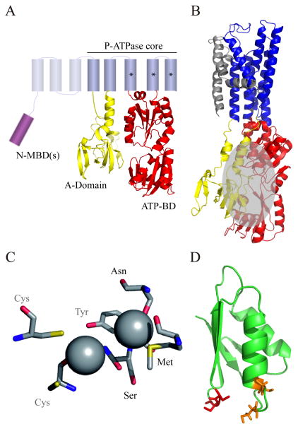
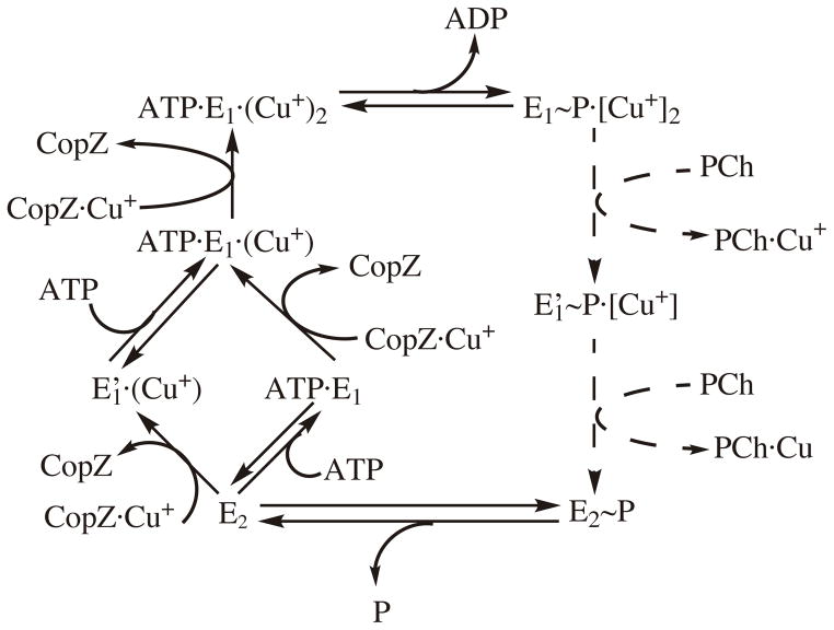
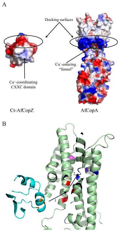
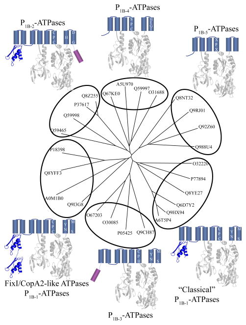
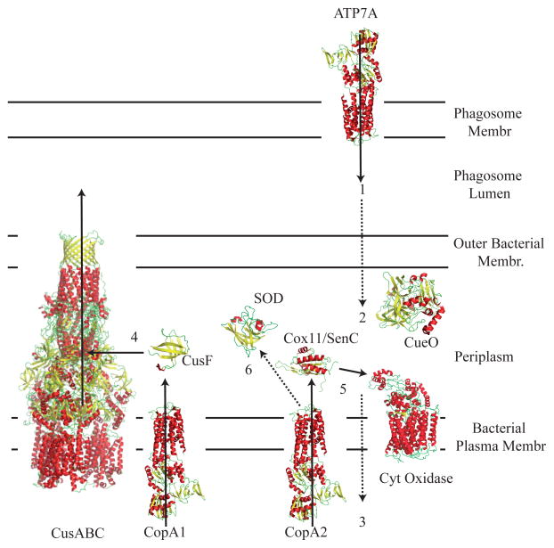
P(1B)-type ATPases are polytopic membrane proteins that couple the hydrolysis of ATP to the efflux of cytoplasmic transition metals. This paper reviews recent progress in our understanding of the structure and function of these proteins in bacteria. These are members of the P-type superfamily of transport ATPases. Cu(+)-ATPases are the most frequently observed and best-characterized members of this group of transporters. However, bacterial genomes show diverse arrays of P(1B)-type ATPases with a range of substrates (Cu(+), Zn(2+), Co(2+)). Furthermore, because of the structural similarities among transitions metals, these proteins can also transport nonphysiological substrates (Cd(2+), Pb(2+), Au(+), Ag(+)). P(1B)-type ATPases have six or eight transmembrane segments (TM) with metal coordinating amino acids in three core TMs flanking the cytoplasmic domain responsible for ATP binding and hydrolysis. In addition, regulatory cytoplasmic metal binding domains are present in most P(1B)-type ATPases. Central to the transport mechanism is the binding of the uncomplexed metal to these proteins when cytoplasmic substrates are bound to chaperone and chelating molecules. Metal binding to regulatory sites is through a reversible metal exchange among chaperones and cytoplasmic metal binding domains. In contrast, the chaperone-mediated metal delivery to transport sites appears as a largely irreversible event. P(1B)-ATPases have two overarching physiological functions: to maintain cytoplasmic metal levels and to provide metals for the periplasmic assembly of metalloproteins. Recent studies have shown that both roles are critical for bacterial virulence, since P(1B)-ATPases appear key to overcome high phagosomal metal levels and are required for the assembly of periplasmic and secreted metalloproteins that are essential for survival in extreme oxidant environments.


Figures

FIGURE 1
FIGURE 1
Structural features of P1B-ATPases. (A) Topology of a typical P1B-ATPase. Asterisks indicate the position of amino acids forming TM-MBS(s). (B) Crystal structure of LpCopA (PDB 3RFU). Cytosolic A-domain and ATP-BD are shown in yellow and red, respectively. The grey helices correspond to MA and MB. The grey globe indicates the predicted N-MBD contact with A-domain and ATP-BD (31). (C) Model of Cu+-ATPase TM-MBSs. The model was built by using SERCA 1SU4 structure as template. N-terminal Cys had to be manually reoriented to coordinate Cu+. (D) Model of the N-MBD of P. aeruginosa CopA2. Modelling was done using Menkes 4th N-MBD structure 1AW0. In orange are indicated the conserved Cys of the CXXC domain common to most Cu+-ATPases N-MBDs. In red is the CC motif specific of FixI/CopA2-like ATPases.
FIGURE 2
FIGURE 2
Catalytic and transport cycle of Cu+-ATPases. Cytoplasmic Cu+ binding to two transmembrane metal binding sites (TM-MBSs) is coupled to ATP hydrolysis and enzyme phosphorylation (E1P(Cu+)2). Subsequently, the enzyme undergoes a conformational change (to E2P) leading to the TM-MBSs opening to the extracellular/periplasmic compartment with the consequent metal release. Enzyme dephosphorylation allows the return to the E1 form with TM-MBSs facing the cytoplasm. It is relevant that the E2→E1 transition is accelerated by ATP (or ADP) acting with low affinity; i.e., a modulatory mode. Note the irreversibility of the Cu+ transfer from Cu.CopZ to TM-MBS and, that binding of ATP is required to full occupancy of the transport site. Discontinued lines indicate proposed steps in the cycle. PCh indicates a hypothetical periplasmic Cu+-chaperone/acceptor.
FIGURE 3
FIGURE 3
Cu+ transit from the Cu+ chaperone to the extracellular space/lumen/periplasm. (A) Electrostatic map of modelled Archaeoglobus fulgidus C-terminal domain of the Cu+ chaperone CopZ (using E. hirae CopZ 1CPZ as template) and A. fulgidus CopA (using L. pneumophila CopA 3RFU as model). Red indicates negative charges and blue positive ones. (B) Metal transport pathway from CopZ to CopA pre-release sites. A. fulgidus Ct-CopZ is shown in cyan and A. fulgidus CopA in green. Cu+ is transferred from the CXXC domain (orange) in the chaperone to the pre-docking amino acids in the ATPase (red) (1). Subsequently, the metal reaches one of the two TM-MBSs (blue and purple) (2) indistinctively. Only when ATP is bound to the ATP-BD, the second TM-MBS is occupied. This triggers ATP hydrolysis and release of Cu+ which is facilitated by amino acid/s in the luminal side of the ATPase (pink) (3).
FIGURE 4
FIGURE 4
Subfamilies of P1B-ATPases. Sequences used for the tree were: Symbiobacterium thermophilum Q67KE0, M. tuberculosis A5U970, Synechocystis sp. PCC 6803 Q59997, Bacillus subtilis O31688, Corynebacterium glutamicum Q8NT32, Streptomyces coelicolor Q9RJ01, Sinorhizobium meliloti Q92Z60, Mesorhizobium loti Q988U4, B. subtilis O32220, M. tuberculosis P77894, Brucella melitensis Q8YE27, Erwinina carotovora Q6D7Y2, P. aeruginosa Q9HX93, Klebsiella pneumonia A6T5P4, Lactococcus lactis Q9CH87, E. hirae P05435, A. fulgidus O30085, Aquifex aeolicus O67203, P. aeruginosa Q9I3G8, Gramella forsetii A0M1B0, B. melitensis Q8YFF3, S. meliloti P18398, Helicobacter pylori Q59465, Synechocystis sp. PCC 6803 Q59998, E. coli P37617, S. enterica Q8Z255.
FIGURE 5
FIGURE 5
Hypothetical Cu+ homeostasis in the phagocytic cell/microbe interface. Cu+ is introduced in the phagosome by ATP7A (1) and it crosses the outer membrane reaching the periplasm, where some is oxidized to Cu2+ by CueO (2). However, some Cu+still crosses to the bacterial cytoplasm (3). Excess cytosolic Cu+ is transported back to the periplasm by CopA1-like ATPases, where it binds to periplasmic Cu+-chaperones such as CusF, which transfers Cu+ to CusABC-like transporters to be translocated towards the extracellular space (4). Cu+ is also used to synthetize Cu-proteins. CopA2-like transporters are responsible for this, transferring Cu+ to other periplasmic Cu+ chaperones, such as SenC, which would subsequently donate the metal to cytochrome oxidases (5). Other periplasmic Cu+ chaperones must exist to transfer the metal to other periplasmic apoproteins, such as Cu, Zn superoxide dismutases (6).

References

    1. Fraústro da Silva JJR, Williams RJP. The biological chemistry of the elements. 2. Oxford Unviversity Press; New York: 2001.
    1. Shi W, Zhan C, Ignatov A, Manjasetty BA, Marinkovic N, Sullivan M, Huang R, Chance MR. Metalloproteomics: high-throughput structural and functional annotation of proteins in structural genomics. Structure. 2005;13:1473–1486. - PubMed
    1. Cvetkovic A, Menon AL, Thorgersen MP, Scott JW, Poole FL, 2nd, Jenney FE, Jr, Lancaster WA, Praissman JL, Shanmukh S, Vaccaro BJ, Trauger SA, Kalisiak E, Apon JV, Siuzdak G, Yannone SM, Tainer JA, Adams MW. Microbial metalloproteomes are largely uncharacterized. Nature. 2010;466:779–782. - PubMed
    1. Macomber L, Imlay JA. The iron-sulfur clusters of dehydratases are primary intracellular targets of copper toxicity. Proc Natl Acad Sci USA. 2009;106:8344–8349. - PMC - PubMed
    1. Goldstein S, Meyerstein D, Czapski G. The Fenton reagents. Free Radical Biol Med. 1993;15:435–445. - PubMed

Publication types

MeSH terms