. 2011 Oct 14;147(2):306-19.
doi: 10.1016/j.cell.2011.08.046.

Targeting the SH2-kinase interface in Bcr-Abl inhibits leukemogenesis

Affiliations

Targeting the SH2-kinase interface in Bcr-Abl inhibits leukemogenesis

Florian Grebien et al. Cell. .

Abstract

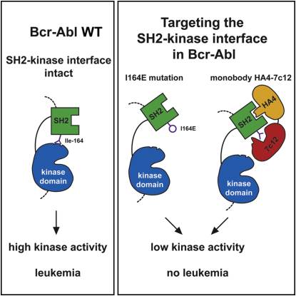
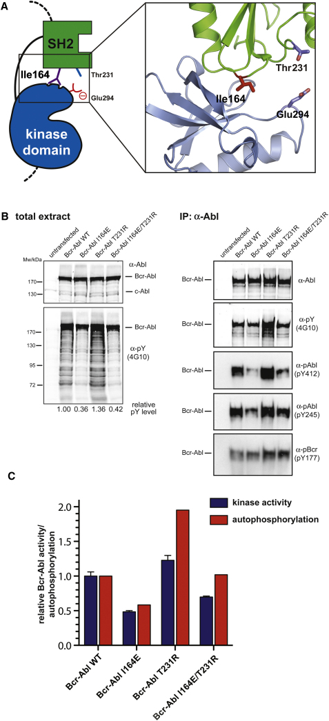
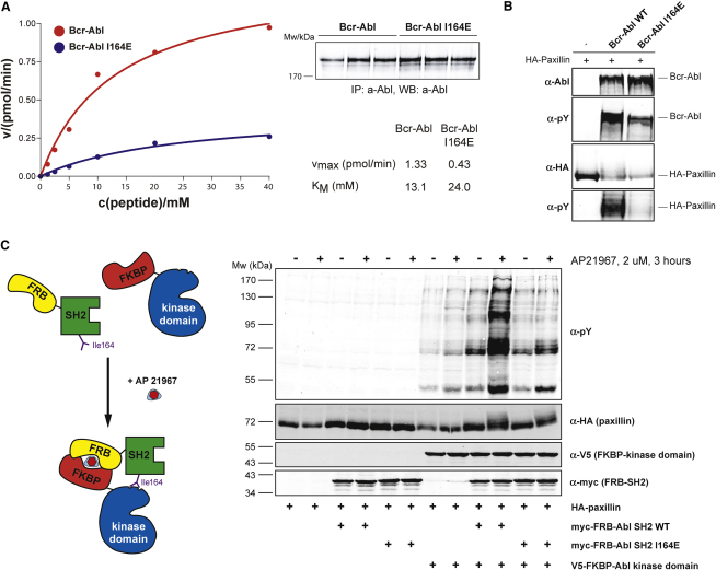
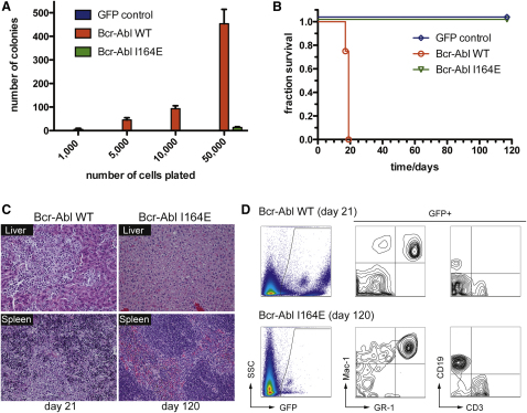
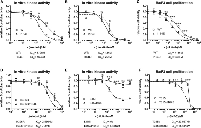
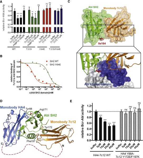
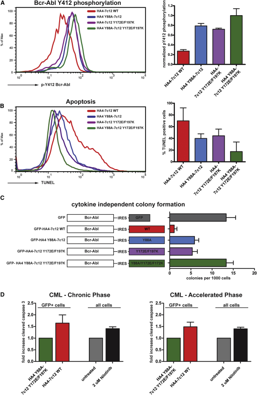
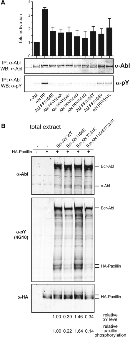
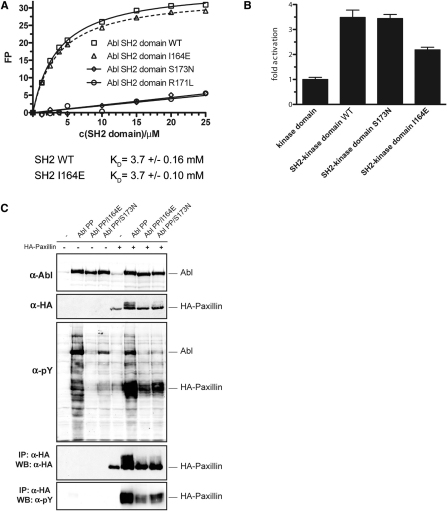
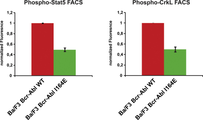
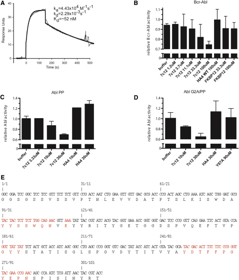
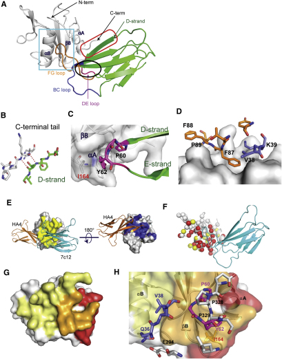
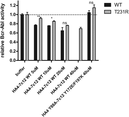
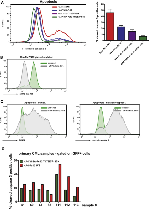
Chronic myelogenous leukemia (CML) is caused by the constitutively active tyrosine kinase Bcr-Abl and treated with the tyrosine kinase inhibitor (TKI) imatinib. However, emerging TKI resistance prevents complete cure. Therefore, alternative strategies targeting regulatory modules of Bcr-Abl in addition to the kinase active site are strongly desirable. Here, we show that an intramolecular interaction between the SH2 and kinase domains in Bcr-Abl is both necessary and sufficient for high catalytic activity of the enzyme. Disruption of this interface led to inhibition of downstream events critical for CML signaling and, importantly, completely abolished leukemia formation in mice. Furthermore, disruption of the SH2-kinase interface increased sensitivity of imatinib-resistant Bcr-Abl mutants to TKI inhibition. An engineered Abl SH2-binding fibronectin type III monobody inhibited Bcr-Abl kinase activity both in vitro and in primary CML cells, where it induced apoptosis. This work validates the SH2-kinase interface as an allosteric target for therapeutic intervention.


Figures

None
Graphical abstract
Figure 1
Figure 1
Structure-Function Analysis of the SH2-Kinase Domain Interface in Bcr-Abl (A) Cartoon diagram of the active SH2-kinase domain unit of Abl. Critical residues involved in the SH2-kinase domain interface that are discussed in the text are highlighted. (B) HEK293 cells were transiently transfected with Bcr-Abl WT, I164E, T231R, or I164E/T231R. Total protein extracts (left panels) and Abl immunoprecipitates (right panels) were analyzed by immunoblotting using the indicated antibodies. (C) Abl immunoprecipitates were assayed for catalytic activity using an optimal Abl substrate peptide, and total autophosphorylation was quantified. Kinase activity and autophosphorylation of Bcr-Abl WT were set to 1. The bar graph shows averages ± standard deviation (SD) from two independent experiments done in triplicate. See also Figure S1.
Figure 2
Figure 2
The SH2-Kinase Domain Interface in Bcr-Abl Is Both Necessary and Sufficient for High Catalytic Activity of the Enzyme (A) Immunoprecipitated Bcr-Abl WT and I164E proteins were assayed for catalytic activity in the presence of the indicated concentrations of an optimal Abl substrate peptide containing a single tyrosine phosphorylation site. Averages of three replicates are plotted. The KM and vmax values were calculated after fitting the data to the Michaelis-Menten equation. (B) Bcr-Abl WT and I164E constructs were coexpressed with HA-tagged paxillin in HEK293 cells, and total protein extracts were analyzed by immunoblotting using the indicated antibodies. (C) The indicated constructs were transiently transfected in HEK293 cells along with HA-paxillin. FRB-FKBP dimerization was induced by treating cells with AP21967. Total cell lysates were analyzed by immunoblotting using the indicated antibodies. See also Figure S2.
Figure 3
Figure 3
Bcr-Abl I164E Is Not Leukemogenic in a Bcr-Abl Mouse Bone Marrow Transplantation Model (A) Indicated amounts of primary murine bone marrow cells expressing Bcr-Abl WT, Bcr-Abl I164E, or GFP were seeded in semisolid medium. Cytokine-independent colonies were scored after 8 days. Error bars represent SD. (B) Equal amounts of primary murine Lin c-Kit+ Sca-1+ cells expressing Bcr-Abl WT, Bcr-Abl I164E, or GFP were injected in lethally irradiated recipient mice (n = 4). Overall survival of transplanted mice was monitored over 120 days. (C) Sections from liver (top panels) and spleen (bottom panels) of representative Bcr-Abl WT and I164E transplanted animals were stained with hematoxylin/eosin. (D) Peripheral blood samples were prepared at indicated time points after transplantation and stained with the indicated antibodies and analyzed by FACS.
Figure 4
Figure 4
Bcr-Abl I164E Renders Ba/F3 or UT-7 Cell Lines Factor Independent and Differentially Impacts Cellular Signaling Pathways (A) Expression levels and cellular activity of Bcr-Abl WT and I164E transduced Ba/F3 cells. (B) Proliferation of Bcr-Abl WT, I164E, or GFP control Ba/F3 cells or UT-7 cells was measured in the presence of the indicated concentrations of IL-3 or GM-CSF. (C and D) Activation levels of selected components of different cellular signaling pathways by Bcr-Abl WT versus I164E were investigated by immunoblotting of total lysates from Ba/F3 (B) and U937 cells (C) stably expressing Bcr-Abl WT or Bcr-Abl I164E using the indicated antibodies. See also Figure S3.
Figure 5
Figure 5
Bcr-Abl I164E Sensitized WT and Imatinib-Resistant Bcr-Abl Forms to TKI Inhibition (A, B, D, and E) Kinase activity of the indicated immunoprecipitated Bcr-Abl constructs was assayed in the presence of the indicated concentrations of imatinib (A), dasatinib (B), or nilotinib (D and E). For each dataset, the activities of the untreated samples were set to 1. Each data point in the graphs represents the averages ± SD from two independent experiments done in duplicate. (C and F) Viability of Ba/F3 cells expressing Bcr-Abl WT, I164E, T315I, and T315I/I164E was measured in the presence of the indicated concentrations of nilotinib (C) or GNF-2 (F) and normalized to 1 for each dataset. For all panels, significance levels are indicated (ns: not significant, p < 0.05, ∗∗p < 0.01, ∗∗∗p < 0.001). Each data point in the graphs represents the averages ± SD from one representative experiment done in sextuplicate.
Figure 6
Figure 6
Targeting the SH2-Kinase Domain Interface with the Engineered Monobody Protein 7c12 Leads to Bcr-Abl Inhibition (A) In vitro kinase activity of immunoprecipitated Bcr-Abl WT, I164E, T315I, and T315I/I164E proteins was assayed in the presence of the indicated concentrations of recombinant 7c12. FKBP12 was used as a control recombinant protein. Activity of each Bcr-Abl mutant-buffer control was normalized to 1. Significance levels are indicated (ns: not significant, p < 0.05, ∗∗p < 0.01, ∗∗∗p < 0.001). The bar graph shows averages ± SD from at least three independent experiments done in triplicate. See also Figure S4. (B) Competition phage ELISA of the 7c12 monobody. Phages displaying the 7c12 monobody were bound to GST-Abl SH2 WT or I164E protein. Unbound phages were exposed to immobilized Abl SH2 domain and detected using an anti-phage antibody. Values are normalized to the intensity of the ELISA signal in the absence of a competitor. (C) Crystal structure of the complex of the monobody 7c12-Abl SH2 domain complex (PDB entry 3T04). Ile164 is part of the interface and shown in red. Lower panel: Superposition of the 7c12-Abl SH2 complex and the active Abl structure. The surface of the Abl kinase domain is shown to highlight the incompatibility of simultaneous binding of 7c12 and the Abl kinase domain to the SH2 domain. See Table S1 and Figure S5 for details. (D) Superposition of the 7c12-Abl SH2 domain complex (PDB entry 3T04) and the HA4-Abl SH2 complex (PDB entry 3K2M) crystal structures. HA4 and 7c12 are shown in blue and orange, respectively. The Gly-Ser linker between HA4 and 7c12 is indicated as a dotted red line. Arg171 in the Abl SH2 domain is shown to indicate the location of the phosphotyrosine-binding pocket. Residues Tyr88, Tyr172, and Phe197 were mutated to abolish binding to the Abl SH2 domain. See also Figure S5. (E) In vitro kinase activity of immunoprecipitated Bcr-Abl WT protein was assayed in the presence of the indicated concentrations of recombinant HA4-7c12 WT or the nonbinding Y88A/Y172E/F197K mutant protein. Significance levels are indicated (ns: not significant, p < 0.05, ∗∗p < 0.01, ∗∗∗p < 0.001). The bar graph shows averages ± SD from one representative experiment done in triplicate. See also Figure S6.
Figure 7
Figure 7
The Tandem Monobody HA4-7c12 Strongly Inhibits Cellular Bcr-Abl Kinase Activity and Induces Apoptosis in CML Cell Lines and Primary Cells (A) K562 cells were transiently transfected with the indicated HA4-7c12-GFP fusion constructs, and Bcr-Abl Tyr412 phosphorylation was measured using flow cytometry. Left panel: Histograms of Bcr-Abl pY412 fluorescence intensities of the GFP-positive fraction of cells transfected with the indicated constructs. Right panel: Quantitation of Bcr-Abl pY412 fluorescence. The intensities were normalized to K562 cells treated with nilotinib (complete inhibition—arbitrarily set to 0; Figure S5) and cells transfected with the nonbinding mutant HA4 Y88A-7c12 Y172E/F197K (no inhibition—arbitrarily set to 1) (n = 3). (B) K562 cells treated as described in (C) were analyzed for apoptosis using TUNEL staining. Left panel: Histograms of TUNEL fluorescence of the GFP-positive fraction of cells transfected with the indicated constructs. Right panel: Quantitation of TUNEL-positive cells (n = 3). The intensities were normalized to K562 cells treated with nilotinib (arbitrarily set to 100%; Figure S7C) and mock-transfected cells (arbitrarily set to 0%) (n = 3). (C) Primary murine bone marrow cells were transduced with the indicated retroviral constructs. GFP-positive cells were seeded in semisolid medium, and cytokine-independent colonies were scored. (D) Primary cells from CML patients from chronic phase (left panel) or accelerated phase (right panel) were transduced with lentiviruses encoding HA4-7c12 WT or HA4 Y88A-7c12 Y172E/F197K. Parallel cultures from the same patients were treated with nilotinib for 5 days. Apoptosis was measured after 5 days via cleaved caspase staining by FACS. Levels of apoptosis in the GFP-positive fraction of cells transduced with HA4 Y88A-7c12 Y172E/F197K or in untreated cells were normalized to 1. p values: Chronic phase (five patients): 0.0007; accelerated phase (two patients): 0.011; one-way ANOVA). In all panels, error bars represent SD. See Table S2 and Figure S7 for details.
Figure S1
Figure S1
Effects of Mutations of Ile164 and/or Thr231 in the SH2-Kinase Interface on Abl Kinase Activity and Phosphorylation of the Abl Substrate Paxillin, Related to Figure 1 (A) The indicated mutations of Ile164 were introduced in the constitutive active mutant Abl PP and the resulting constructs were transiently expressed in HEK293 cells. Constructs were immunoprecipitated and subjected to in vitro kinase assays in the presence of an optimal Abl substrate peptide containing a single tyrosine phosphorylation site (upper panel) and western blotting using the indicated antibodies (lower panels). Mutation of Ile164 to all residues tested led to a significant reduction of in vitro kinase activity and tyrosine phosphorylation, indicating that the effect is not specifically restricted to the I164E mutation. Error bars represent SD. (B) Bcr-Abl WT, Bcr-Abl I164E, Bcr-Abl T231R, and Bcr-Abl I164E/T231R constructs were coexpressed with HA-tagged paxillin in HEK293 cells and total protein extracts were analyzed by immunoblotting using the indicated antibodies.
Figure S2
Figure S2
The I164E Mutation Does Not Interfere with the Phosphotyrosine-Binding Capability of the Abl SH2 Domain, Related to Figure 2 (A) Fluorescence polarization binding assay of recombinant WT and mutant Abl SH2 domains to a fluorescently labeled tyrosine phosphorylated peptide (EPPVpYANLS). The recombinant Abl SH2 domain harboring the I164E mutation was found to bind a phosphorylated peptide with equal affinity as the wt Abl SH2 domain. In contrast, Abl SH2 domains bearing the FLVRES mutants R171L or S173N were unable to bind tyrosine phosphorylated peptides. (B) HEK293 cells were transfected with expression constructs comprising the Abl kinase domain alone or the SH2-kinase domain module of c-Abl (Abl SH2-kinase domain WT, S173N, I164E). Constructs were immunoprecipitated and subjected to in vitro kinase assays in the presence of an optimal Abl substrate peptide containing a single tyrosine phosphorylation site. In line with previous results, the SH2 domain had a positive role on the kinase activity, as we observed higher kinase activity of the Abl SH2-kinase domain construct as compared to the Abl kinase domain alone, and introduction of the I164E reverted this effect. However, the phosphotyrosine-binding mutant S173N did not show significantly different kinase activity as compared to the WT Abl SH2-kinase domain construct. As the kinase substrate peptide used in this assay contains only one tyrosine residue, no (processive phosphorylation) effects dependent on the phosphotyrosine-binding capability of the Abl SH2 domain are observed. Together, these data rule out that the I164E mutation in the Abl SH2 domain leads to reduced kinase activity by interfering with phosphotyrosine binding of the SH2 domain. Furthermore, this experiment shows that the ability of the Abl SH2 domain to bind phosphotyrosine does not influence the in vitro kinase activity of the Abl kinase. Error bars represent SD. (C) HEK293 cells were transiently transfected with the indicated c-Abl mutants in the presence or absence of a HA-tagged form of the Abl substrate paxillin. Total cell lysates were subjected to immunoblot analysis using antibodies against Abl, HA and phosphotyrosine. HA-tagged paxillin was immunoprecipitated from the same lysates and subjected to immunoblotting using antibodies against phosphotyrosine and HA. The Abl substrate paxillin contains multiple tyrosine residues that can serve as substrates for Abl. Thus, basal phosphorylation of one tyrosine residue might exert a positive feedback to Abl activity that is mediated by the Abl SH2 domain by binding the first phosphotyrosine and thereby positioning another unphosphorylated tyrosine close to the active site of the kinase. This phenomenon is referred to as processive phosphorylation and is dependent on the capability of the SH2 domain to bind to tyrosine-phosphorylated substrates. Paxillin was efficiently tyrosine phosphorylated by the constitutively active form of Abl (Abl PP) in a processive way (phosphorylation on multiple tyrosine residues as indicated by the shift of the HA-reactive bands to higher molecular weight species). Both Abl PP I164E and Abl PP S173N were able to phosphorylate paxillin at basal levels but neither of the mutants induced efficient processive phosphorylation of this substrate. This phenotype can be explained by the inability of the S173N mutant to bind to tyrosine-phosphorylated proteins, while in the case of the I164E mutant, this could be attributed to the lower overall activity of the kinase caused by the loss of the positive allosteric effect of the SH2 domain, which is independent of phosphotyrosine binding. In addition, this may indicate that for the recognition of Abl substrates with multiple phosphorylation sites the correct positioning of the SH2 domain appears to be of equal importance.
Figure S3
Figure S3
Bcr-Abl I164E-Expressing Ba/F3 Cells Show Reduced Tyrosine Phosphorylation of Stat5 and CrkL, Related to Figure 4 Ba/F3 cells expressing Bcr-Abl WT or Bcr-Abl I164E were fixed and stained with antibodies against Stat5 phosphorylated on Tyr694 (BD Biosciences) (left panel) or CrkL phosphorylated on Tyr207 (Cell Signaling) (right panel) and analyzed by flow cytometry. Mean fluorescence intensities of samples from Bcr-Abl wt-expressing cells were normalized to 1. Error bars represent SD.
Figure S4
Figure S4
Binding Parameters, Inhibition of Active Forms of Abl, Binding Properties, and Sequence of the SH2 Domain Monobody 7c12, Related to Figure 6 (A) Surface plasmon resonance trace for the Abl SH2 domain binding to immobilized 7c12. Parameters for the Langmuir fit of binding data are provided, and the black lines show the best fit. (B–D) In vitro kinase activity of immunoprecipitated Bcr-Abl and the indicated constitutive active Abl mutant proteins (B, Bcr-Abl, C, Abl PP, D, Abl G2A-PP) was assayed in the presence of the indicated concentrations of 7c12, showing a dose-dependent inhibition of kinase activity of 7c12. FKBP12 or HA4 were used as control recombinant proteins. (E) Nucleotide and amino acid sequence of 7c12. Positions that were randomized in the phage-display library that was used to select for Abl SH2 binders are indicated in red. Error bars represent SD.
Figure S5
Figure S5
Crystal Structure of the 7c12sm-Abl SH2 Domain Complex and Comparison to the Abl SH2-Kinase Domain Interface, Related to Figure 6 (A) The asymmetric unit of the 2.10 Å structure consists of a single copy of the 7c12sm-Abl SH2 complex. 7c12sm is a variant of 7c12 containing surface mutations that enhance solubility (see Extended Experimental Procedures). The Abl SH2 domain is shown as the white cartoon diagram. The N terminus and C terminus of the Abl SH2 domain are labeled, as are Abl SH2 helices αA and αB, and β strand B. 7c12sm is shown as a green cartoon diagram, with the BC, DE, and FG loops colored in blue, pink, and orange, respectively. The D strand of 7c12sm is indicated in green lettering and by the green arrow. An intramolecular interaction between 7c12sm strand D and the C-terminal tail of the SH2 domain is surrounded by the red oval, and shown in close-up in (B), where hydrogen bonds are shown as dotted red lines. The hairpin formed by strands D and E of 7c12sm, along with the DE loop, is circled in black, and shown in close-up in (C). Monobody residues are shown with black labels. I164 of the Abl SH2 domain is labeled in red and shown in stick form. (D) Close-up of the interactions highlighted in the cyan box in (A). Selected 7c12sm BC and FG loop residues are shown as stick models colored as in (A) and labeled in black. The SH2 domain is shown as a white surface model. (E) Orthogonal views of HA4-Abl SH2 and 7c12sm-Abl SH2 complexes superimposed according to the coordinates of the SH2 domain common to both structures. HA4 is shown as the orange cartoon model, 7c12sm as the cyan cartoon model. The Abl SH2 domain is shown as a white surface model, with additional coloring as follows: HA4 interface residues (defined as Abl SH2 domain residues within 5 Å of a monobody) are colored in blue and 7c12sm interface residues are colored in yellow. (F) NMR epitope mapping of the 7c12sm interaction interface. 7c12sm is shown as cyan. Abl SH2 is shown as the white cartoon model, with individual residues as colored spheres. Red, yellow, and white spheres indicate the Cα atom positions for residues whose NMR signals are strongly affected, weakly affected, and not affected, respectively, by the binding of 7c12sm. (G) Abl SH2 is shown as a transparent white surface model with additional coloring as follows: red: surfaces contacted by the kinase domain; yellow: surfaces contacted by 7c12sm; orange: surfaces contacted by both 7c12sm and the kinase domain in their respective structures. (H) Close-up of (G) emphasizing the interface contacted by both the kinase domain and 7c12sm. The SH2 domain is also shown as a ribbon diagram beneath the transparent surface. Selected residues are highlighted as stick models. Kinase domain residues are shown with carbon atoms in white, nitrogen in blue, and oxygen in red and are labeled in black. 7c12-BC loop Q36 and V38 are labeled in blue, DE loop P60 and Y62 are in pink. Abl SH2 I164 is labeled in red, and shown as orange sticks. The αA, αB, and βB secondary structure elements of the Abl SH2 domain are labeled in black.
Figure S6
Figure S6
The T231R Mutation in the SH2-Kinase Interface Partially Blocks Inhibition of Bcr-Abl Activity by the HA4-7c12 Tandem Monobody, Related to Figure 6 In vitro kinase activity of immunoprecipitated Bcr-Abl WT and T231R proteins was assayed in the presence of the indicated concentrations of recombinant HA4-7c12. The nonbinding mutant HA4 Y88A-7c12 Y173E/F197K was used as a control. Activity of each Bcr-Abl mutant-buffer control was normalized to 1. Significance levels are indicated (ns: not significant, p < 0.05, ∗∗p < 0.01). Error bars represent SD.
Figure S7
Figure S7
The Tandem Monobody HA4-7c12 Induces Apoptosis in CML Cell Lines and Primary Cells from CML Patients, Related to Figure 7 (A) K562 cells were transiently transfected with the indicated HA4-7c12-GFP fusion constructs and the levels of cleaved caspase 3 were measured 48 hr later using flow cytometry. Left panel: Histograms of fluorescence intensities of the GFP-positive fraction of cells transfected with the indicated constructs. Right panel: Quantitation of cleaved caspase-3-positive cells. The percentages were normalized to K562 cells treated with 3 μM Nilotinib for 24 hr (arbitrarily set to 100%, see C, right panel) and mock-transfected cells (no apoptosis—arbitrarily set to 0%) (n = 3). Error bars represent SD. (B) K562 cells were treated with 1 μM nilotinib for 3 hr or left untreated and Bcr-Abl activation loop phosphorylation (P-Tyr412) was measured by flow cytometry. (C) K562 cells were treated with 1 μM nilotinib for 24 hr or left untreated and apoptosis was measured by flow cytometry using TUNEL (left panel) or cleaved caspase 3 staining (right panel) according the manufacturer's instructions. (D) HA4-7c12 WT and the nonbinding mutant HA4 Y88A-7c12 Y173E/F197K were expressed in primary cells from CML patients (see Table S2 for patient characteristics) using lentiviral constructs expressing GFP as a marker. Seven days later, cells were fixed and analyzed for cleaved caspase 3-positive cells by FACS. Shown are percentages of apoptotic cells from the GFP-positive population.

Comment in

References

    1. Belshaw P.J., Ho S.N., Crabtree G.R., Schreiber S.L. Controlling protein association and subcellular localization with a synthetic ligand that induces heterodimerization of proteins. Proc. Natl. Acad. Sci. USA. 1996;93:4604–4607. - PMC - PubMed
    1. Daley G.Q., Van Etten R.A., Baltimore D. Induction of chronic myelogenous leukemia in mice by the P210bcr/abl gene of the Philadelphia chromosome. Science. 1990;247:824–830. - PubMed
    1. De Keersmaecker K., Versele M., Cools J., Superti-Furga G., Hantschel O. Intrinsic differences between the catalytic properties of the oncogenic NUP214-ABL1 and BCR-ABL1 fusion protein kinases. Leukemia. 2008;22:2208–2216. - PubMed
    1. Deininger M., Buchdunger E., Druker B.J. The development of imatinib as a therapeutic agent for chronic myeloid leukemia. Blood. 2005;105:2640–2653. - PubMed
    1. Filippakopoulos P., Kofler M., Hantschel O., Gish G.D., Grebien F., Salah E., Neudecker P., Kay L.E., Turk B.E., Superti-Furga G. Structural coupling of SH2-kinase domains links Fes and Abl substrate recognition and kinase activation. Cell. 2008;134:793–803. - PMC - PubMed

Supplemental References

    1. Collaborative Computational Project, Number 4. (1994). The CCP4 suite: programs for protein crystallography. Acta Crystallogr. D Biol. Crystallogr. 50, 760–763. - PubMed
    1. Barilá, D., and Superti-Furga, G. (1998). An intramolecular SH3-domain interaction regulates c-Abl activity. Nat. Genet. 18, 280–282. - PubMed
    1. Davis, I.W., Leaver-Fay, A., Chen, V.B., Block, J.N., Kapral, G.J., Wang, X., Murray, L.W., Arendall, W.B., 3rd, Snoeyink, J., Richardson, J.S., and Richardson, D.C. (2007). MolProbity: all-atom contacts and structure validation for proteins and nucleic acids. Nucleic Acids Res. 35 (Web Server issue), W375–W383. - PMC - PubMed
    1. DeLano, W.L. (2002). The PyMOL Molecular Graphics System. http://pymol.sourceforge.net.
    1. Filippakopoulos, P., Kofler, M., Hantschel, O., Gish, G.D., Grebien, F., Salah, E., Neudecker, P., Kay, L.E., Turk, B.E., Superti-Furga, G., et al. (2008). Structural coupling of SH2-kinase domains links Fes and Abl substrate recognition and kinase activation. Cell 134, 793–803. - PMC - PubMed

Publication types

MeSH terms