. 2011 Oct 14;147(2):358-69.
doi: 10.1016/j.cell.2011.09.028.

A long noncoding RNA controls muscle differentiation by functioning as a competing endogenous RNA

Affiliations

A long noncoding RNA controls muscle differentiation by functioning as a competing endogenous RNA

Marcella Cesana et al. Cell. .

Erratum in

  • Cell. 2011 Nov 11;147(4):947

Abstract

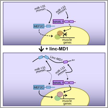
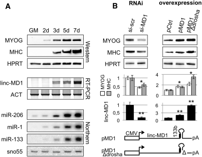
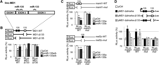
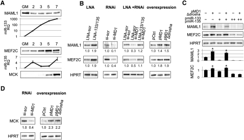
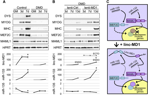
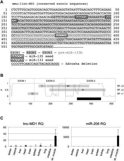
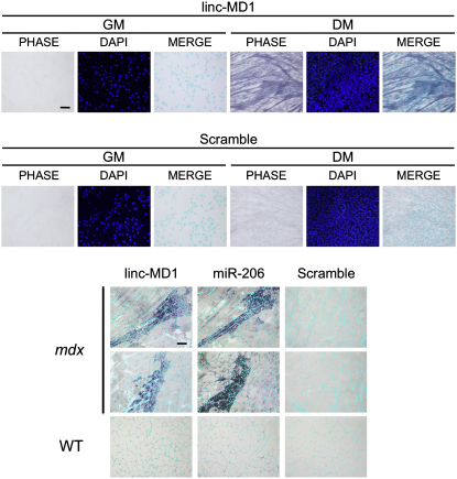
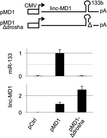
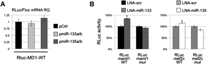
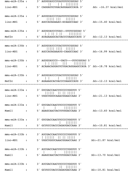
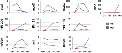
Recently, a new regulatory circuitry has been identified in which RNAs can crosstalk with each other by competing for shared microRNAs. Such competing endogenous RNAs (ceRNAs) regulate the distribution of miRNA molecules on their targets and thereby impose an additional level of post-transcriptional regulation. Here we identify a muscle-specific long noncoding RNA, linc-MD1, which governs the time of muscle differentiation by acting as a ceRNA in mouse and human myoblasts. Downregulation or overexpression of linc-MD1 correlate with retardation or anticipation of the muscle differentiation program, respectively. We show that linc-MD1 "sponges" miR-133 and miR-133 [corrected] to regulate the expression of MAML1 and MEF2C, transcription factors that activate muscle-specific gene expression. Finally, we demonstrate that linc-MD1 exerts the same control over differentiation timing in human myoblasts, and that its levels are strongly reduced in Duchenne muscle cells. We conclude that the ceRNA network plays an important role in muscle differentiation.


Figures

None
Graphical abstract
Figure 1
Figure 1
Muscle-Specific lincRNA Profiling (A) Human miRNA genomic location relative to their host genes. (B) Schematic representation of the murine miR-206/133b genomic locus. Upper panel shows transcriptional start sites (TSS, indicated by arrows) mapped through 5′ RACE analysis in differentiated myoblasts. The genomic structure of the identified linc-MD1 and the exon-intron lengths are shown (conserved exons in black, nonconserved 3′-portions in dash) as well as pre-miRNA sequences. Lower panel shows conserved regions among vertebrates together with E-boxes and regulatory elements (DIST, PROX and pA signals). (C) RT-PCR for linc-MD1, pri-miR-206, and pri-miR-133b expression performed in mouse myoblasts in growth (GM) or differentiation (DM) conditions. The total, nuclear (nuc), cytoplasmic (cyt), polyadenylated (pA+) and nonpolyadenylated (pA-) RNA fractions are shown. The same analysis was also performed in primary satellite cells in GM and DM conditions and in mouse embryonic fibroblasts (MEFs) infected with a MyoD expressing lentivirus (MyoD) or control (gfp). HPRT mRNA was used as endogenous control. (D) Northern blot analysis for linc-MD1 in the poly-A+ fraction from mouse myoblasts in GM and DM conditions. (E) RT-PCR for linc-MD1, pri-miR-206, pri-miR-133b performed on RNA from liver (LIV), heart (HEA), tibialis anterior (TIB) and soleus (SOL) of wild-type (WT) and mdx mice. (F) In situ analysis with a DIG-labeled linc-MD1 probe in C2 myoblasts in GM and DM. (G) In situ analysis with DIG-labeled linc-MD1 and miR-206 probes in wild-type (WT) and mdx gastrocnemius cryosections; DAPI staining (light blue) is also shown. Original magnification is 20×. The scale bar represents 100 μm. Additional fields and controls are show in Figure S2.
Figure 2
Figure 2
Transcriptional and Epigenetic Regulation of miR-206/133b Genomic Locus (A) Promoter activity assay. Upper panel shows that distal (box D) and proximal (box P) regions were cloned upstream of murine pre-miR-223 and tested in C2 myoblasts in GM and DM conditions. A region between miR-206 and miR-133b was used as negative control (dashed box). miR-223 expression was measured by northern blot (Figure S3) and qRT-PCR. U6 snRNA was used as endogenous control. miR-223 relative quantification (RQ) is shown with respect to control vector in GM set to a value of 1. Lower panel shows that the same regions were cloned, alone or in combinations, in a firefly luciferase reporter construct (FLuc) and tested in GM and DM conditions. A luciferase construct (RLuc) was transfected as control. Luciferase activity, from three independent experiments, was measured as FLuc/RLuc RQ shown with respect to the negative control vector in GM set to a value of 1. (B) ChIP analysis for MyoD enrichment on distal (DIST), proximal (PROX) and negative control (CTRL) regions performed on chromatin extracted from C2 myoblasts in GM and DM conditions. Amplifications of IgG control immunoprecipitations (IgG) and 10% of input chromatin (Inp) are shown. (C) Upper panel is a schematic representation of the miR-206/133b genomic locus. Capital letters indicate regions analyzed in ChIP experiments. Location of regulatory regions (DIST, PROX and pA), pre-miRNAs (206, 133b) and linc-MD1 are shown along the locus as in Figure 1B. Lower panel displays ChIP analyses for RNA polymerase II (RNAP II), H3K9ac, H3K27me3, and H3K4me3 performed on chromatin extracted from myoblasts in GM and DM. Values derived from three independent experiments were normalized for background signals (IgG) and expressed as percentage of Input chromatin (% Inp). Unless specifically indicated, statistical significance was calculated with respect to GM conditions. Data are shown as mean ± SD. One asterisk indicates p < 0.05; two asterisks: p < 0.01.
Figure 3
Figure 3
Tridimensional Architecture of miR-206/133b Genomic Locus (A) Upper panel shows StyI restriction sites (vertical lines), regulatory regions (DIST, PROX, pA), pre-miRNAs (206, 133b), and linc-MD1. Capital letters (A–I, X, and Y) indicate the position of StyI sites analyzed in 3C experiments. A–I sites identify the same regions analyzed in ChIP experiments in Figure 2C. A common reverse primer identifying the proximal region (X) was used in combination with different reverse primers. Lower panel shows 3C analysis performed on chromatin extracted from C2 myoblasts in GM and DM conditions and from fibroblast cell cultures. Crosslinking frequencies relative to X region were measured both by semiquantitative PCR (data not shown) and qRT-PCR. Data from multiple experiments were normalized for primer amplification efficiency and reported with respect to X-B interaction in GM set to a value of 1. Statistical significance was calculated with respect to GM conditions. (B) The same 3C analysis was performed in liver (LIV), heart (HEA), tibialis anterior (TIB), soleus (SOL) of wild-type (WT), and mdx animals. For each set of interactions analyzed, crosslinking frequencies relative to X region derived from multiple experiments were reported with respect to WT HEA interaction set to a value of 1. Statistical significance was calculated with respect to WT HEA tissue. Data are shown as mean ± SD. One asterisk indicates p < 0.05; two asterisks: p < 0.01. (C) Schematic representation of the interactions detected upon induction of differentiation.
Figure 4
Figure 4
Modulation of Linc-MD1 Expression Affects Myogenesis (A) RNA and protein samples were extracted from myoblasts in proliferation (GM) and after shift to differentiation medium (DM) for the indicated times. Upper panels show western blot analysis for myogenin (MYOG), myosin heavy chain (MHC), and HPRT. Middle panels show RT-PCR analysis for linc-MD1 and Actin mRNA (ACT) expression. Lower panels show northern blots for miR-206, miR-1, miR-133, and snoRNA55. (B) Left panel: siRNAs for linc-MD1 (si-MD1) or scramble control (si-scr) transfected in C2 myoblasts and maintained in DM for 5 days. Right panel: linc-MD1 overexpression was obtained by transfection of linc-MD1 (pMD1) and linc-MD1-Δdrosha (pMD1-Δdrosha) constructs (see schematic representation below) together with a control GFP cDNA (pCtrl). Samples were collected 4 days after induction of differentiation. Densitometric analysis, normalized for HPRT, is shown below. Lower panels display the levels of linc-MD1, normalized for HPRT, measured by qRT-PCR. Data are shown with respect to control experiments set to a value of 1. Data are shown as mean ± SD. One asterisk indicates: p < 0.05; two asterisks: p < 0.01.
Figure 5
Figure 5
linc-MD1 Acts as a Natural Decoy for miR-135 and miR-133 (A) Positions of miR-135 and miR-133 binding sites on linc-MD1. (B) linc-MD1 (RLuc-MD1-WT) and mutant derivatives devoid of miR-133 or miR-135 binding sites (RLuc-MD1-Δ133 and Rluc-MD1-Δ135) were cloned downstream the Renilla luciferase coding region (RLuc). These constructs were cotransfected in C2 myoblasts together with plasmids expressing miR-135 (pmiR-135a/b) or miR-133 (pmiR-133a/b) or with a control vector (pCtrl). (C) The 3′UTR of MAML1 and of MEF2C were cloned downstream RLuc (RLuc-maml1-WT, RLuc-mef2c-WT) together with mutant derivatives lacking miRNA recognition sequences (RLuc-maml1-mut and RLuc-mef2c-mut). These constructs were cotransfected in C2 myoblasts with plasmids expressing miR-133 (pmiR-133a/b) or miR-135 (pmiR-135a/b) or with a control vector (pCtrl). (D) RLuc-maml1-WT, RLuc-mef2c-WT and their corresponding mutant derivatives (-mut) were transfected in C2 myoblasts with pMD1-Δdrosha or its mutant derivatives depleted in either miR-135 (pMD1-Δdrosha-Δ135) or miR-133 (pMD1-Δdrosha-Δ133) recognition elements. A GFP construct was used as control (pCtrl). For miRNA overexpression experiments, unless specifically stated, a mix of a and b pre-miRNA expression plasmids was used. For all the experiments, histograms indicate the values of luciferase measured 24 hr after transfection. Data, derived from three independent experiments, are shown with respect to RLuc control vector set to a value of 100%. Data are shown as mean ± SD. One asterisk indicates p < 0.05.
Figure 6
Figure 6
linc-MD1 Modulates MAML1 and MEF2C Expression in Muscle (A) Western blot analysis for MAML1, MEF2C, and MCK, in growth medium (GM) and at different days upon shift to differentiation conditions (DM). The relative expression of miR-133 and miR-135 is also reported. (B) MAML1 and MEF2C levels upon modulation of miRNA and linc-MD1 levels in C2 myoblasts maintained in DM for 5 days. Modulation was obtained with: anti-miR-133 and anti-miR-135 LNAs, RNAi against linc-MD1, and linc-MD1 overexpression (pMD1 and pMD1-Δdrosha). Scrambled LNA (LNA-scr) and siRNA (si-scr) were used as control. HPRT was used as a loading control. Below each panel, relative quantifications with respect to control samples set to a value of 1 are displayed. (C) Western blot for the same proteins from C2 myoblasts transfected with different combinations of pMD1-Δdrosha and pmiR-133/pmiR-135 expression plasmids. (+) corresponds to 1.5 μg pMD1-Δdrosha and to 50 ng of pmiR-133/miR-135, while (++) corresponds to 300 ng of pmiR-133/pmiR-135. Control myoblasts were transfected with a GFP plasmid (−). The values, derived from densitometric analysis, are reported with respect to mock samples set to a value of 1. Data are shown as mean ± SD. One asterisk indicates p < 0.05. (D) MCK measured in cells treated with the indicated LNA or siRNAs as in panel (B).
Figure 7
Figure 7
linc-MD1 Is Conserved in Humans, and It Improves Differentiation of Duchenne Myoblasts (A) Myoblasts derived from healthy (Control) and Duchenne Muscular Dystrophy (DMD) individuals were induced to differentiate for the indicated times and RNA and protein samples were collected. Western blot analysis for the indicated proteins was performed in parallel with qRT-PCR for the expression of linc-MD1, miR-133 and miR-135. (B) DMD myoblasts were infected with lentiviral vectors carrying a control GFP-expressing cassette (lenti-Ctrl) or the linc-MD1-Δdrosha construct under the control of the CMV promoter (lenti-MD1). Cells were induced to differentiate for the indicated times and RNA and protein samples were collected. Western blot analysis for the indicated proteins was performed in parallel with qRT-PCR for the expression of linc-MD1, miR-133 and miR-135. Data are shown as mean ± SD. One asterisk indicates p < 0.05. (C) Schematic representation of the circuitry linking linc-MD1, miR-135, miR-133, and muscle differentiation.
Figure S1
Figure S1
Related to Figure 1 (A) Murine linc-MD1 with exons identification, miRNA binding sites and sequence deleted in Δdrosha constructs. The nucleotide sequence refers to the portion of the transcript which is conserved between human and mouse. Two 3′-end isoforms mapping at two pA-sites have been mapped in a non conserved region at 1060 and 1450 nt downstream the indicated sequence (Figure 1B). (B) Conservation pattern of the linc-MD1 transcript expressed as probability of negative selection taken from the Mammal Cons track. RF+1, +2 and +3 refer to the reading frames, gray boxes indicate the position of ORFs in number of triplets. The mature transcript contains four open reading frames (defined as any sequence starting with AUG and containing at least 10 non terminator codons) ranging in size from 11 to 91 triplets. None of the AUG shows a Kozak consensus (Kozak 1989), nor are the sequences more or less conserved than the surrounding regions. (C) linc-MD1 and miR-206 relative quantifications in the indicated tisses. Values are normalized for HPRT or U6 snRNA respectively and shown with respect to testes sample set to a value of 1.
Figure S2
Figure S2
Related to Figure 1 (A) In situ analysis for linc-MD1, in C2 myoblasts in GM and DM. DAPI staining is also shown alone (DAPI) or together with DIG staining (MERGE). (B) In situ analysis for linc-MD1, miR-206 in tibialis cryosections from WT and mdx animals. DAPI staining is also shown in light blue. Original magnification X20, scale bar 100 μm. In both analyses scramble oligo is used as control.
Figure S3
Figure S3
Related to Figure 2 Northern blot analysis for murine mir-223 expression in C2 myoblasts transfected with D-miR-223 and P-miR-223 constructs and maintained in GM or DM conditions. Ctrl plasmid contains a region between miR-206 and miR-133b, used as negative control. miR-16 was used as loading control.
Figure S4
Figure S4
Related to Figure 4 Schematic representation of pMD-1 and pMD-1-Δdrosha constructs used in linc-MD1 overexpression experiments. Histograms show miR-133 and linc-MD1 RNA relative expression measured by qRT-PCR normalized for U6 snRNA or HPRT respectively and shown with respect to pCtrl set to a value of 1.
Figure S5
Figure S5
Related to Figure 5 (A) RLuc/FLuc mRNA ratio measured by qRT-PCR in C2 myoblasts transfected with RLuc-MD1-WT construct together with plasmids expressing miR-133a/b, miR-135a/b or control. Values are normalized for HPRT and shown with respect to pCtrl set to a value of 1. (B) The constructs described in Figure 5 were cotransfected in C2 myoblasts with scamble LNA (LNA-scr) or LNA for the indicated miRNA. Histograms indicate the values of renilla luciferase measured 48 hr after transfection in DM. Data are shown with respect to the values of RLuc control vector set to a value of 100%. One asterisk: p < 0.05.
Figure S6
Figure S6
Related to Figure 5 Base pairing of the miRNAs with their predicted binding sites on linc-MD1 and on their mRNA targets. ΔG values were obtained from miRanda (Enright et al., 2003)
Figure S7
Figure S7
Related to Figure 7 qRT-PCR for the indicated mRNAs and miRNAs in satellite cells purified from WT or mdx mice differentiated for the indicated hours. Values were normalized for HPRT or U6 snRNA respectively and shown with respect to GM set to a value of 1.

Comment in

  • RNA: a new layer of regulation.
    David R. David R. Nat Rev Mol Cell Biol. 2011 Nov 3;12(12):766. doi: 10.1038/nrm3225. Nat Rev Mol Cell Biol. 2011. PMID: 22048709 No abstract available.
  • ceRNAs: miRNA target mimic mimics.
    Rubio-Somoza I, Weigel D, Franco-Zorilla JM, García JA, Paz-Ares J. Rubio-Somoza I, et al. Cell. 2011 Dec 23;147(7):1431-2. doi: 10.1016/j.cell.2011.12.003. Cell. 2011. PMID: 22196719 No abstract available.

References

    1. Abou El Hassan M., Bremner R. A rapid simple approach to quantify chromosome conformation capture. Nucleic Acids Res. 2009;37:e35. - PMC - PubMed
    1. Amaral P.P., Mattick J.S. Noncoding RNA in development. Mamm. Genome 19, 454–492. Genome Res. 2008;17:556–565. - PubMed
    1. Ballarino M., Pagano F., Girardi E., Morlando M., Cacchiarelli D., Marchioni M., Proudfoot N.J., Bozzoni I. Coupled RNA processing and transcription of intergenic primary microRNAs. Mol. Cell. Biol. 2009;29:5632–5638. - PMC - PubMed
    1. Beltran M., Puig I., Peña C., García J.M., Alvarez A.B., Peña R., Bonilla F., de Herreros A.G. A natural antisense transcript regulates Zeb2/Sip1 gene expression during Snail1-induced epithelial-mesenchymal transition. Genes Dev. 2008;22:756–769. - PMC - PubMed
    1. Buckingham M., Vincent S.D. Distinct and dynamic myogenic populations in the vertebrate embryo. Curr. Opin. Genet. Dev. 2009;19:444–453. - PubMed

Supplemental References

    1. Ashburner, M., Ball, C.A., Blake, J.A., Botstein, D., Butler, H., Cherry, J.M., Davis, A.P., Dolinski, K., Dwight, S.S., Eppig, J.T., et al. (2000). Gene ontology: tool for the unification of biology. The Gene Ontology Consortium. Nat. Genet. 25, 25–29. - PMC - PubMed
    1. Barrett, T., Troup, D.B., Wilhite, S.E., Ledoux, P., Evangelista, C., Kim, I.F., Tomashevsky, M., Marshall, K.A., Phillippy, K.H., Sherman, P.M., et al. (2011). NCBI GEO: archive for functional genomics data sets–10 years on. Nucleic Acids Res. 39 (Database issue), D1005–D1010. - PMC - PubMed
    1. Cardinali, B., Castellani, L., Fasanaro, P., Basso, A., Alemà, S., Martelli, F., and Falcone, G. (2009). Microrna-221 and microrna-222 modulate differentiation and maturation of skeletal muscle cells. PLoS ONE 4, e7607. - PMC - PubMed
    1. Denti, M.A., Rosa, A., D'Antona, G., Sthandier, O., De Angelis, F.G., Nicoletti, C., Allocca, M., Pansarasa, O., Parente, V., Musarò, A., et al. (2006). Body-wide gene therapy of Duchenne Muscular Dystrophy in the mdx mouse model. Proc. Natl. Acad. Sci. USA 103, 3758–3763. - PMC - PubMed
    1. Friedman, R.C., Farh, K.K., Burge, C.B., and Bartel, D.P. (2009). Most Mammalian mRNAs Are Conserved Targets of MicroRNAs. Genome Res. 19, 92–105. - PMC - PubMed

Publication types

MeSH terms