. 2011 Nov 9;19(11):1691-700.
doi: 10.1016/j.str.2011.09.017. Epub 2011 Oct 13.

Structure of a Blinkin-BUBR1 complex reveals an interaction crucial for kinetochore-mitotic checkpoint regulation via an unanticipated binding Site

Affiliations

Structure of a Blinkin-BUBR1 complex reveals an interaction crucial for kinetochore-mitotic checkpoint regulation via an unanticipated binding Site

Victor M Bolanos-Garcia et al. Structure. .

Erratum in

  • Structure. 2011 Dec 7;19(12):1895

Abstract

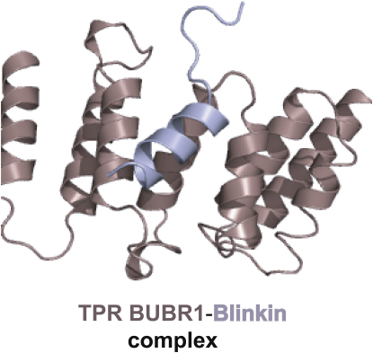
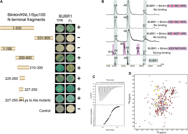
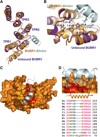
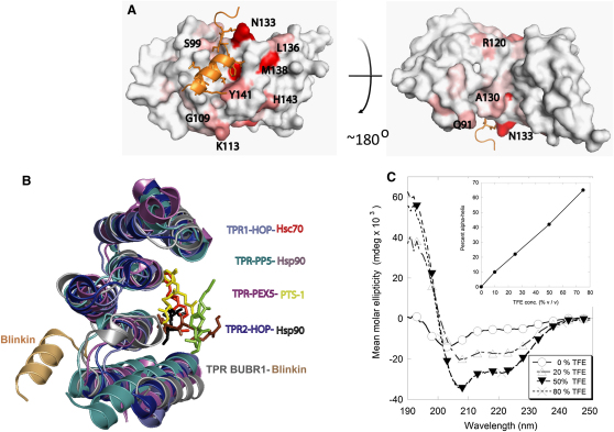
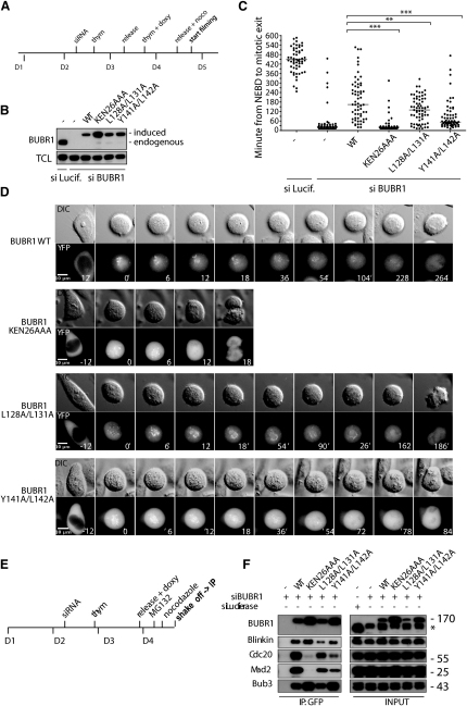
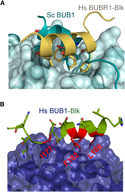
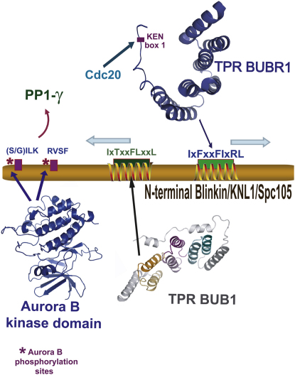
The maintenance of genomic stability relies on the spindle assembly checkpoint (SAC), which ensures accurate chromosome segregation by delaying the onset of anaphase until all chromosomes are properly bioriented and attached to the mitotic spindle. BUB1 and BUBR1 kinases are central for this process and by interacting with Blinkin, link the SAC with the kinetochore, the macromolecular assembly that connects microtubules with centromeric DNA. Here, we identify the Blinkin motif critical for interaction with BUBR1, define the stoichiometry and affinity of the interaction, and present a 2.2 Å resolution crystal structure of the complex. The structure defines an unanticipated BUBR1 region responsible for the interaction and reveals a novel Blinkin motif that undergoes a disorder-to-order transition upon ligand binding. We also show that substitution of several BUBR1 residues engaged in binding Blinkin leads to defects in the SAC, thus providing the first molecular details of the recognition mechanism underlying kinetochore-SAC signaling.


Figures

None
Graphical abstract
Figure 1
Figure 1
Mapping the BUBR1-Blinkin Interaction (A) BUBR1 interacts with N-terminal Blinkin fragments in a Y2H assay. Strains Y187[pGBKT7-bait] and GOLD[pGADT7-target] were mated and X-α-Gal activity assays performed on colonies grown on SD/-Ade/-His/-Leu/-Trp plates. Colony growth requires the activation of ade and his reporter genes and the blue-producing X-α-Gal reaction requires the activation of the mel1 reporter gene to express the secreted reporter enzyme α-galactosidase. (B) Nano-ESI MS of synthetic peptides that mimic Blinkin S208-K226 confirm the interaction while peptides harbouring site-specific substitutions indicate the hydrophobic residues I213, F215, F218 and I219 are critical for binding BUBR1. (C) ITC data shows the affinity of the interaction (Kd = 9 μM); ΔH and ΔS of −2.7 kcal mol-1 and −64 kcal mol-1, respectively. Saturation of BUBR157-220 with Blinkin peptide S208-K226 was achieved when BUBR157-220 at concentration 27 μM was used to titrate against 200 μM of the peptide. (D) An overlay of the 2D 1H-15N HSQC spectra of BUBR1-Blinkin chimera (red) and free BUBR1 after addition of up to 2 moles of Blinkin peptide to one mol of protein. All structured amides from BUBR1 occupy an identical position in the chimera. Some extra peaks from the additional residues in the chimera are indicated: (blue, residue number shown) residues from Blinkin, which become structured upon binding and (orange) unstructured residues from the linker and extreme N terminus of Blinkin (see also Figure S1).
Figure 2
Figure 2
The 2.2 Å Crystal Structure of the BUBR1-Blinkin Complex Reveals an Unexpected Mode of Binding (A) Superposition of the free (magenta) and Blinkin-bound TPR BUBR1 crystal structures (orange) reveals that little conformational changes occur upon ligand binding. A closeup of TPR2 BUBR1, where the slight conformational changes that result from Blinkin binding are most noticeable. (B) Surface representation of TPR BUBR1; the hydrophobic residues relevant for the interaction of this protein with Blinkin (blue ribbon) are highlighted in red. (C) Blinkin residues I213, F215, F218 and I219 bind a shallow hydrophobic groove. (D) Amino acid sequence alignment of the Blinkin BUBR1 binding region reveals a high conservation of these residues in higher organisms. Each molecule representation was generated with Pymol (DeLano, 2002). The aligned sequences are from Homo sapiens (Hs); Rattus norvegicus (Rn); Mus musculus (Mm); Pan troglodytes (Pt); Callithrix jacchus (Cj); Equus caballus (Ec); Bos taurus (Bt); Ailuropoda melanoleuca (Am); Canis familiaris (Cf); Oryctolagus cuniculus (Oc) and Monodelphis domestica (Md); Drosophila melanogaster (Dm); and Saccharomyces cerevisiae (Sc).
Figure 3
Figure 3
Blinkin Shows a Novel BUBR1 Binding Motif (A) NMR chemical shift changes are consistent with the location of the peptide in the chimera. BUBR1-Blinkin chimera is shown as a surface representation with chemical shift changes upon binding of Blinkin peptide to native BUBR1 indicated as a white-red ramp: white, no change/data not available; red, largest shift change upon peptide binding. The largest changes in chemical environment are on the convex face and are consistent with the location of the peptide in the crystal, although some shifts propagate through to the concave face (see also Figure S1). (B) Blinkin binds a region that differs from that observed in TPR-peptide complexes of high structure similarity. (C) Far-UV circular dichroism spectra reveal a predominantly disorder structure of Blinkin peptide S208-K226 in aqueous solutions. Gradual addition of TFE resulted in a dramatic conformational change and the formation of a predominantly α-helical structure.
Figure 4
Figure 4
The Interaction between BUBR1 and Blinkin Is Required for a Functional SAC (A) Synchronization protocol for RNAi depletion of BUBR1 and rescue with mutants. Time is shown in days (D). (B) Western blot for BUBR1 showing depletion of endogenous BUBR1 and level of expression of the different mutants (see also Figure S3). (C) Plot showing time spent in mitosis in the presence of 100 nM nocodazole with each dot representing a single cell analyzed by time-lapse microscopy and bar indicating the median. Time is scored as the time from nuclear envelope breakdown (NEBD) to time of mitotic exit and for each condition 60 cells have been analyzed. The statistical significance is indicated above as determined by the Mann-Whitney test. The time spent in mitosis in the Luciferase control is underestimated as 49 out of 60 cells did not exit mitosis during filming. (D) Representative still images from the different cell lines analyzed. Time is in minutes and t = 0 is at NEBD. (E) Synchronization protocol used for the immunodepletion experiment. (F) Immunopurified BUBR1 complexes analyzed for the presence of Blinkin, Cdc20, Mad2, and Bub3. In the input the asterisks indicates an unspecific band from previous probing of the blot.
Figure 5
Figure 5
BUB1 and Blinkin/KNL1/Spc105 Complexes Seem to Show a Similar Mode of Interaction (A) Superposition of the crystal structures of yeast BUB1 and BUBR1-Blinkin showing that the intermolecular interactions observed in the former molecule occur in a similar fashion as those defining the BUBR1-Blinkin complex. (B) Structure model of human TPR BUB1 (blue) in complex with a putative Blinkin/KNL1 BUB1 binding motif residues E172-H189, predicts that hydrophobic residues I177, F182, L183, and L186 (shown in stick representation) and possibly T179 (not shown) play an important role in the interaction.
Figure 6
Figure 6
Functional Implications of the BUB1/BUBR1-Blinkin Interaction Binding to the kinetochore stimulates BUBR1 kinase activity, which results in prolonged mitotic arrest. Blinkin N-terminal residues defining the I177xTxxFLxxL186 motif are predicted to constitute a putative BUB1 binding site. The conserved Blinkin motif I213xFxxFIxRL222 is responsible of binding BUBR1 in a process that involves important disorder to order transitions. Thus, local conformational changes upon BUB1 and/or BUBR1 binding (blue arrows) are likely to affect other interactions involving this Blinkin region. For instance, N-terminal Blinkin contains a conserved motif that binds protein phosphatase 1 γ (PP1 γ), an interaction that is required for the targeting of PP1 to the outer kinetochore. The Blinkin-PP1 γ interaction is disrupted by Aurora B-dependent phosphorylation of Blinkin, which targets residues of two conserved motifs (S/GILK) and (RVSF). The diversity of key protein-protein interactions mediated by Blinkin evidences the complexity of the molecular mechanisms mediating kinetochore-mitotic checkpoint signaling.

References

    1. Bolanos-Garcia V.M., Blundell T.L. BUB1 and BUBR1: multifaceted kinases of the cell cycle. Trends Biochem. Sci. 2011;36:141–150. - PMC - PubMed
    1. Bolanos-Garcia V.M., Wu Q., Ochi T., Chirgadze D.Y., Sibanda B.L., Blundell T.L. Spatial and temporal organisation of multiprotein assemblies: achieving sensitive control in information-rich cell regulatory systems. Phil. Trans. R. Soc. A. 2011 in press. - PubMed
    1. Bolanos-Garcia V.M., Kiyomitsu T., D'Arcy S., Chirgadze D.Y., Grossmann J.G., Matak-Vinkovic D., Venkitaraman A.R., Yanagida M., Robinson C.V., Blundell T.L. The crystal structure of the N-terminal region of BUB1 provides insight into the mechanism of BUB1 recruitment to kinetochores. Structure. 2009;17:105–116. - PMC - PubMed
    1. Blundell T.L., Bolanos-Garcia V., Chirgadze D.Y., Harmer N.J., Lo T., Pellegrini L., Sibanda B.L. Asymmetry in the multiprotein systems of molecular biology. Struct. Chem. 2002;13:405–412.
    1. CCP4 Collaborative Computational Project. Acta Crystallogr. D Biol. Crystallogr. 1994;50:760.

Publication types

MeSH terms

Associated data