Review
. 2011 Oct 12;19(10):1370-80.
doi: 10.1016/j.str.2011.08.009.

Emerging models of glutamate receptor ion channel structure and function

Affiliations
Review

Emerging models of glutamate receptor ion channel structure and function

Mark L Mayer. Structure. .

Abstract

Excitatory synaptic transmission in the brain is mediated by ligand-gated ion channels (iGluRs) activated by glutamate. Distinct from other neurotransmitter receptors, the extracellular domains of iGluRs are loosely packed assemblies with two clearly distinct layers, each of which has both local and global 2-fold axes of symmetry. By contrast, the iGluR transmembrane segments have 4-fold symmetry and share a conserved pore loop architecture found in tetrameric voltage-gated ion channels. The striking layered architecture of iGluRs revealed by the 3.6 Å resolution structure of an AMPA receptor homotetramer likely arose from gene fusion events that occurred early in evolution. Although this modular design has greatly facilitated biophysical and structural studies on individual iGluR domains, and suggested conserved mechanisms for iGluR gating, recent work is beginning to reveal unanticipated diversity in the structure, allosteric regulation, and assembly of iGluR subtypes.


Figures

Figure 1
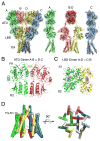
Figure 1. Structure of the rat GluA2 AMPA receptor
(A) Structure of the GluA2 tetramer viewed from the side, with the A–D subunits colored green, red, yellow and blue respectively; as shown by the dissection at right the A and C subunit pairs show a continuous molecular envelope, while the B and D subunits adopt a different conformation with a wide separation between the ATD and LBD layers. (B) The AB and DC subunit ATD dimers have structures essentially identical to that for the isolated GluA2 ATD expressed as a soluble protein, with intersubunit contacts mediated by both the upper (R1) and lower (R2) lobes. (C) The AD and CB subunit LBD dimers also adopt the same conformation found for crystal structures for the antagonist bound soluble GluA2 LBD construct, with intermolecular contacts mediated exclusively by the upper (S1) lobes. (D) The ion channel viewed from the side (left) and after rotation by 90° (right) with the pre-M1 cuff helices colored grey.
Figure 2
Figure 2. Symmetry mismatches in the GluA2 tetramer
Cα trace viewed from the side (left) and top (right) showing the global and local 2-fold symmetry axes in the ATD and LBD layers, and the 4-fold symmetry axis in the ion channel TM layer. Anomalous difference electron density maps contoured at 5 σ reveal the positions of Hg atoms bound to Cys residues with 2-fold symmetry in the ATD and LBD layers, and 4-fold symmetry to the TM layer, for a complex with a Hg derivative of the competitive antagonist ZK 200775 (adapted with permission from Sobolevsky et al., 2009 Fig. 1). Of interest, the Hg atoms bound in the LBD layer, as well as in the ATD and TM layers, were contaminants in the antagonist preparation, and at this contour level the Hg atom on labeled antagonist is not visible, suggesting either partial hydrolysis of the Hg derivative or local disorder.
Figure 3
Figure 3. A gallery of iGluR ATD dimer and tetramer structures
(A) An ATD dimer from the full length GluA2 structure (3KG2) and the isolated ATDs of GluA1 (3SAJ), GluA2 (2WJW, 3H5V & 3HSY), GluA3 (3O21), GluR6 (3H6G), GluR7 (3OLZ), KA2 (3OM0 & 3OM1) and a GluR6/KA2 heterodimer assembly (3QLU). Transparent representations are used for the 2nd and 3rd copies of structures for which more than one dimer assembly was present in the asymmetric unit, and for structures solved by different groups, revealing small changes in subunit orientation resulting principally from rotations parallel to the 2-fold axis of symmetry. (B) ATD homotetramer assemblies generated by crystallographic symmetry operations for GluA2, GluR6 and GluR7. (C) The GluR6/KA2 ATD tetramer assembly (3QLV) showing formation of the dimer of dimers interface by the GluR6 subunits. (D) Stereo view of the GluR6/KA2 ATD heterodimer showing packing between helices B and C in the upper lobes; the S-loop projects into the dimer interface; below this a conserved aromatic residue projects into a hydrophobic slot on the dimer partner.
Figure 4
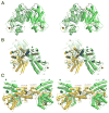
Figure 4. NMDA receptor ATD dimer and tetramer structures
(A) Stereoview of the rat GluN1 ATD homodimer (3Q41), with the two subunits colored in pale and bright green; the N and C termini marked by blue and red spheres; and the S-loop colored dark blue. (B) Stereoview of the GluN1/GluN2B heterodimer rotated by approximately 90° from the view in A, with the GluN1 subunit colored pale green, the GluN2B subunit colored gold, and the S-loop which includes an α-helix colored dark blue, with the rat GluN1 ATD homodimer drawn in transparent shading after superposition using the heterodimer GluN1 subunit coordinates. (C) Stereoview of the GluN1/GluN2B ATD asymmetric unit contents showing two copies of the ATD heterodimer, with a tetramer formed by crystal contacts mediated by the GluN2B subunits, colored as in B.
Figure 5
Figure 5. NMDA receptor binding sites for allosteric modulators
(A) Cartoons illustrating models proposed for the binding sites and mechanism of inhibition of NMDA receptors by Zn and ifenprodil. Based on homology with LIVBP it was proposed that Zn2+ and ifenprodil would bind deep in the cleft of the NR2 subunit ATD stabilizing a closed cleft conformation. (B) Crystal structures of the GluN1/GluN2B ATD heterodimer complex with ifenprodil (3QEL) and the GluN2B ATD monomer in complex with Zn2+ (3JPY) drawn with transparent shading reveal a more complex binding mechanism. Ifenprodil binds within the interface between the GluN1 and GluN2B subunits, not in the cleft as previously assumed. Zn2+ binds at the lateral edge of the interdomain cleft in a solvent exposed cleft, and is not buried deep within the ATD clamshell.

References

    1. Abuin L, Bargeton B, Ulbrich MH, Isacoff EY, Kellenberger S, Benton R. Functional architecture of olfactory ionotropic glutamate receptors. Neuron. 2011;69:44–60. - PMC - PubMed
    1. Armstrong N, Gouaux E. Mechanisms for activation and antagonism of an AMPA-sensitive glutamate receptor: Crystal structures of the GluR2 ligand binding core. Neuron. 2000;28:165–181. - PubMed
    1. Armstrong N, Mayer M, Gouaux E. Tuning activation of the AMPA-sensitive GluR2 ion channel by genetic adjustment of agonist-induced conformational changes. Proc Natl Acad Sci USA. 2003;100:5736–5741. - PMC - PubMed
    1. Ayalon G, Segev E, Elgavish S, Stern-Bach Y. Two regions in the N-terminal domain of ionotropic glutamate receptor 3 form the subunit oligomerization interfaces that control subtype-specific receptor assembly. J Biol Chem. 2005;280:15053–15060. - PubMed
    1. Benton R, Vannice KS, Gomez-Diaz C, Vosshall LB. Variant ionotropic glutamate receptors as chemosensory receptors in Drosophila. Cell. 2009;136:149–162. - PMC - PubMed

Publication types

MeSH terms

LinkOut - more resources