Review
. 2011 Oct 12;19(10):1381-93.
doi: 10.1016/j.str.2011.09.001.

Advances in structural and functional analysis of membrane proteins by electron crystallography

Affiliations
Review

Advances in structural and functional analysis of membrane proteins by electron crystallography

Goragot Wisedchaisri et al. Structure. .

Abstract

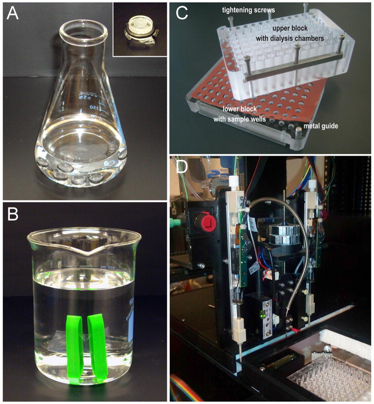
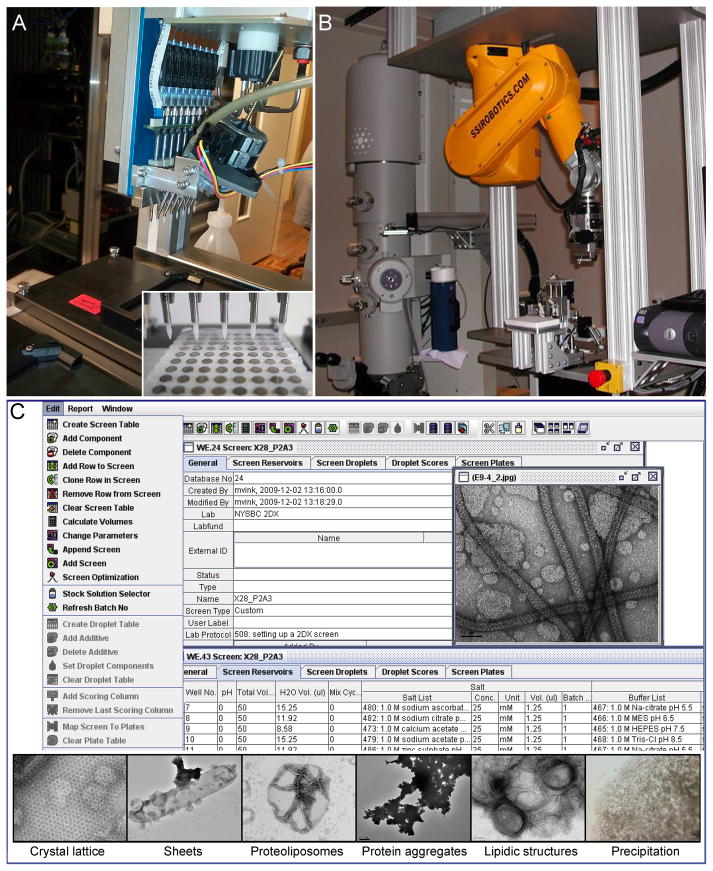
Electron crystallography is a powerful technique for the study of membrane protein structure and function in the lipid environment. When well-ordered two-dimensional crystals are obtained the structure of both protein and lipid can be determined and lipid-protein interactions analyzed. Protons and ionic charges can be visualized by electron crystallography and the protein of interest can be captured for structural analysis in a variety of physiologically distinct states. This review highlights the strengths of electron crystallography and the momentum that is building up in automation and the development of high throughput tools and methods for structural and functional analysis of membrane proteins by electron crystallography.


Figures

Figure 1
Figure 1. Gallery of selected membrane protein structures determined by electron crystallography
(top, left to right) The light-driven proton pump bacteriorhodopsin, represented by the first reported atomic structure (PDB 1BRD (Henderson et al., 1990)); the plant light-harvesting complex from photosystem II (Kuhlbrandt et al., 1994); aquaporin-1 as a representative of the aquaporin family of water channels (PDB 1FQY (Murata et al., 2000)); the membrane associated glutathione transferases, represented by microsomal glutathione S-transferase 1 (PDB 2H8A (Holm et al., 2006)); and the acetylcholine receptor (PDB 2BG9 (Unwin, 2005)). (bottom, left to right) The H+, K+ -ATPase (PDB 2XZB (Abe et al., 2011)); the major facilitator superfamily, represented by the oxalate sugar transporter (Hirai et al., 2002); the sodium coupled proton antiporter, NhaA (PDB 3FI1 (Appel et al., 2009)); the small multidrug transporter, EmrE (PDB 2I68 (Fleishman et al., 2006)); and gap junctions, represented by connexin-26 (PDB 3IZ1 (Oshima et al., 2011)).
Figure 2
Figure 2. Dialysis and high-throughput robotics for 2D crystallization
(A) Slow dialysis using dialysis buttons. Membrane protein/detergent/lipid mixtures are prepared in the 50μl buttons and sealed with a dialysis membrane. The detergent is gradually removed from the mixture by slow dialysis against buffer lacking the detergent. Inset, closeup view of an assembled dialysis button. (B) Dialysis tubing. Membrane protein/detergent/lipid mixture is placed in the dialysis tube between two plastic clamps that are assembled at either end. This set up is submerged in a buffer lacking the detergent and provides a faster dialysis rate than the buttons presented in panel A. (C) 96-well dialysis block for high-throughput crystallization screening. The samples of membrane protein/detergent/lipid mixtures are placed in the lower block. The dialysis membrane is sandwiched in between the lower block and the upper block together with silicone sheets to prevent leakage. The dialysis buffers are added to the upper block. Reprinted from (Vink et al., 2007). Copyright (2007) with permission from Elsevier. (D) The 2DX robot for automated addition of cyclodextrin for detergent removal (Iacovache et al., 2010). A dispenser adds cyclodexdrin to the samples of membrane protein/detergent/lipid mixtures in a 96-well microplate. The robot is equipped a light scattering detector to measure turbidity as an indicator of reconstitution and crystal growth. Picture courtesy of Prof. Andreas Engel (Case Western Reserve University).
Figure 3
Figure 3. Automated grid preparation, screening and crystal evaluation
(A) Automatic grid staining robot (Coudray et al., 2011). The multi-channel pipetting system in the robot allows sample deposit, blotting, washing and staining of EM grids in a 96-well microplate format (inset). Picture courtesy of Prof. Andreas Engel (Case Western Reserve University). Inset reprinted from (Coudray et al., 2011). Copyright (2007) with permission from Elsevier. (B) Robotic grid loading system. An EM grid is first picked up from a 96-well format grid tray by a vacuum system and transferred to a grid holder. Subsequently, the robot automatically loads the grid holder into the electron microscope. The operation of the robotic arm is controlled by software. Reprinted from (Cheng et al., 2007). Copyright (2007) with permission from Elsevier. (C) 2D crystallization data management and evaluation. The Laboratory Information Management System (LIMS) software archives EM images with the corresponding crystallization conditions and allows image viewing through the graphical user interface. Reprinted from (Hu et al., 2010). Copyright (2007) with permission from Elsevier. (Inset) Crystallization results are evaluated and sorted into different categories such as crystal lattice, planar sheets and tubular vesicles, proteoliposomes, protein aggregates, lipidic structures, and macroscopic precipitation. Reprinted from (Kim et al., 2010). Copyright (2010) with kind permission from Springer Science+Business Media B. V.
Figure 4
Figure 4. Newly developed electron crystallographic data processing suites
(A) Graphical user interface of the 2dx program. Fourier transform of 2D crystal images can be indexed by automated lattice determination in 2dx_image and merged in 2dx_merge modules. Image parameters are displayed in the interface and can be easily entered or modified. The reconstructed projection map can be viewed through the interface. Reprinted from (Gipson et al., 2007a). Copyright (2007) with permission from Elsevier. (B) Graphic user interface of the IPLT program. Both image and electron diffraction data can be processed with IPLT. Several functions in IPLT include lattice search in a power spectrum or diffraction pattern, lattice refinement based on 2D Gaussian profile fitting, integration of diffraction peaks, tilt geometry determination, and import/export of the CCP4 mtz file format. Image courtesy of Prof. Andreas Engel (Case Western Reserve University).
Figure 5
Figure 5. Advances in strategies for structure determination by electron crystallography
(A) Structure determination of H+, K+-ATPase from very small patches of coherent arrays. (left) Negatively stained mosaic 2D crystal of the H+, K+-ATPase shows small coherent areas of well-ordered lattice (arrows) as well as vesicle aggregation (arrowhead). These 2D crystals are not suitable for electron diffraction but could be used for structural analysis by imaging. (middle) The density map of the H+, K+-ATPase 2D crystal after image processing shows that the 2D crystals consist of two membrane layers (bar lines). One αβ-protomer (dark blue) is shown in the dashed box. Reprinted by permission from Macmillan Publishers Ltd. from (Abe et al., 2009), Copyright (2009). (right) Model of the H+, K+-ATPase αβ-protomer in ribbon representation. The transmembrane domain is indicated by the bar lines. (B) Fragment-based phase extension method for phasing high-resolution electron diffraction. The example of AQP0 at 1.9 Å is presented. (top) Phase data at 6 Å resolution served as the starting point for fragment positioning. The σA-weight 2Fobs-Fcalc density maps (with the corresponding protein models overlaid) were gradually improved at the end of cycles 1 and 2 of the phase extension procedures allowing model building and refinement. (bottom) The starting map at 6 Å resolution did not reveal densities for the lipid or water molecules, but after cycles 1 and 2 as phases were extended to 1.9 Å resolution, the density for lipid and water molecules became apparent and well-defined. In the final map, the densities for the lipid and water molecules became more accurate and appeared similar to the previously published study (PDB 2B6O (Gonen et al., 2005)). Reprinted from (Wisedchaisri and Gonen, 2011). Copyright (2011) with permission from Elsevier.
Figure 6
Figure 6. Unraveling membrane transport mechanisms by electron crystallography at increasing resolutions
(A) Substrate specific transport by the trimeric E. coli sugar transporter (GalP) in crystalline vesicles. (left, top) Electron micrograph of GalP crystalline vesicles. (left, bottom) 2D-projection structure of GalP at 18 Å reveals a lattice composed of GalP trimers. (right) The crystalline vesicles were shown to selectively transport glucose over lactose in assays using the fluorescent glucose analogue 2-NBDG (Zheng et al., 2010). Reprinted from (Zheng et al., 2010). Copyright (2010) with permission from Elsevier. (B) Gating mechanism of the acetylcholine (Ach) receptor at millisecond resolution. (left) Tubular crystals of the Ach receptor were embedded on an electron microscopy (EM) grid and sprayed with Ach less then 5 ms before plunge freezing into liquid ethane. (right) The Ach receptor in its closed conformation; colored by secondary structure (Unwin, 2005). Binding of Ach at a conserved site in the extracellular domain (residue in red) induces a rotation of the transmembrane helical domain that opens the channel pore allowing ion conductance. (C) Light driven proton transport in bacteriorhodopsin (bR) determined at atomic resolution. (left) Structural overlay of native bR (white) and a bR mutant that mimics the open state of the proton transport cycle (red) (Subramaniam and Henderson, 2000). (middle) Zoom view of bR with the retinal chromophore (RET) shown in cyan. The acidic residues (E194 and D212 shown as stick representation) and two waters (or hydroxonium ions, shown in purple) involved in the proton transport pathway of bR are highlighted. (right) Positive charges were directly visualized from the |Fo-Fc| difference maps obtained from low and high-resolution diffraction data, shown as red cage (arrow) (Mitsuoka et al., 1999). Reprinted from (Mitsuoka et al., 1999). Copyright (1999) with permission from Elsevier.
Figure 7
Figure 7. Lipid-protein interactions in two-dimensional crystals
(A) Density map of bacteriorhodopsin (bR) showing the native purple membrane lipid bilayer (Mitsuoka et al., 1999) (B) Density map of aquaporin-0 (AQP0) showing the phosphatidylcholine (PC) lipid bilayer (Gonen et al., 2005). A complete annular lipid shell was modeled from the observed electron density for both bR and AQP0. (C) Hexagonal lattice of bR trimers with protein (grey) and lipids (purple). Specifically bound lipids were observed within the trimeric 3-fold axis and at the monomer-monomer interface. (D) Square lattice of AQP0 tetramers with protein (grey) and lipids (purple). Bulk lipids were observed at the 4-fold tetramer interface. (E) Box plot comparing the protein crystallographic B-factors of bR and AQP0 obtained by cryo EM (in blue) (PDB 2AT9 (Mitsuoka et al., 1999) and PDB 2B6O (Gonen et al., 2005)) and X-ray crystallography (in red) (PDB 1C3W (Luecke et al., 1999) and PDB 1YMG (Harries et al., 2004)). Each box indicates the median B-factor and the first and third quartile B-factor values for all atoms of each protein. The whiskers represent the minimum and maximum values. The B-factors obtained by cryo EM are overall lower compared to structures determined by X-ray crystallography (Hite et al., 2008). (F) Comparison of AQP0 monomer (grey) with annular PC lipids (PC1 – 7) (Gonen et al., 2005) and E. coli lipids (EL1 – 7) (Hite et al., 2010). The AQP0 structure is nearly identical in both structures. The PC and EL lipids bind in similar positions, but adopt unique conformations that adapt to the protein surface.

Similar articles

Cited by

References

    1. Abe K, Tani K, Fujiyoshi Y. Conformational rearrangement of gastric H(+), K(+)-ATPase induced by an acid suppressant. Nat Commun. 2011;2:155. - PMC - PubMed
    1. Abe K, Tani K, Nishizawa T, Fujiyoshi Y. Inter-subunit interaction of gastric H+, K+-ATPase prevents reverse reaction of the transport cycle. EMBO J. 2009;28:1637–1643. - PMC - PubMed
    1. Andrews S, Reichow SL, Gonen T. Electron crystallography of aquaporins. IUBMB life. 2008;60:430–436. - PMC - PubMed
    1. Appel M, Hizlan D, Vinothkumar KR, Ziegler C, Kuhlbrandt W. Conformations of NhaA, the Na/H exchanger from Escherichia coli, in the pH-activated and ion-translocating states. Journal of molecular biology. 2009;386:351–365. - PubMed
    1. Cheng A, Leung A, Fellmann D, Quispe J, Suloway C, Pulokas J, Abeyrathne PD, Lam JS, Carragher B, Potter CS. Towards automated screening of two-dimensional crystals. Journal of structural biology. 2007;160:324–331. - PMC - PubMed

Publication types

MeSH terms

LinkOut - more resources