7-Tesla susceptibility-weighted imaging to assess the effects of radiotherapy on normal-appearing brain in patients with glioma
- PMID: 22000750
- PMCID: PMC3268881
- DOI: 10.1016/j.ijrobp.2011.05.046
7-Tesla susceptibility-weighted imaging to assess the effects of radiotherapy on normal-appearing brain in patients with glioma
Abstract
Purpose: To evaluate the intermediate- and long-term imaging manifestations of radiotherapy on normal-appearing brain tissue in patients with treated gliomas using 7T susceptibility-weighted imaging (SWI).
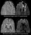
Methods and materials: SWI was performed on 25 patients with stable gliomas on a 7 Tesla magnet. Microbleeds were identified as discrete foci of susceptibility that did not correspond to vessels. The number of microbleeds was counted within and outside of the T2-hyperintense lesion. For 3 patients, radiation dosimetry maps were reconstructed and fused with the 7T SWI data.
Results: Multiple foci of susceptibility consistent with microhemorrhages were observed in patients 2 years after chemoradiation. These lesions were not present in patients who were not irradiated. The prevalence of microhemorrhages increased with the time since completion of radiotherapy, and these lesions often extended outside the boundaries of the initial high-dose volume and into the contralateral hemisphere.
Conclusions: High-field SWI has potential for visualizing the appearance of microbleeds associated with long-term effects of radiotherapy on brain tissue. The ability to visualize these lesions in normal-appearing brain tissue may be important in further understanding the utility of this treatment in patients with longer survival.
Copyright © 2012 Elsevier Inc. All rights reserved.
Figures


References
-
- Karim AB, Afra D, Cornu P, et al. Randomized trial on the efficacy of radiotherapy for cerebral low-grade glioma in the adult: European Organization for Research and Treatment of Cancer Study 22845 with the Medical Research Council study BRO4: an interim analysis. Int J Radiat Oncol Biol Phys. 2002;52(2):316–324. - PubMed
-
- Wallner KE, Galicich JH, et al. Patterns of failure following treatment for glioblastoma multiforme and anaplastic astrocytoma. Int J Radiat Oncol Biol Phys. 1989;16(6):1405–1409. - PubMed
-
- Hochberg FH, Pruitt A. Assumptions in the radiotherapy of glioblastoma. Neurology. 1980;30(9):907–911. - PubMed
-
- Oppitz U, Maessen D, Zunterer H, et al. 3D-recurrence-patterns of glioblastomas after CT-planned postoperative irradiation. Radiother Oncol. 1999;53(1):53–57. - PubMed
-
- Park I, Tamai G, Lee MC, et al. Patterns of recurrence analysis in newly diagnosed glioblastoma multiforme after three-dimensional conformal radiation therapy with respect to pre-radiation therapy magnetic resonance spectroscopic findings. Int J Radiat Oncol Biol Phys. 2007;69(2):381–389. - PMC - PubMed
Publication types
MeSH terms
Grants and funding
LinkOut - more resources
Full Text Sources
Medical

