. 2011 Nov 15;21(5):848-61.
doi: 10.1016/j.devcel.2011.09.007. Epub 2011 Oct 13.

Structural and functional studies of LRP6 ectodomain reveal a platform for Wnt signaling

Affiliations

Structural and functional studies of LRP6 ectodomain reveal a platform for Wnt signaling

Shuo Chen et al. Dev Cell. .

Abstract

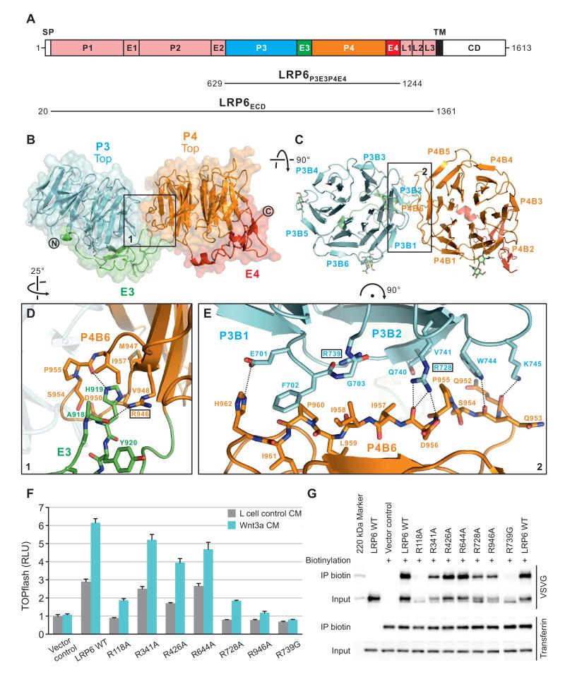
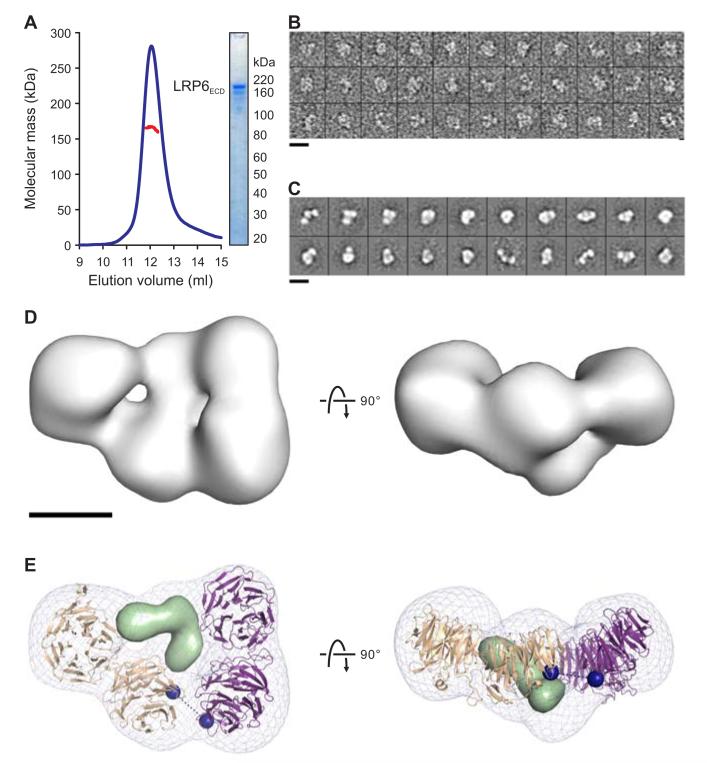
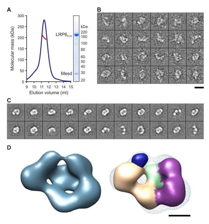
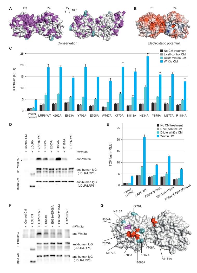
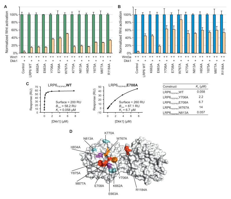
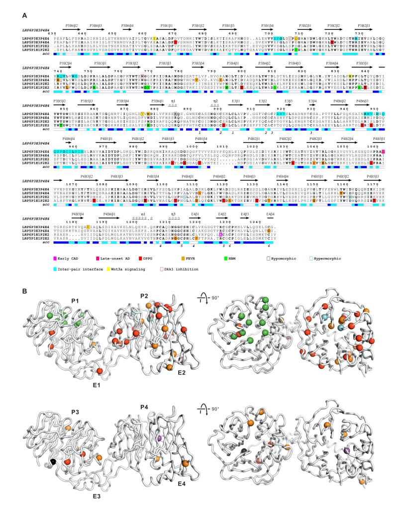
LDL-receptor-related protein 6 (LRP6), alongside Frizzled receptors, transduces Wnt signaling across the plasma membrane. The LRP6 ectodomain comprises four tandem β-propeller-EGF-like domain (PE) pairs that harbor binding sites for Wnt morphogens and their antagonists including Dickkopf 1 (Dkk1). To understand how these multiple interactions are integrated, we combined crystallographic analysis of the third and fourth PE pairs with electron microscopy (EM) to determine the complete ectodomain structure. An extensive inter-pair interface, conserved for the first-to-second and third-to-fourth PE interactions, contributes to a compact platform-like architecture, which is disrupted by mutations implicated in developmental diseases. EM reconstruction of the LRP6 platform bound to chaperone Mesd exemplifies a binding mode spanning PE pairs. Cellular and binding assays identify overlapping Wnt3a- and Dkk1-binding surfaces on the third PE pair, consistent with steric competition, but also suggest a model in which the platform structure supports an interplay of ligands through multiple interaction sites.


Figures

Figure 1
Figure 1. Crystal Structure of LRP6P3E3P4E4
(A) Schematic domain organization of LRP6. SP, signal peptide; TM, transmembrane domain; CD, cytoplasmic domain. Constructs for crystallographic (LRP6P3E3P4E4) and electron microscopic (LRP6ECD) studies are shown. The domain color scheme is used for all figures unless otherwise stated. (B and C) Cartoon representation of the crystal structure of LRP6P3E3P4E4 viewed from one side (B) and the top face (C) of the β-propellers. Disulfide bridges and N-linked glycans are shown in sticks. Blades of the β-propellers are numbered by convention. (D and E) Detailed views of the boxed areas in (B) and (C) highlight contacts between E3 and P4 (D) and between P3 and P4 (E). Interface residues are shown labeled in sticks, and those chosen for mutagenesis boxed. Dashed lines indicate inter-pair hydrogen bonds or salt bridges. (F) Mutations at the inter-pair interfaces of LRP6 affected signaling capabilities variously in Wnt-responsive TOPflash luciferase assay. RLU, relative light unit. Error bars represent standard deviations. (G) LRP6 inter-pair interface mutants express at different levels on the cell surface as demonstrated by cell surface biotinylation. The endogenous transferrin receptor was used as a control for cell surface protein and labeling. See also Figure S1.
Figure 2
Figure 2. LRP6ECD Is a Monomer With a Compact Fold
(A) Multi-angle light scattering (MALS) indicates an experimental molecular mass (red line) of 165.2 ± 2.0 kDa for LRP6ECD (blue line; elution profile in 50 mM NaAc (pH 5.0), 150 mM NaCl, axis not shown) as observed by SDS–PAGE (inset) and in agreement with the calculated molecular mass for a monomer (169.9 kDa). (B) Representative raw images of negatively stained LRP6ECD. (C) Reference-free 2D class averages reveal a skewed horseshoe structure. (D) Two orthogonal views of the 3D reconstruction of LRP6ECD (grey surface) at 25-Å resolution (Figure S2). (E) A pseudo-atomic model for LRP6ECD consisting of the LRP6P3E3P4E4 crystal structure and a homology model for LRP6P1E1P2E2 were fit into the reconstruction (grey mesh), and refined as two rigid bodies. Two possible placements of the model, related by a 180° rotation interchanging the N- and C-termini were scored. Having equivalent correlation coefficients, the one shown here corresponds to the model that minimizes the distance linking the two rigid bodies. Black dashed line connects the two termini (blue spheres). Tandem PE pairs are shown as tan and purple ribbons. Density corresponding to a homology model of the three LDLR type A domains of LRP6 is shown as a light green surface. Scale bars for (B) and (C) (140 Å), and (D) and (E) (50 Å) are indicated. See also Figure S2.
Figure 3
Figure 3. LRP6ECD–Mesd Complex
(A) MALS analysis indicates an experimental molecular mass (red line) of 199.8 ± 5.4 kDa for the LRP6ECD–Mesd complex (blue line; elution profile in 10 mM Tris-HCl (pH 8.0), 150 mM NaCl, axis not shown) as observed by SDS–PAGE (inset) and in agreement with the calculated molecular mass for a 1:1 LRP6ECD–Mesd complex (195.4 kDa). (B) Raw images of the negatively stained LRP6ECD–Mesd complex. (C) Representative class averages of the complex show a skewed ring-like structure of similar dimensions to LRP6ECD alone. (D) 3D reconstruction of the LRP6ECD–Mesd complex at 26 Å resolution (blue surface, left panel) (Figure S3). The apo LRP6ECD reconstruction (density corresponding to tandem PE pairs colored tan and purple while density representing LDLR type A domains in light green) was real-space refined into density for the complex (blue mesh, right panel). The crystal structure of the mouse Mesd core domain (dark blue surface) was rendered at 25 Å resolution and manually placed into the remaining density of the reconstruction. Scale bars for (B) and (C) (140 Å), and (D) and (E) (50 Å) are indicated. See also Figure S3.
Figure 4
Figure 4. Wnt3a Contacts the Top Face of β-propeller 3 of LRP6
(A) LRP6P3E3P4E4 is color-coded by conservation from white (not conserved) to magenta (conserved) based on a sequence alignment across LRP5 and LRP6 in vertebrates with glycans shown in cyan. The left panel has the same view as Figure 1C. (B) The solvent-accessible surface of LRP6P3E3P4E4 is color-coded by electrostatic potential from red (−9 kbT/ec; negatively charged) to blue (9 kbT/ec; positively charged). (C) Responsiveness of LRP6 mutants to Wnt3a in luciferase assays. Error bars represent standard deviations. (D) Binding between representative LRP6 ectodomain mutants and Wnt3a, as assayed by co-immunoprecipitations. (E and F) Luciferase reporter (E) and co-immunoprecipitation (F) assays of LRP6 double or triple mutants corroborated the top face of LRP6 P3 as a Wnt3a-interacting interface. Error bars represent standard deviations. (G) A Wnt3a-binding surface on LRP6 deduced from cellular and binding assays. Residues are colored according to mutational effects. Hypoactive mutations are in red whereas the hyperactive one in magenta. Residues in cyan when mutated had little or no effect on LRP6 signaling capacity. See also Figure S4.
Figure 5
Figure 5. Dkk1 Binding to β-propeller 3 of LRP6 Is Involved in Inhibition of Wnt1 and Wnt3a
(A and B) Mutations on the top face of P3 affect Dkk1-mediated inhibition of Wnt1 (A) and Wnt3a (B) in luciferase reporter assays. RLU was set at 100% in the absence of Dkk1. Error bars represent standard deviations. (C) SPR binding results between Dkk1 and LRP6P3E3P4E4 or its mutants are consistent with those from luciferase reporter assays. RU, response units. (D) Wnt3a and Dkk1 have overlapping binding surfaces on LRP6. Residues important for the activity and binding of both Wnt3a and Dkk1 (red), of Wnt3a only (magenta), of Dkk1 only (orange) and of neither (cyan) are labeled. See also Figure S5.
Figure 6
Figure 6. Disease-Associated LRP Mutations
(A) The amino acid sequence alignment of tandem PE pairs from human LRP6 (P1E1P2E2, residues 20–629; P3E3P4E4, residues 630–1244), LRP5 (P1E1P2E2, residues 32–642; P3E3P4E4, residues 643–1254). LRP5 and LRP6 residues implicated in diseases (Table S1) are indicated. Secondary structure assignments derived from LRP6P3E3P4E4 are displayed above the alignment. The relative accessibility of each residue is indicated below each block: white for buried (<10% exposed); cyan for moderately exposed (10–40% exposed); blue for exposed (>40% exposed). Residues at the inter-PE pair interface observed in the crystal structure and those important for Wnt3a and Dkk1 binding are shown. (B) Cα traces of LRP6P3E3P4E4. Residues implicated in human diseases and mouse models from P1E1P2E2 and P3E3P4E4 are respectively mapped to the equivalent Cα atom positions of two copies of the crystal structure of LRP6P3E3P4E4, shown as spheres and color-coded as in the sequence alignment (A). See also Table S1.

Comment in

Similar articles

Cited by

References

    1. Ai M, Holmen SL, Van Hul W, Williams BO, Warman ML. Reduced affinity to and inhibition by DKK1 form a common mechanism by which high bone mass-associated missense mutations in LRP5 affect canonical Wnt signaling. Molecular and cellular biology. 2005;25:4946–4955. - PMC - PubMed
    1. Aricescu AR, Lu W, Jones EY. A time- and cost-efficient system for high-level protein production in mammalian cells. Acta Crystallogr D Biol Crystallogr. 2006;62:1243–1250. - PubMed
    1. Bafico A, Liu G, Yaniv A, Gazit A, Aaronson SA. Novel mechanism of Wnt signalling inhibition mediated by Dickkopf-1 interaction with LRP6/Arrow. Nat Cell Biol. 2001;3:683–686. - PubMed
    1. Balemans W, Patel N, Ebeling M, Van Hul E, Wuyts W, Lacza C, Dioszegi M, Dikkers FG, Hildering P, Willems PJ, et al. Identification of a 52 kb deletion downstream of the SOST gene in patients with van Buchem disease. J Med Genet. 2002;39:91–97. - PMC - PubMed
    1. Binnerts ME, Tomasevic N, Bright JM, Leung J, Ahn VE, Kim KA, Zhan X, Liu S, Yonkovich S, Williams J, et al. The first propeller domain of LRP6 regulates sensitivity to DKK1. Mol Biol Cell. 2009;20:3552–3560. - PMC - PubMed

Publication types

MeSH terms

Substances

Associated data

LinkOut - more resources