. 2011 Nov 15;21(5):862-73.
doi: 10.1016/j.devcel.2011.09.003. Epub 2011 Oct 13.

Structural basis of Wnt signaling inhibition by Dickkopf binding to LRP5/6

Affiliations

Structural basis of Wnt signaling inhibition by Dickkopf binding to LRP5/6

Victoria E Ahn et al. Dev Cell. .

Abstract

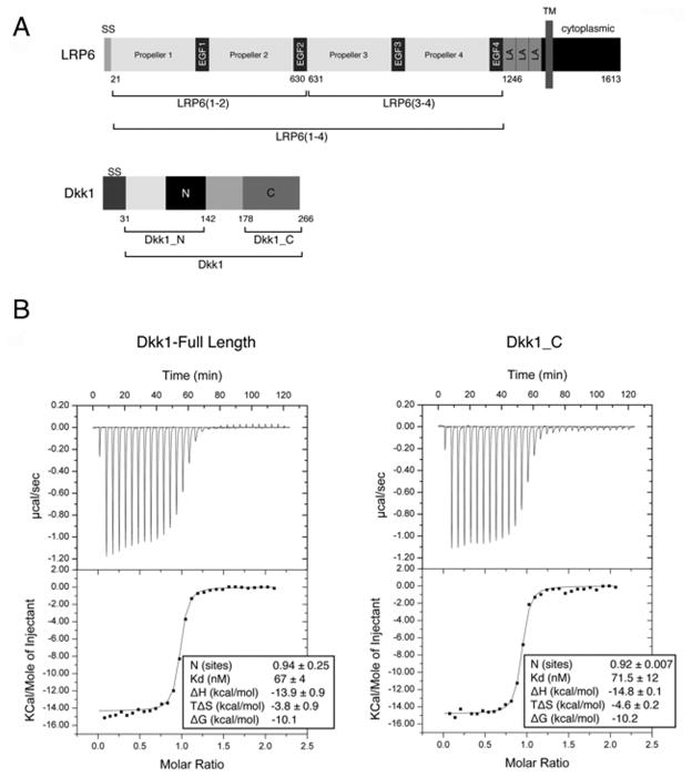
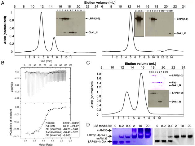
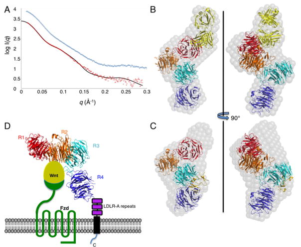
LDL receptor-related proteins 5 and 6 (LRP5/6) are coreceptors for Wnt growth factors, and also bind Dkk proteins, secreted inhibitors of Wnt signaling. The LRP5/6 ectodomain contains four β-propeller/EGF-like domain repeats. The first two repeats, LRP6(1-2), bind to several Wnt variants, whereas LRP6(3-4) binds other Wnts. We present the crystal structure of the Dkk1 C-terminal domain bound to LRP6(3-4), and show that the Dkk1 N-terminal domain binds to LRP6(1-2), demonstrating that a single Dkk1 molecule can bind to both portions of the LRP6 ectodomain and thereby inhibit different Wnts. Small-angle X-ray scattering analysis of LRP6(1-4) bound to a noninhibitory antibody fragment or to full-length Dkk1 shows that in both cases the ectodomain adopts a curved conformation that places the first three repeats at a similar height relative to the membrane. Thus, Wnts bound to either portion of the LRP6 ectodomain likely bear a similar spatial relationship to Frizzled coreceptors.


Figures

Figure 1
Figure 1. Dkk1_C mediates binding to LRP6(3-4)
(A) Primary structures of human LRP6 and Dkk1. The conserved cysteine-rich N- and C-terminal domains of Dkk1 are denoted “N” and “C”. SS, signal sequence; LA, LDLR type A repeat, TM, transmembrane segment. Boundaries of constructs used in this study are indicated below each protein. (B) ITC binding of LRP6(3-4) to either full length Dkk1 (left) or Dkk1_C (right). See also Table S1.
Figure 2
Figure 2. Structures of LRP6(3-4) and Dkk1_C
(A) The LRP6(3-4) structure, viewed down the pseudo-six fold symmetry axis of propeller 3 (top) or towards the side (bottom). Blades are numbered in repeat 3, and individual strands are labeled in blade 1. (B) Closeup of the repeat 3-4 interface. Interacting side chains are shown in stick representation. Polar interactions are shown with dashed lines. Repeat 4 residue labels are underlined. The complete list of interactions is given in Table S2. See also Figure S1. (C) Ribbon representation of Dkk1_C. Disulfide bridges are show in green. The loop between β-strands 5 and 6 is disordered. See also Figure S2 (D) Superposition of human Dkk1_C structure (gold) with the NMR solution structure of mouse Dkk2_C (blue) (PDB entry 2JTK).
Figure 3
Figure 3. Dkk1_C interacts with both copies of LRP6(3-4) in the asymmetric unit
(A) Overall structure of the asymmetric unit. LRP6(3-4) copy A is shown in green and copy B in cyan, with repeats 3 and 4 respectively shown in darker and ligher shades. The single copy of Dkk1_C is shown in gold. (B) Electrostatic surface of LRP6(3-4) copy B bound to Dkk1_C. Red represents regions of negative charge, blue positive, contoured from −5 to +5 kBT/e. (C,D) Closeup view of the LRP6(3-4) copy B interface with Dkk1_C, looking down the pseudo-six fold axis of LRP6 propeller 3 (C) or viewed from the side (D). Interacting residues are shown in stick representation. Polar interactions are shown with dashed lines. Dkk1 residue labels are italicized. The complete list of interactions is given in Table S3. Glu708 and Asp811 are highlighted in red ovals. See also Figures S1, S2 and S5. The copy A interface is shown in Figure S3 and contacts listed in Table S4.
Figure 4
Figure 4. Effect of Dkk1 and LRP6 mutations on Wnt3a activity
LSL cells were treated with Wnt3a conditioned media plus Dkk1_C, wild-type Dkk1 or a mutant. The activity from treatment with Wnt3a CM is taken to be 100%. Error bars denote standard deviation. (A) Dkk1 mutants. (B) LRP6(3-4) mutants.
Figure 5
Figure 5. Dkk1_N and Dkk1_C bind to distinct regions of the LRP6 ectodomain
(A) LRP6(1-2) at 2.5 μM was mixed with 12.5 μM Dkk1_N (left) or 12.5 μM Dkk1_C (right) and run on a Superdex 200 column. An anti-His6 western blot was used to visualize the proteins in the indicated fractions. Dkk1_C transfers poorly for western blotting, as seen by the difference in band intensities versus Dkk1_N even though the same amount of protein is present in both experiments. The overexposed western blot of lanes 3-6 shown in the Dkk1_C experiment demonstrates that no Dkk1_C co-eluted with LRP6(1-2). (B) ITC measurement of Dkk1_N binding to LRP6(1-2). See also Figure S4 for alternative binding assay. (C) LRP6(1-2) at 2.5 μM was mixed with 2.5 μM Dkk1_N and 12.5 μM Fab135 and run on a Superdex 200 column. Fractions were analyzed by SDS-PAGE. The upper gel was stained with Coomassie blue to visualize LRP6(1-2) and Fab135; the lower gel was analyzed by western blot with anti-His6 to visualize LRP6(1-2) and Dkk1. (D) mAb135 and Dkk1 can both bind to LRP6(1-4). LRP6(1-4)–Dkk1 complex at 2 μM was incubated with the indicated concentration of mAb135, run on native PAGE, and analyzed by Coomassie blue staining (left) and western blot with anti-Dkk1 (right). The antibody shifts the LRP6(1-4)–Dkk1 complex upward to the middle band, which still contains Dkk1 as shown by the western blot.
Figure 6
Figure 6. SAXS reconstructions of LRP6(1-4) bound to Fab135 or Dkk1
(A) SAXS data for the LRP6(1-4)–Fab135 (red) and LRP6(1-4)–Dkk1 (blue) complexes, offset on the y axis for clarity. The solid line shows the scattering curve calculated from the LRP6(1-4)–Fab135 model shown in (B), χ2=3.1. See also Figure S6. (B) Ab initio reconstruction (grey spheres) of the LRP6(1-4)–Fab135 complex, with a model of the LRP6(1-4)–Fab135 superimposed. LRP6 repeats 1, 2, 3 and 4 are shown in red, orange, teal, and blue, respectively, and the Fab is shown in yellow. (C) Ab initio reconstruction of the LRP6(1-4)–Dkk1 complex. The model of the LRP6(1-4) region shown in (B) is superimposed on the envelope. The position of Dkk1_C after superposition of the LRP6(3-4)–Dkk1_C crystal structure on the SAXS model is shown in gold. (D) Alternative view of LRP6(1-4) SAXS-derived model, oriented with repeats 1-3 roughly coplanar and the C-terminus of repeat 4 near the bottom, illustrating that the Wnt-binding regions of the receptor likely have similar heights with respect to the membrane and Fzd. Wnt, Fzd, and the three LDLR-A repeats, the transmembrane anchor, and the cytoplasmic domain of LRP5/6 are shown schematically.

Comment in

References

    1. Adams PD, Grosse-Kunstleve RW, Hung LW, Ioerger TR, McCoy AJ, Moriarty NW, Read RJ, Sacchettini JC, Sauter NK, Terwilliger TC. PHENIX: building new software for automated crystallographic structure determination. Acta Crystallogr D Biol Crystallogr. 2002;58:1948–1954. - PubMed
    1. Ai M, Holmen SL, Van Hul W, Williams BO, Warman ML. Reduced affinity to and inhibition by DKK1 form a common mechanism by which high bone mass-associated missense mutations in LRP5 affect canonical Wnt signaling. Mol Cell Biol. 2005;25:4946–4955. - PMC - PubMed
    1. Bafico A, Liu G, Yaniv A, Gazit A, Aaronson SA. Novel mechanism of Wnt signalling inhibition mediated by Dickkopf-1 interaction with LRP6/Arrow. Nat Cell Biol. 2001;3:683–686. - PubMed
    1. Balemans W, Devogelaer JP, Cleiren E, Piters E, Caussin E, Van Hul W. Novel LRP5 missense mutation in a patient with a high bone mass phenotype results in decreased DKK1-mediated inhibition of Wnt signaling. J Bone Miner Res. 2007;22:708–716. - PubMed
    1. Binnerts ME, Tomasevic N, Bright JM, Leung J, Ahn VE, Kim KA, Zhan X, Liu S, Yonkovich S, Williams J, et al. The first propeller domain of LRP6 regulates sensitivity to DKK1. Mol Biol Cell. 2009;20:3552–3560. - PMC - PubMed

Publication types

MeSH terms

Substances

LinkOut - more resources