. 2012 Jan;340(1):134-42.
doi: 10.1124/jpet.111.184192. Epub 2011 Oct 14.

Acetaminophen-induced hepatotoxicity and protein nitration in neuronal nitric-oxide synthase knockout mice

Affiliations

Acetaminophen-induced hepatotoxicity and protein nitration in neuronal nitric-oxide synthase knockout mice

Rakhee Agarwal et al. J Pharmacol Exp Ther. 2012 Jan.

Abstract

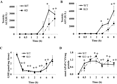
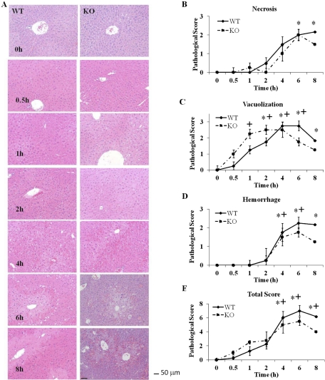
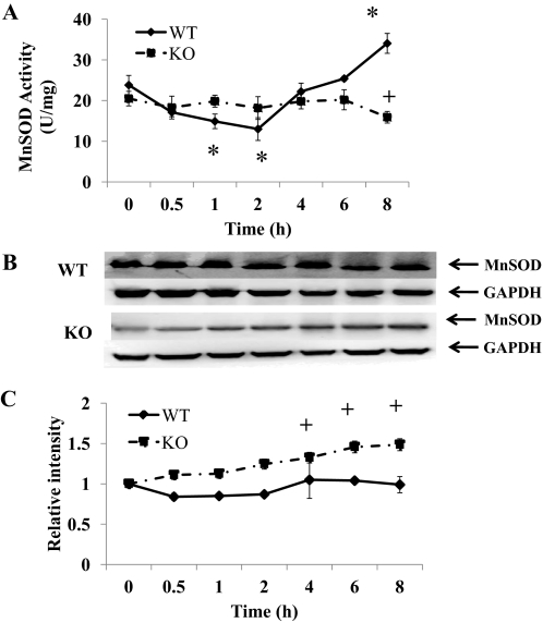
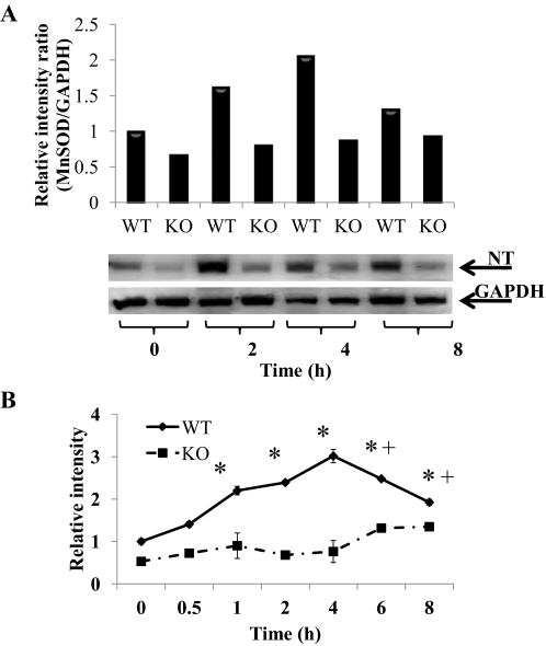
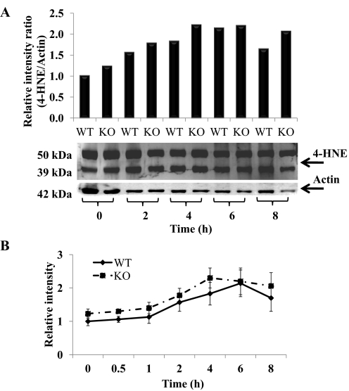
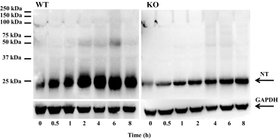
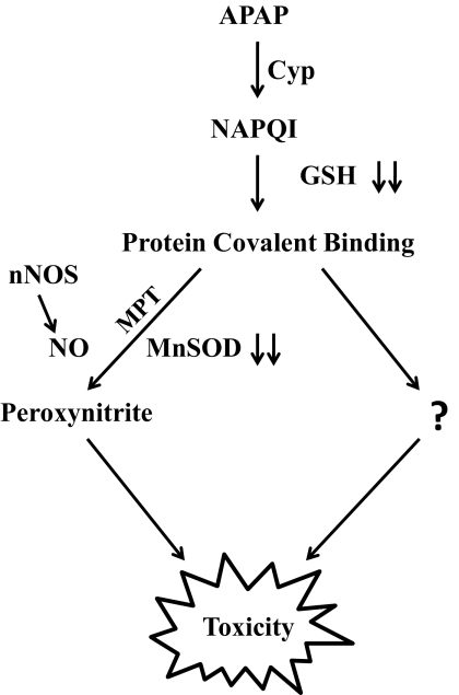
In overdose acetaminophen (APAP) is hepatotoxic. Toxicity occurs by metabolism to N-acetyl-p-benzoquinone imine, which depletes GSH and covalently binds to proteins followed by protein nitration. Nitration can occur via the strong oxidant and nitrating agent peroxynitrite, formed from superoxide and nitric oxide (NO). In hepatocyte suspensions we reported that an inhibitor of neuronal nitric-oxide synthase (nNOS; NOS1), which has been reported to be in mitochondria, inhibited toxicity and protein nitration. We recently showed that manganese superoxide dismutase (MnSOD; SOD2) was nitrated and inactivated in APAP-treated mice. To understand the role of nNOS in APAP toxicity and MnSOD nitration, nNOS knockout (KO) and wild-type (WT) mice were administered APAP (300 mg/kg). In WT mice serum alanine aminotransferase (ALT) significantly increased at 6 and 8 h, and serum aspartate aminotransferase (AST) significantly increased at 4, 6 and 8 h; however, in KO mice neither ALT nor AST significantly increased until 8 h. There were no significant differences in hepatic GSH depletion, APAP protein binding, hydroxynonenal covalent binding, or histopathological assessment of toxicity. The activity of hepatic MnSOD was significantly lower at 1 to 2 h in WT mice and subsequently increased at 8 h. MnSOD activity was not altered at 0 to 6 h in KO mice but was significantly decreased at 8 h. There were significant increases in MnSOD nitration at 1 to 8 h in WT mice and 6 to 8 h in KO mice. Significantly more nitration occurred at 1 to 6 h in WT than in KO mice. MnSOD was the only observed nitrated protein after APAP treatment. These data indicate a role for nNOS with inactivation of MnSOD and ALT release during APAP toxicity.


Figures

Fig. 1.
Fig. 1.
Time course for the effect of APAP on serum ALT, serum AST, hepatic GSH, and formation of APAP-cysteine adducts in WT and KO mice. Mice (n = 4) were treated with a 300 mg/kg dose of APAP and sacrificed at the indicated times, and serum and liver were collected. The 0 time is the saline-treated control mice in both WT and KO. A, ALT levels in serum (hepatotoxicity). B, AST levels in serum (hepatotoxicity). C, GSH levels in liver. D, APAP-cysteine adducts in liver. The data are presented as mean ± S.E. * and + indicate significant difference of WT and KO from their respective saline controls, and ▴ indicates a significant difference between WT and KO at a particular time point, p ≤ 0.05. Only two animals were used for study at 0.5 h in the KO group.
Fig. 2.
Fig. 2.
Histopathological analysis for hepatic necrosis in livers from APAP-treated WT and KO mice. Livers from mice (n = 4) were treated with acetaminophen (300 mg/kg) or saline control. A, hematoxylin and eosin staining. Liver sections were prepared at the indicated times and stained with hematoxylin and eosin. One representative picture is shown per time point. Magnification, 200×. Bar equals 50 μm. B, necrosis. C, vacuolization. D, hemorrhage. E, total score. The data are presented as mean ± S.E. * and + indicate significant difference of WT and KO from their respective saline controls at a particular time point, p ≤ 0.05.
Fig. 3.
Fig. 3.
Effect of APAP on MnSOD in livers of WT and KO mice. Mice (n = 4) were treated with 300 mg/kg dose of APAP and sacrificed at the indicated times, and liver was collected. The 0 time is the saline-treated control mice in both WT and KO. A, effect on MnSOD activity. B, Western blot analysis of combined homogenates for MnSOD in livers of WT and KO mice. GAPDH was used as a loading control. C, time course for MnSOD protein in livers of WT and KO mice. Western blot analysis was performed on each homogenate by using MnSOD antibody. GAPDH was used as a loading control. The data are presented as mean ± S.E. of the relative density of the 24-kDa MnSOD protein divided by the density of 38-kDa GAPDH protein. * and + indicate significant differences of WT and KO from respective saline, and ▴ indicates significant differences between WT and KO at particular time points, p ≤ 0.05.
Fig. 4.
Fig. 4.
Effect of APAP on nitration of MnSOD protein in livers of WT and KO mice. Mice (n = 4) were treated with 300 mg/kg dose of APAP and sacrificed at the indicated times, and liver was collected. The 0 time is the saline-treated control mice in both WT and KO. A, nitrated MnSOD (NT) levels in liver of WT and KO mice at 0, 2, 4, and 8 h. GAPDH was used as a loading control. The data are presented as the relative density of the 24-kDa MnSOD protein divided by the density of 38-kDa GAPDH protein. Representative gels are shown above the density values. B, time course for nitration of MnSOD protein in livers of WT and KO mice. GAPDH was used as a loading control. The data are presented as mean ± S.E. of the relative density of the 24-kDa MnSOD protein divided by the density of 38-kDa GAPDH protein. * and + indicate significant differences of WT and KO from respective saline control, and ▴ indicates significant differences between WT and KO at particular time points, p ≤ 0.05.
Fig. 5.
Fig. 5.
Hepatic lipid peroxidation in APAP-treated WT and KO mice. Hepatic tissue was collected at the indicated time points from APAP-treated WT and KO mice. The 0 time is the saline-treated control mice in both WT and KO mice. Hepatic lipid peroxidation was assessed by 4-HNE Western blotting. A, 4-HNE levels in livers of WT and KO mice at 0, 2, 4, 6, and 8 h. Actin was used as a loading control. The data are presented as the relative ratio of the combined density of 50- and 39-kDa HNE proteins divided by the density of 42-kDa actin protein. Representative gels are shown above the density values. B, time course for lipid peroxidation in livers of WT and KO mice. Actin was used as a loading control. The data are presented as mean ± S.E. of the ratio of relative combined density of 50- and 39-kDa HNE proteins divided by the density of 42-kDa actin protein.
Fig. 6.
Fig. 6.
Nitration of proteins in liver homogenates of APAP-treated WT (left) and KO (right) mice. Mice (n = 4) were treated with a 300 mg/kg dose of APAP and sacrificed at the indicated times, and livers were collected. The 0 time is the saline-treated control mice in both WT and KO mice. Hepatic homogenates were analyzed by Western blotting for the presence of nitrotyrosine (NT) protein adducts by using anti-3-nitrotyrosine antibody. GAPDH was used as a loading control. One representative gel is shown.
Fig. 7.
Fig. 7.
Postulated mechanism of APAP toxicity.

References

    1. Abdelmegeed MA, Moon KH, Chen C, Gonzalez FJ, Song BJ. (2010) Role of cytochrome P450 2E1 in protein nitration and ubiquitin-mediated degradation during acetaminophen toxicity. Biochem Pharmacol 79:57–66 - PMC - PubMed
    1. Agarwal R, MacMillan-Crow LA, Rafferty TM, Saba H, Roberts DW, Fifer EK, James LP, Hinson JA. (2011) Acetaminophen-induced hepatotoxicity in mice occurs with inhibition of activity and nitration of mitochondrial manganese superoxide dismutase. J Pharmacol Exp Ther 337:110–116 - PMC - PubMed
    1. Amimoto T, Matsura T, Koyama SY, Nakanishi T, Yamada K, Kajiyama G. (1995) Acetaminophen-induced hepatic injury in mice: the role of lipid peroxidation and effects of pretreatment with coenzyme Q10 and α-tocopherol. Free Radic Biol Med 19:169–176 - PubMed
    1. Boobis AR, Seddon CE, Nasseri-Sina P, Davies DS. (1990) Evidence for a direct role of intracellular calcium in paracetamol toxicity. Biochem Pharmacol 39:1277–1281 - PubMed
    1. Boobis AR, Tee LB, Hampden CE, Davies DS. (1986) Freshly isolated hepatocytes as a model for studying the toxicity of paracetamol. Food Chem Toxicol 24:731–736 - PubMed

Publication types

MeSH terms