

External pallidal stimulation improves parkinsonian motor signs and modulates neuronal activity throughout the basal ganglia thalamic network
- PMID: 22001773
- PMCID: PMC3536483
- DOI: 10.1016/j.expneurol.2011.09.031
External pallidal stimulation improves parkinsonian motor signs and modulates neuronal activity throughout the basal ganglia thalamic network
Abstract
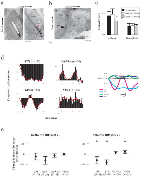
Deep brain stimulation (DBS) of the internal segment of the globus pallidus (GPi) and the subthalamic nucleus (STN) are effective for the treatment of advanced Parkinson's disease (PD). We have shown previously that DBS of the external segment of the globus pallidus (GPe) is associated with improvements in parkinsonian motor signs; however, the mechanism of this effect is not known. In this study, we extend our findings on the effect of STN and GPi DBS on neuronal activity in the basal ganglia thalamic network to include GPe DBS using the 1-methyl-4-phenyl-1.2.3.6-tetrahydropyridine (MPTP) monkey model. Stimulation parameters that improved bradykinesia were associated with changes in the pattern and mean discharge rate of neuronal activity in the GPi, STN, and the pallidal [ventralis lateralis pars oralis (VLo) and ventralis anterior (VA)] and cerebellar [ventralis lateralis posterior pars oralis (VPLo)] receiving areas of the motor thalamus. Population post-stimulation time histograms revealed a complex pattern of stimulation-related inhibition and excitation for the GPi and VA/VLo, with a more consistent pattern of inhibition in STN and excitation in VPLo. Mean discharge rate was reduced in the GPi and STN and increased in the VPLo. Effective GPe DBS also reduced bursting in the STN and GPi. These data support the hypothesis that therapeutic DBS activates output from the stimulated structure and changes the temporal pattern of neuronal activity throughout the basal ganglia thalamic network and provide further support for GPe as a potential therapeutic target for DBS in the treatment of PD.
Copyright © 2011 Elsevier Inc. All rights reserved.
Figures
Similar articles
-
Modulation of Neuronal Activity in the Motor Thalamus during GPi-DBS in the MPTP Nonhuman Primate Model of Parkinson's Disease.Brain Stimul. 2017 Jan-Feb;10(1):126-138. doi: 10.1016/j.brs.2016.10.005. Epub 2016 Oct 11. Brain Stimul. 2017. PMID: 27839724 Free PMC article.
-
Subthalamic nucleus stimulation modulates thalamic neuronal activity.J Neurosci. 2008 Nov 12;28(46):11916-24. doi: 10.1523/JNEUROSCI.2027-08.2008. J Neurosci. 2008. PMID: 19005057 Free PMC article.
-
Effect of globus pallidus internus stimulation on neuronal activity in the pedunculopontine tegmental nucleus in the primate model of Parkinson's disease.Exp Neurol. 2012 Jan;233(1):575-80. doi: 10.1016/j.expneurol.2011.07.007. Epub 2011 Jul 29. Exp Neurol. 2012. PMID: 21821025 Free PMC article.
-
Pathophysiology of Parkinson's disease: the MPTP primate model of the human disorder.Ann N Y Acad Sci. 2003 Jun;991:199-213. doi: 10.1111/j.1749-6632.2003.tb07477.x. Ann N Y Acad Sci. 2003. PMID: 12846988 Review.
-
Functional organization of the basal ganglia: therapeutic implications for Parkinson's disease.Mov Disord. 2008;23 Suppl 3:S548-59. doi: 10.1002/mds.22062. Mov Disord. 2008. PMID: 18781672 Review.
Cited by
-
Therapeutic mechanisms of high-frequency stimulation in Parkinson's disease and neural restoration via loop-based reinforcement.Proc Natl Acad Sci U S A. 2015 Feb 10;112(6):E586-95. doi: 10.1073/pnas.1406549111. Epub 2015 Jan 26. Proc Natl Acad Sci U S A. 2015. PMID: 25624501 Free PMC article.
-
Neural targets for relieving parkinsonian rigidity and bradykinesia with pallidal deep brain stimulation.J Neurophysiol. 2012 Jul;108(2):567-77. doi: 10.1152/jn.00039.2012. Epub 2012 Apr 18. J Neurophysiol. 2012. PMID: 22514292 Free PMC article.
-
Pallidostriatal Projections Promote β Oscillations in a Dopamine-Depleted Biophysical Network Model.J Neurosci. 2016 May 18;36(20):5556-71. doi: 10.1523/JNEUROSCI.0339-16.2016. J Neurosci. 2016. PMID: 27194335 Free PMC article.
-
Deep brain stimulation in the globus pallidus externa promotes sleep.Neuroscience. 2016 May 13;322:115-20. doi: 10.1016/j.neuroscience.2016.02.032. Epub 2016 Feb 22. Neuroscience. 2016. PMID: 26917269 Free PMC article.
-
Identification of a direct GABAergic pallidocortical pathway in rodents.Eur J Neurosci. 2015 Mar;41(6):748-59. doi: 10.1111/ejn.12822. Epub 2015 Jan 8. Eur J Neurosci. 2015. PMID: 25581560 Free PMC article.
References
-
- Anderson ME, Postupna N, Ruffo M. Effects of high-frequency stimulation in the internal globus pallidus on the activity of thalamic neurons in the awake monkey. J Neurophysiol. 2003;89:1150–1160. - PubMed
-
- Asdourian D, Lentz SI, Kelland MD. Motor effects of globus pallidus stimulation in the rat: lesions to corticofugal fibers block the motor effects. Behav Brain Res. 1991;44:185–193. - PubMed
-
- Dostrovsky JO, Levy R, Wu JP, Hutchison WD, Tasker RR, Lozano AM. Microstimulation-induced inhibition of neuronal firing in human globus pallidus. J Neurophysiol. 2000;84:570–574. - PubMed
-
- Elder CM, Hashimoto T, Zhang J, Vitek JL. Chronic implantation of deep brain stimulation leads in animal models of neurological disorders. J Neurosci Methods. 2005;142:11–16. - PubMed
-
- Hashimoto T, Elder C, Vitek J. A template subtraction method for stimulus artifact removal in high-frequency deep brain stimulation. J Neurosci Methods. 2002;113:181–186. - PubMed
Publication types
MeSH terms
Grants and funding
LinkOut - more resources
Full Text Sources
Other Literature Sources
Miscellaneous
