. 2012 Jan;142(1):21-24.e7.
doi: 10.1053/j.gastro.2011.09.050. Epub 2011 Oct 14.

Tamoxifen induces rapid, reversible atrophy, and metaplasia in mouse stomach

Affiliations

Tamoxifen induces rapid, reversible atrophy, and metaplasia in mouse stomach

Won Jae Huh et al. Gastroenterology. 2012 Jan.

Abstract

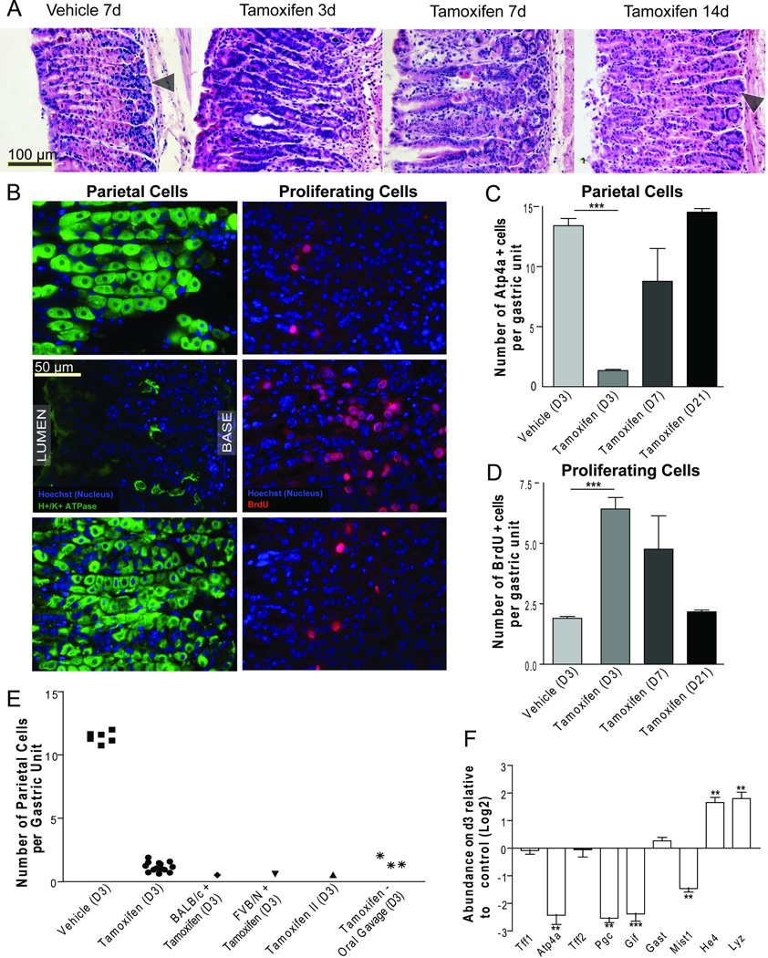
Tamoxifen, a selective estrogen receptor modulator, is widely used in research and clinically in patients. We find that treatment of normal mice with a single ≥3 mg/20 g body weight dose of tamoxifen leads to apoptosis of >90% of all gastric parietal cells (PCs) and metaplasia of zymogenic chief cells within 3 days. Remarkably, gastric histology returns to nearly normal by 3 weeks. Tamoxifen toxicity occurs by oral and intraperitoneal administration, in both sexes, in multiple strains, and does not depend on estrogen, though acid secretion inhibition is partially protective. Thus, substantial gastric toxicity is a heretofore unappreciated tamoxifen side effect.


Conflict of interest statement

The authors disclose no conflicts of interest.

Figures

Fig. 1
Fig. 1
A) H&E of wild-type mice following intraperitoneal (i.p.) injection of vehicle at day 7 or 5mg/20g body weight tamoxifen at 3, 7, and 14 days following injection. B) Immunofluorescence (green: anti-ATP4A; red:anti-BrdU) at d3, quantified in C,D. E) Quantification of mean PCs/unit/individual mouse by H&E; unless otherwise indicated, mice were C57/B6 strain; “Tamoxifen II”: tamoxifen from another supplier. F) Whole stomach qRT-PCR (expressed as Log2 scale. *P<0.05;**P<0.01;***P<0.001)
Fig. 2
Fig. 2
A) Nuclear LacZ labeled PCs following tamoxifen treatment. B) Top: TdT-mediated dUTP nick-end labeling shows dying PCs (arrowheads). Below: Cytochrome C staining is punctate, consistent with mitochondrial localization in vehicle-treated (below left) and dispersed throughout the cytoplasm tamoxifen-treated PCs (below right) C) Cleaved caspase 3 western blot with tubulin loading control. D) At 2.5d following tamoxifen, PCs show chromatin condensation (arrows with black outline), consistent with early apoptosis. E) Another degenerating PC exhibits mitochondria ranging in morphology from normal (dashed arrow) to electron-dense-inclusion-containing (white solid arrow) to electron-dense and degenerating (arrowheads).

References

    1. Feil R, et al. Proc Natl Acad Sci U S A. 1996;93:10887–10890. - PMC - PubMed
    1. Jordan VC. Cancer. 1992;70:977–982. - PubMed
    1. Love RR, et al. N Engl J Med. 1992;326:852–856. - PubMed
    1. Chandanos E, et al. Br J Cancer. 2006;95:118–122. - PMC - PubMed
    1. Matsuyama Y, et al. Ann Oncol. 2000;11:1537–1543. - PubMed

Publication types

MeSH terms