. 2011 Dec 16;286(50):43013-25.
doi: 10.1074/jbc.M111.261271. Epub 2011 Oct 14.

Complex regulation of p73 isoforms after alteration of amyloid precursor polypeptide (APP) function and DNA damage in neurons

Affiliations

Complex regulation of p73 isoforms after alteration of amyloid precursor polypeptide (APP) function and DNA damage in neurons

Samir Benosman et al. J Biol Chem. .

Abstract

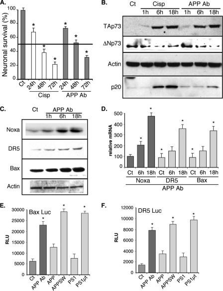
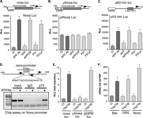
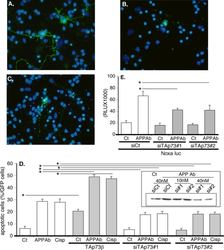
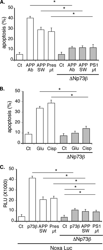
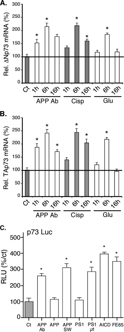
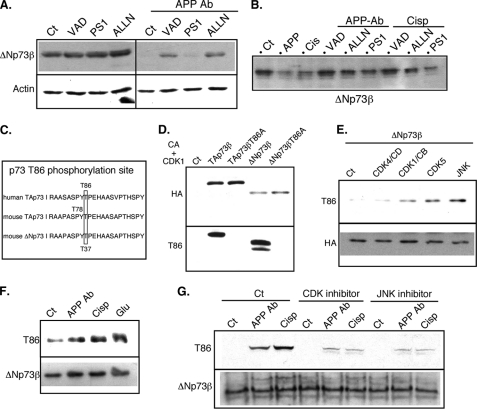
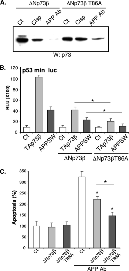
Genetic ablations of p73 have shown its implication in the development of the nervous system. However, the relative contribution of ΔNp73 and TAp73 isoforms in neuronal functions is still unclear. In this study, we have analyzed the expression of these isoforms during neuronal death induced by alteration of the amyloid-β precursor protein function or cisplatin. We observed a concomitant up-regulation of a TAp73 isoform and a down-regulation of a ΔNp73 isoform. The shift in favor of the pro-apoptotic isoform correlated with an induction of the p53/p73 target genes such as Noxa. At a functional level, we showed that TAp73 induced neuronal death and that ΔNp73 has a neuroprotective role toward amyloid-β precursor protein alteration or cisplatin. We investigated the mechanisms of p73 expression and found that the TAp73 expression was regulated at the promoter level. In contrast, regulation of ΔNp73 protein levels was regulated by phosphorylation at residue 86 and multiple proteases. Thus, this study indicates that tight transcriptional and post-translational mechanisms regulate the p73 isoform ratios that play an important role in neuronal survival.


Figures

FIGURE 1.
FIGURE 1.
Cisplatin and APP alteration induce a shift in p73β isoform protein levels in cortical neurons leading to the activation of p53 target genes. A, primary cultures of mouse cortical neurons were treated for the indicated time with cisplatin (Cisp, 20 μm) or the APP-directed antibody (APP Ab, 5 μg/ml). Neuronal survival was evaluated using an MTT test. Results are presented in % compared with the control condition (Ct). Bars represent means ± S.D. of 8 wells from one experiment out of four independent experiments. The thick black line corresponds to 50% of viability (IC50). B and C, cortical neurons were treated for the indicated time with cisplatin (20 μm) or the APP Ab (5 μg/ml). Cells were lysed, and proteins (75 μg) were separated on a 10% SDS-PAGE and immunoblotted with the TA73A, CO73A, actin, p20 (active fragment of Caspase 3), Noxa, DR5, or Bax antibodies. D, cortical neurons were treated for the indicated time with the APP Ab (5 μg/ml). Cells were lysed, and total RNA were extracted and reverse-transcribed (1 μg). Quantitative PCR for Noxa, DR5, Bax, and TBP genes was performed. Bars correspond to means of normalized (TBP) and calibrated (no APP Ab = 100) values with standard deviation of triplicates from one experiment out of three independent experiments. E and F, cortical neurons were transfected with luciferase reporter genes containing either the Bax or the DR5 promoter. Where indicated, cells were treated with the APP Ab antibody (5 μg/ml) or cotransfected with expression vectors coding for either APP or PS1 variants (wild type or harboring mutations found in Alzheimer patients). Bars represent means ± S.D. (relative light units (RLU)) of 3 wells from one experiment out of three independent experiments. * indicates statistically significant differences tested by a one-way analysis of variance followed by a Neuman-Keuls test by pairs (p < 0.05).
FIGURE 2.
FIGURE 2.
Alteration of function of APP leads to induction of Noxa through TAp73. A–C, cortical neurons were transfected with luciferase reporter genes containing either the Noxa promoter (wild type, noxa Luc; mutated in the p53-binding sites, μtnoxa Luc) or a chimerical promoter containing p53-binding sites (p53RE) in front of the c-fos promoter (p53 min Luc). Where indicated, cells were treated with the APP Ab antibody (5 μg/ml) or cotransfected with expression vectors coding for either APP or PS1 variants. Bars represent means ± S.D. (relative light units (RLU)) of 3 wells from one experiment out of three independent experiments. D, chromatin immunoprecipitation of p73 on the Noxa promoter. Control (−) or APP Ab-treated (5 μg/ml, +) cortical neuron lysates were used for chromatin immunoprecipitation with control (normal IgG) or TA73A antibodies. Agarose gel showing PCR amplification (35 cycles) of a Noxa promoter fragment using inputs (1% of chromatin used for ChIP) or ChIPs as templates. E, cortical neurons were transfected with luciferase reporter genes containing either the Noxa promoter (wild type, noxa Luc; mutated in the p53-binding sites, μtnoxa Luc) or a chimerical promoter containing p53-binding sites in front of the c-fos promoter (p53 min Luc). Cells were cotransfected with an empty vector (Ct) or with an expression vector coding for TAp73β. Bars represent means ± S.D. (relative light units (RLU)) of 3 wells from one experiment out of three independent experiments. F, N2A neuroblastoma cells were transfected with an empty vector (Ct) or an expression vector encoding TAp73β. After 36 h of expression, cells were lysed, and total RNA were extracted and reverse-transcribed (1 μg). Quantitative PCR for Noxa, DR5, Bax, and TBP genes were performed. Bars correspond to means of normalized (TBP) and calibrated (Ct = 100) values with standard deviation of triplicates from one experiment out of three independent experiments. * indicates statistically significant differences tested by a one-way analysis of variance followed by a Neuman-Keuls test by pairs (p < 0.05).
FIGURE 3.
FIGURE 3.
TAp73β induces pro-apoptotic gene expression and neuronal apoptosis. A–D, cortical neurons were transfected with a GFP expression vector and either with the TAp73β expression vector or TAp73 siRNA duplexes (siTAp73#1 or siTAp73#2). After 24 h, cells were left untreated (Ct) or treated with APP Ab (5 μg/ml) or cisplatin (Cisp) (20 μm) for 24 h. Cells were then fixed, stained with Hoechst, and observed with a fluorescence microscope. A shows two GFP-positive control cells (untreated). B shows a dead GFP-positive cell treated with APP Ab. C shows a dead cortical neuron transfected with TAp73β. D, graph represents the percentage of dead cells among the GFP-positive cells counted. Bars correspond to means of three wells of a representative experiment with S.D. The inset illustrates the silencing of TAp73β by TAp73 siRNAs (Western blot with TA73A and actin antibodies). E, cortical neurons were cotransfected with the noxa Luc reporter construct and either control (siCt) or TAp73 siRNAs for 36 h. Then cells were treated with APP Ab for 8 h or not (Ct). The siRNA concentration used was 40 nm. Bars represent means ± S.D. (relative light units (RLU)) of 3 wells from one experiment out of three independent experiments. * indicates statistically significant difference tested by a one-way analysis of variance followed by a Neuman-Keuls test by pairs (p < 0.05).
FIGURE 4.
FIGURE 4.
Overexpression of ΔNp73 antagonizes neuronal apoptosis induced by various kinds of neurotoxic stresses. A and B, cortical neurons were cotransfected with the indicated combination of ΔNp73β, mutated APP (APP SW), or mutated presenilin 1 (Pres μt) and GFP expression vectors. After 24 h of expression, cells were left untreated (Ct) or treated with APP Ab (5 μg/ml), cisplatin (Cisp, 20 μm) or glutamate (Glu, 250 μm) for 24 h. Cells were then fixed, stained with Hoechst, and observed with a fluorescence microscope. Bars represent the percentage of dead cells among the GFP-positive cells counted. Bars are means of 3 wells of a representative experiment with S.D. C, cortical neurons were cotransfected with the noxaLuc reporter gene and the indicated combination of TAp73β, ΔNp73β, mutated APP, or mutated presenilin 1 expression vectors. Bars represent means ± S.D. (relative light units (RLU)) of 3 wells from one experiment out of three independent experiments. * indicates statistically significant differences tested by a one-way analysis of variance followed by a Neuman-Keuls test by pairs (p < 0.05).
FIGURE 5.
FIGURE 5.
Neurotoxic stresses induce both TAp73 and ΔNp73 isoforms mRNAs. A and B, cortical neurons were left untreated (Ct) or treated with the APP Ab (5 μg/ml), cisplatin (Cisp, 20 μm) or glutamate (Glu, 250 μm) for the indicated time before preparation of total mRNAs. After reverse transcription, real time PCRs were performed using TBP and either ΔNp73 (A) or TAp73 (B) isoform-specific primers. For standardization, TBP values were used. Graphs represent the percentage of induction relative to the control condition (Ct). Bars are means of three wells of a representative experiment with S.D. C, cortical neurons were transfected with the p73-Luc reporter construct along with different expression vectors (empty, Ct; encoding APP, wild type (APP), mutated as in Alzheimer patients (APPSW) or the AICD cytoplasmic fragment (AICD); encoding the APP cofactor FE65 (FE65)) or treated with the APP-directed antibody (5 μg/ml). Graphs represent means ± S.D. (relative light units (RLU)) of 3 wells from one experiment out of three independent experiments. * indicates statistically significant differences (analysis of variance, Neuman-Keuls multiple comparison test, p < 0.05).
FIGURE 6.
FIGURE 6.
Neurotoxic stresses lead to ΔNp73 degradation by proteases and phosphorylation at threonine 86. A, cortical neurons were left untreated or treated for 18 h with APP Ab (5 μg/ml) in the presence or absence of protease inhibitors (VAD, 50 μm; PS1, 25 μm; ALLN, 15 μm). Cells were lysed, and proteins (75 μg) were separated on a 10% SDS-PAGE and immunoblotted with the p73 CO73A and actin antibodies. B, overexpressed ΔNp73β was immunoprecipitated from N2A protein extracts and incubated with extracts of cortical treated with the APP-directed antibody (APP Ab, 5 μg/ml) or cisplatin (Cisp, 20 μm) in the presence or absence of protease inhibitors. Proteins were then separated on a 10% SDS-PAGE and immunoblotted with an HA-directed antibody. C, schematic representation and comparison between mouse and human sequences for TAp73 and ΔNp73. D, TAp73β wild type, TAp73β mutated in the Thr-86 site (TAp73βT86A), ΔNp73β wild type, and ΔNp73β mutated in the Thr-86 site (ΔNp73βT86A) were expressed in N2A cells. Proteins were extracted with a TEGN buffer containing no phosphatase inhibitor, and p73 proteins were immunoprecipitated as described. Immunoprecipitated p73 proteins were submitted to an in vitro kinase assay using a purified cyclin A-CDK1 complex (CA+CDK1) before SDS-PAGE and successive immunoblotting with the phospho-specific Thr-86 antibody (Thr-86, lower panel) and the HA antibody (upper panel) to detect the level of immunoprecipitated p73 proteins. E, N2A cells were cotransfected with HA-ΔNp73β, cyclin D (CD), and CDK4, cyclin B (CB), and CDK1, CDK5, or JNK expression vectors as indicated. HA-immunoprecipitated p73 proteins were separated by SDS-PAGE and successively immunoblotted with the anti-phospho-Thr-86 antibody (Thr-86, upper panel) and the HA antibody (lower panel). F and G, cortical neurons were treated with the APP-directed antibody (APP Ab, 5 μg/ml), cisplatin (Cisp, 20 μm), or glutamate (Glu, 250 μm) for 24 h. Co-treatments with CDK inhibitor (roscovitine, 5 μm) or JNK inhibitor (PB1557, 5 μm) was performed in G. p73 proteins were immunoprecipitated from the protein extract (2 mg) using the COP73A p73 antibody, separated by SDS-PAGE, and successively immunoblotted using the anti-phospho-Thr-86 antibody (Thr-86, upper panel) and the COP73A antibody (ΔNp73β, lower panel) to check the levels of immunoprecipitated p73 proteins. Images presented are from a representative experiment out of at least three independent experiments.
FIGURE 7.
FIGURE 7.
Thr-86 phosphorylation regulates and destabilizes ΔNp73 proteins. A, overexpressed ΔNp73β and ΔNp73βT86A were immunoprecipitated from N2A cellular extracts and incubated with extracts of cortical neurons left untreated (Ct), treated with the APP-directed antibody (APP Ab, 5 μg/ml), or cisplatin (Cisp, 20 μm). Proteins were then separated on a 10% SDS-PAGE and immunoblotted with a p73 antibody. B, cortical neurons were cotransfected with the p53 min luciferase reporter construct, and the expression vectors coding for APP SW, TAp73β, and ΔNp73β (wild type or T86A mutant) as indicated. Bars represent means ± S.D. (relative light units (RLU)) of 3 wells from one experiment out of three independent experiments. C, cortical neurons were cotransfected with combination of ΔNp73β or ΔNp73βT86A and GFP expression vectors. After 24 h of expression, cells were treated with APP Ab (5 μg/ml) for 24 h. Cells were then fixed, stained with Hoechst, and observed with a fluorescence microscope. Graphs represent the percentage of apoptotic cells among the GFP-positive cells counted. Bars are means of three wells of a representative experiment with S.D. * indicates statistically significant differences (analysis of variance, Neuman-Keuls Multiple comparison test, p < 0.05).

References

    1. Prives C., Hall P. A. (1999) J. Pathol. 187, 112–126 - PubMed
    1. Menendez D., Inga A., Resnick M. A. (2009) Nat. Rev. Cancer 9, 724–737 - PubMed
    1. Eizenberg O., Faber-Elman A., Gottlieb E., Oren M., Rotter V., Schwartz M. (1996) Mol. Cell. Biol. 16, 5178–5185 - PMC - PubMed
    1. Ferreira A., Kosik K. S. (1996) J. Cell Sci. 109, 1509–1516 - PubMed
    1. Migliorini D., Lazzerini Denchi E., Danovi D., Jochemsen A., Capillo M., Gobbi A., Helin K., Pelicci P. G., Marine J. C. (2002) Mol. Cell. Biol. 22, 5527–5538 - PMC - PubMed

Publication types

MeSH terms