

A novel disrupter of telomere silencing 1-like (DOT1L) interaction is required for signal transducer and activator of transcription 1 (STAT1)-activated gene expression
- PMID: 22002246
- PMCID: PMC3308833
- DOI: 10.1074/jbc.M111.284190
A novel disrupter of telomere silencing 1-like (DOT1L) interaction is required for signal transducer and activator of transcription 1 (STAT1)-activated gene expression
Abstract
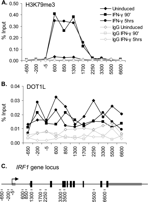
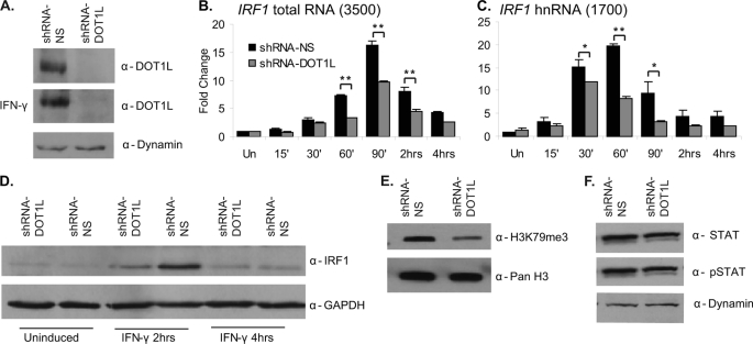
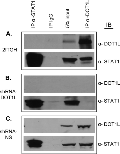
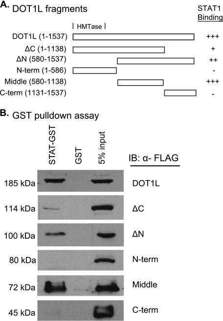
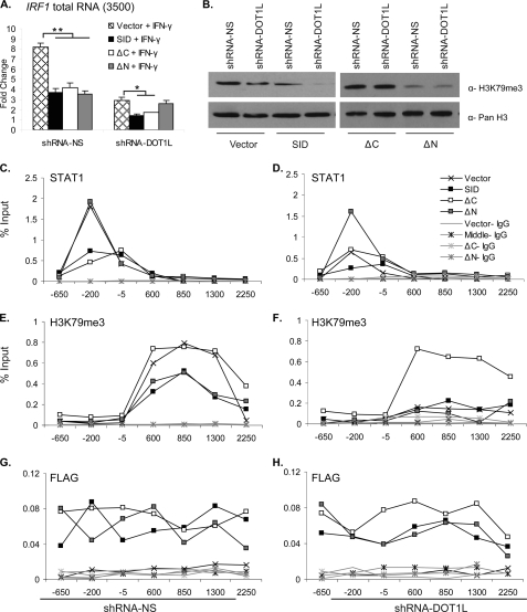
JAK-STAT-activated gene expression is both rapid and transient and requires dynamic post-translational modification of the chromatin template. Previously, we showed that following IFN-γ treatment, trimethylation of histone H3 at lysine 79 (H3K79me3) is rapidly and highly induced in the 5'-end of the STAT1-dependent gene interferon regulatory factor 1 (IRF1), but the role of this histone modification was unexplored. Here we report that DOT1L, the non-SET domain containing methyltransferase that modifies Lys-79, is localized across IRF1 in the uninduced state and is not further recruited by IFN-γ induction. RNAi-mediated depletion of DOT1L prevents the induction of H3K79me3 and lowers the transcription of IRF1 2-fold, as expected. Surprisingly, STAT1 binding to its DNA recognition element near the IRF1 promoter is diminished 2-fold in the DOT1L-depleted cell line. In vivo and in vitro protein interaction assays reveal a DOT1L-STAT1 interaction. Domain mapping identifies the middle region of DOT1L (amino acids 580-1183) as the STAT1 interaction domain. Overexpression of the DOT1L STAT1 interaction domain represses IRF1 transcription (2-fold) and interferes with STAT1 DNA binding at IRF1 and endogenous DOT1L histone methyltransferase activity. Collectively, our findings reveal a novel STAT1-DOT1L interaction that is required for the regulation JAK-STAT-inducible gene expression.
Figures

Similar articles
-
Menin and RNF20 recruitment is associated with dynamic histone modifications that regulate signal transducer and activator of transcription 1 (STAT1)-activated transcription of the interferon regulatory factor 1 gene (IRF1).Epigenetics Chromatin. 2010 Sep 8;3(1):16. doi: 10.1186/1756-8935-3-16. Epigenetics Chromatin. 2010. PMID: 20825659 Free PMC article.
-
IRF1 supports DNA binding of STAT1 by promoting its phosphorylation.Immunol Cell Biol. 2018 Nov;96(10):1095-1103. doi: 10.1111/imcb.12185. Epub 2018 Jun 28. Immunol Cell Biol. 2018. PMID: 29893425
-
Distinct modes of action applied by transcription factors STAT1 and IRF1 to initiate transcription of the IFN-gamma-inducible gbp2 gene.Proc Natl Acad Sci U S A. 2007 Feb 20;104(8):2849-54. doi: 10.1073/pnas.0610944104. Epub 2007 Feb 9. Proc Natl Acad Sci U S A. 2007. PMID: 17293456 Free PMC article.
-
The histone methyltransferase Dot1/DOT1L as a critical regulator of the cell cycle.Cell Cycle. 2014;13(5):726-38. doi: 10.4161/cc.28104. Epub 2014 Feb 6. Cell Cycle. 2014. PMID: 24526115 Free PMC article. Review.
-
A Positive Feedback Amplifier Circuit That Regulates Interferon (IFN)-Stimulated Gene Expression and Controls Type I and Type II IFN Responses.Front Immunol. 2018 May 28;9:1135. doi: 10.3389/fimmu.2018.01135. eCollection 2018. Front Immunol. 2018. PMID: 29892288 Free PMC article. Review.
Cited by
-
Use of biotinylated plasmid DNA as a surrogate for HSV DNA to identify proteins that repress or activate viral gene expression.Proc Natl Acad Sci U S A. 2012 Dec 18;109(51):E3549-57. doi: 10.1073/pnas.1218783109. Epub 2012 Dec 5. Proc Natl Acad Sci U S A. 2012. PMID: 23223531 Free PMC article.
-
IDH1 regulates human erythropoiesis by eliciting chromatin state reprogramming.Elife. 2025 Apr 29;13:RP100406. doi: 10.7554/eLife.100406. Elife. 2025. PMID: 40299922 Free PMC article.
-
Transcription-associated histone modifications and cryptic transcription.Biochim Biophys Acta. 2013 Jan;1829(1):84-97. doi: 10.1016/j.bbagrm.2012.08.008. Epub 2012 Sep 7. Biochim Biophys Acta. 2013. PMID: 22982198 Free PMC article. Review.
-
Rare de novo gain-of-function missense variants in DOT1L are associated with developmental delay and congenital anomalies.Am J Hum Genet. 2023 Nov 2;110(11):1919-1937. doi: 10.1016/j.ajhg.2023.09.009. Epub 2023 Oct 11. Am J Hum Genet. 2023. PMID: 37827158 Free PMC article.
-
Gain-of-function mutations in the catalytic domain of DOT1L promote lung cancer malignant phenotypes via the MAPK/ERK signaling pathway.Sci Adv. 2023 Jun 2;9(22):eadc9273. doi: 10.1126/sciadv.adc9273. Epub 2023 May 31. Sci Adv. 2023. PMID: 37256945 Free PMC article.
References
MeSH terms
Substances
LinkOut - more resources
Full Text Sources
Molecular Biology Databases
Research Materials
Miscellaneous
