

Temperature-scan cryocrystallography reveals reaction intermediates in bacteriophytochrome
- PMID: 22002602
- PMCID: PMC3337037
- DOI: 10.1038/nature10506
Temperature-scan cryocrystallography reveals reaction intermediates in bacteriophytochrome
Abstract
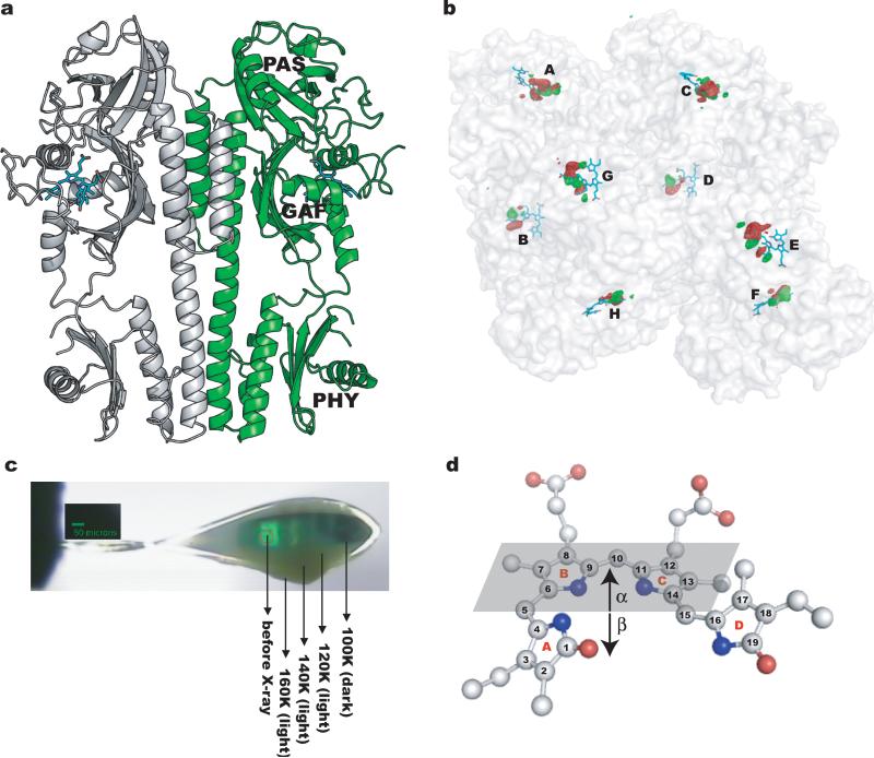
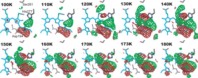
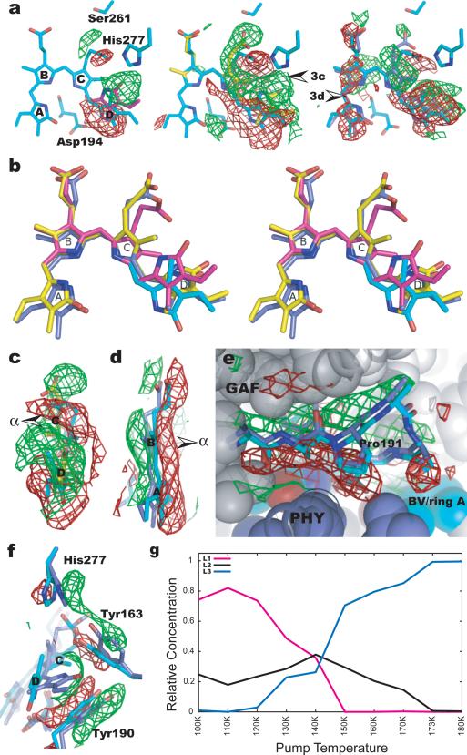
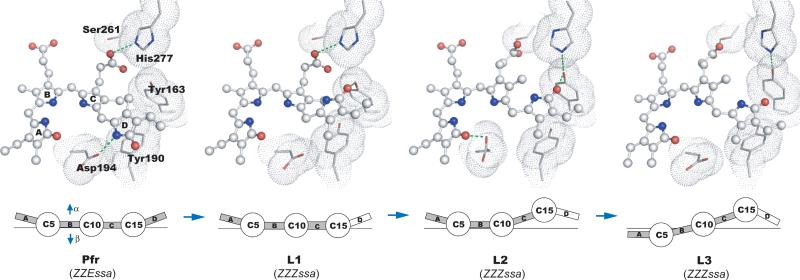
Light is a fundamental signal that regulates important physiological processes such as development and circadian rhythm in living organisms. Phytochromes form a major family of photoreceptors responsible for red light perception in plants, fungi and bacteria. They undergo reversible photoconversion between red-absorbing (Pr) and far-red-absorbing (Pfr) states, thereby ultimately converting a light signal into a distinct biological signal that mediates subsequent cellular responses. Several structures of microbial phytochromes have been determined in their dark-adapted Pr or Pfr states. However, the structural nature of initial photochemical events has not been characterized by crystallography. Here we report the crystal structures of three intermediates in the photoreaction of Pseudomonas aeruginosa bacteriophytochrome (PaBphP). We used cryotrapping crystallography to capture intermediates, and followed structural changes by scanning the temperature at which the photoreaction proceeded. Light-induced conformational changes in PaBphP originate in ring D of the biliverdin (BV) chromophore, and E-to-Z isomerization about the C(15) = C(16) double bond between rings C and D is the initial photochemical event. As the chromophore relaxes, the twist of the C(15) methine bridge about its two dihedral angles is reversed. Structural changes extend further to rings B and A, and to the surrounding protein regions. These data indicate that absorption of a photon by the Pfr state of PaBphP converts a light signal into a structural signal via twisting and untwisting of the methine bridges in the linear tetrapyrrole within the confined protein cavity.
© 2011 Macmillan Publishers Limited. All rights reserved
Figures
References
-
- Montgomery BL, Lagarias JC. Phytochrome ancestry: sensors of bilins and light. Trends Plant Sci. 2002;7:357–66. - PubMed
-
- Wagner JR, Brunzelle JS, Forest KT, Vierstra RD. A light-sensing knot revealed by the structure of the chromophore-binding domain of phytochrome. Nature. 2005;438:325–31. - PubMed
Publication types
MeSH terms
Substances
Associated data
- Actions
- Actions
- Actions
- Actions
Grants and funding
LinkOut - more resources
Full Text Sources
Other Literature Sources
Molecular Biology Databases
Research Materials
