. 2012 Aug;28(6):1567-75.
doi: 10.1007/s10554-011-9954-7. Epub 2011 Oct 15.

A novel iterative reconstruction algorithm allows reduced dose multidetector-row CT imaging of mechanical prosthetic heart valves

Affiliations

A novel iterative reconstruction algorithm allows reduced dose multidetector-row CT imaging of mechanical prosthetic heart valves

Jesse Habets et al. Int J Cardiovasc Imaging. 2012 Aug.

Abstract

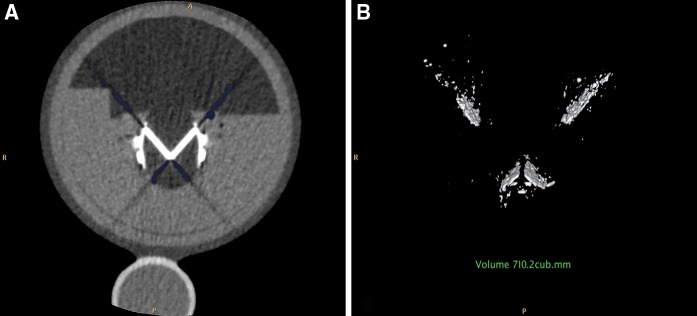
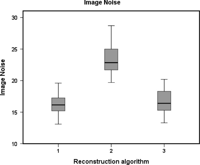
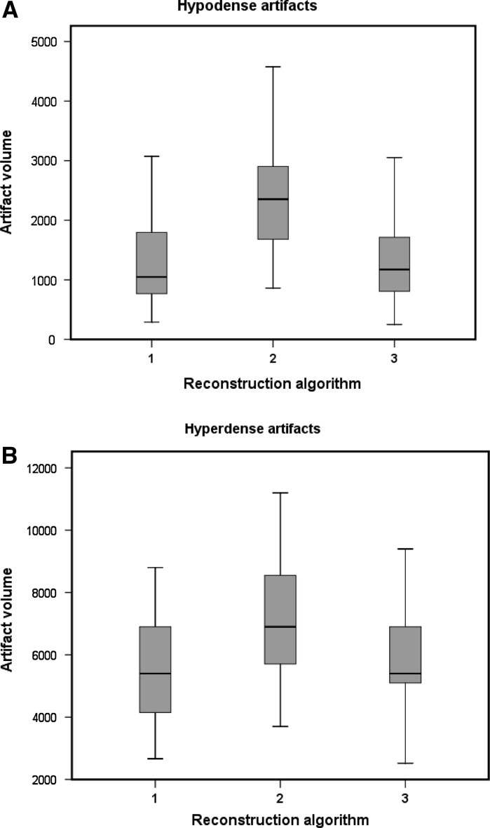
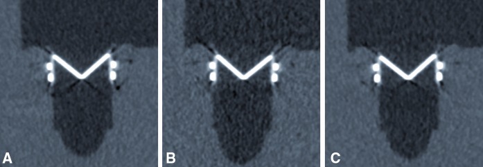
Multidetector-row CT is promising for prosthetic heart valve (PHV) assessment but retrospectively ECG-gated scanning has a considerable radiation dose. Recently introduced iterative reconstruction (IR) algorithms may enable radiation dose reduction with retained image quality. Furthermore, PHV image quality on the CT scan mainly depends on extent of PHV artifacts. IR may decrease streak artifacts. We compared image noise and artifact volumes in scans of mechanical PHVs reconstructed with conventional filtered back projection (FBP) to lower dose scans reconstructed with IR. Four different PHVs (St. Jude, Carbomedics, ON-X and Medtronic Hall) were scanned in a pulsatile in vitro model. Ten retrospectively ECG-gated CT scans were performed of each PHV at 120 kV, 600 mAs (high-dose CTDI(vol) 35.3 mGy) and 120 kV, 300 mAs (low-dose CTDI(vol) 17.7 mGy) on a 64 detector-row scanner. Diastolic and systolic images were reconstructed with FBP (high and low-dose) and the IR algorithm (low-dose only). Hypo- and hyperdense artifact volumes were determined using two threshold filters. Image noise was measured. Mean hypo- and hyperdense artifact volumes (mm(3)) were 1,235/5,346 (high-dose FBP); 2,405/6,877 (low-dose FBP) and 1,218/5,333 (low-dose IR). Low-dose IR reconstructions had similar image noise compared to high-dose FBP (16.5 ± 1.7 vs. 16.3 ± 1.6, mean ± SD, respectively, P = 1.0). IR allows ECG-gated PHV imaging with similar image noise and PHV artifacts at 50% less dose compared to conventional FBP in an pulsatile in vitro model.


Figures

Fig. 1
Fig. 1
Photograph of in vitro pulsatile model in the 64 slice CT-scanner (Brilliance 64, Philips Medical Systems, Cleveland, Ohio)
Fig. 2
Fig. 2
MDCT image of ON-X Bileaflet PHV. a MDCT image with measured artifacts (blue color), b Frontal plane with 3D volume PHV hypodense artifacts including PHV artifact measurement
Fig. 3
Fig. 3
MDCT image of St Jude bileaflet PHV. Image noise (SD) is measured with a circular region of interest in a homogenous part of the polymethyl methacrylate (PMMA) structure. Av (average image signal), HU Hounsfield Units, ED external diameter (mm)
Fig. 4
Fig. 4
Boxplots of image noise for different reconstruction algorithms. 1 120 kV, 600 mAs FBP reconstruction 2 120 kV, 300 mAs FBP reconstruction 3 120 kV, 300 mAs iDose level 4
Fig. 5
Fig. 5
Boxplots of hypodense (a) and hyperdense (b) PHV artifacts for different reconstruction algorithms. 1 120 kV, 600 mAs FBP reconstruction, 2 120 kV, 300 mAs FBP reconstruction, 3 120 kV, 300 mAs iDose level 4
Fig. 6
Fig. 6
MDCT image of ON-X Bileaflet PHV. a Frontal plane 120 kV, 600 mAs reconstructed with FBP, b Frontal plane 120 kV 300 mAs reconstructed with FBP, c Frontal plane 120 kV 300 mAs reconstructed with iDose level 4
Fig. 7
Fig. 7
MDCT image of ON-X Bileaflet PHV. a Frontal plane 120 kV, 600 mAs reconstructed with FBP, b Frontal plane 120 kV, 300 mAs reconstructed with iDose level 4, c Frontal plane with 3D volume PHV hypodense artifacts (120 kV, 600 mAs, BFP), d Frontal plane with 3D volume PHV hypodense artifacts (120kV, 300mAs, iDose level 4)

References

    1. Tsai IC, Lin YK, Chang Y, et al. Correctness of multi-detector-row computed tomography for diagnosing mechanical prosthetic heart valve disorders using operative findings as a gold standard. Eur Radiol. 2009;19:857–867. doi: 10.1007/s00330-008-1232-2. - DOI - PubMed
    1. Symersky P, Budde RP, de Mol BA, et al. Comparison of multidetector-row computed tomography to echocardiography and fluoroscopy for evaluation of patients with mechanical prosthetic valve obstruction. Am J Cardiol. 2009;104:1128–1134. doi: 10.1016/j.amjcard.2009.05.061. - DOI - PubMed
    1. Konen E, Goitein O, Feinberg MS, et al. The role of ECG-gated MDCT in the evaluation of aortic and mitral mechanical valves: initial experience. Am J Roentgenol. 2008;191:26–31. doi: 10.2214/AJR.07.2951. - DOI - PubMed
    1. Feuchtner GM, Stolzmann P, Dichtl W, et al. Multislice computed tomography in infective endocarditis: comparison with transesophageal echocardiography and intraoperative findings. J Am Coll Cardiol. 2009;53:436–444. doi: 10.1016/j.jacc.2008.01.077. - DOI - PubMed
    1. Habets J, Budde RP, Symersky P, et al. Diagnostic evaluation of left-sided prosthetic heart valve dysfunction. Nat Rev Cardiol. 2011;8:466–478. doi: 10.1038/nrcardio.2011.71. - DOI - PubMed

Publication types

LinkOut - more resources