. 2012 Apr;470(4):1204-12.
doi: 10.1007/s11999-011-2128-z. Epub 2011 Oct 15.

Surgical technique: Methods for removing a Compress® compliant prestress implant

Affiliations

Surgical technique: Methods for removing a Compress® compliant prestress implant

Geoffrey D Abrams et al. Clin Orthop Relat Res. 2012 Apr.

Abstract

Background: The Compress® device uses a unique design using compressive forces to achieve bone ingrowth on the prosthesis. Because of its design, removal of this device may require special techniques to preserve host bone. DESCRIPTION OF TECHNIQUES: Techniques needed include removal of a small amount of bone to relieve compressive forces, use of a pin extractor and/or Kirschner wires for removal of transfixation pins, and creation of a cortical window in the diaphysis to gain access to bone preventing removal of the anchor plug.

Methods: We retrospectively reviewed the records of 63 patients receiving a Compress® device from 1996 to 2011 and identified 11 patients who underwent subsequent prosthesis removal. The minimum followup was 1 month (average, 20 months; range, 1-80 months). The most common reason for removal was infection (eight patients) and the most common underlying diagnosis was osteosarcoma (five patients). Three patients underwent above-knee amputation, whereas the others (eight patients) had further limb salvage procedures at the time of prosthesis removal.

Results: Five patients had additional unplanned surgeries after explantation. Irrigation and débridement of the surgical wound was the most common unplanned procedure followed by latissimus free flap and hip prosthesis dislocation. At the time of followup, all patients were ambulating on either salvaged extremities or prostheses.

Conclusion: Although removal of the Compress® device presents unique challenges, we describe techniques to address those challenges.


Figures
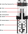
Fig. 1
Fig. 1
A component diagram of the Compress® device shows Belleville washers stacked in series generating compressive load. (Reprinted with permission from Avedian RS, Goldsby RE, Kramer MJ, O’Donnell RJ. Effect of chemotherapy on initial compressive osseointegration of tumor endoprostheses. Clin Orthop Relat Res. 2007;459:48–53.)
Fig. 2
Fig. 2
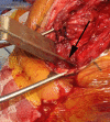
An intraoperative photograph shows the proximal femoral bone with hypertrophic ingrowth into the porous surface interface of the device (arrow), which is attached to a distal femoral replacement prosthesis.
Fig. 3
Fig. 3
This photograph taken in the laboratory shows the underside of the spindle, revealing the compression nut (arrow).
Fig. 4
Fig. 4
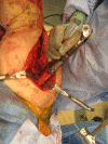
An intraoperative photograph shows the use of a pencil tip burr to remove approximately 3 mm of interface bone to relieve compressive forces when the nut is not accessible.
Fig. 5
Fig. 5
An intraoperative photograph shows the use of the threaded extraction device (arrow) to remove the transverse pins.
Fig. 6
Fig. 6
An intraoperative photograph shows additional technique of transverse pin removal by tapping them through with the use of a wire. Protection of soft tissue structures on the backside of the pins is obtained using a Cobb elevator.
Fig. 7
Fig. 7
An intraoperative photograph shows application of a vise grip to the traction bar (arrow) in preparation to manually remove the anchor plug using a mallet against the vise grip.
Fig. 8
Fig. 8
From left to right, shown are the anchor plug, trephine, and bushing for the traction bar, which are included in the Biomet Compress® revision tray. (Published with permission from Biomet Inc, Warsaw, IN, USA.)
Fig. 9
Fig. 9
An intraoperative photograph shows the creation of a bone window (arrow) to remove a fixed anchor plug. The cortical holes where the previous transfixation pins were have been removed. This gives direct access to the intramedullary bone so it can be removed under direct vision (Fig. 10).
Fig. 10
Fig. 10
An intraoperative photograph shows the creation of a cortical window and removal of bone between the centering sleeve and anchor plug (arrow) to facilitate anchor plug removal.
Fig. 11
Fig. 11
An intraoperative photograph shows extraction of the anchor plug of a tibial prosthesis using a tamp and mallet after creation of a cortical window over the anchor plug.
Fig. 12A–C
Fig. 12A–C
(A) AP and (B) lateral radiographs were obtained of the knee of a patient after the index Compress® placement. The distance between the centering sleeve and the traction bar (indicated by the arrows) can be seen. (C) An AP radiograph obtained of the knee of the same patient at 6 months followup shows narrowing of the space between the centering sleeve and traction bar (arrow), indicating loosening and ingrowth failure. This patient underwent removal of the device for aseptic loosening.

References

    1. Bickels J, Wittig JC, Kollender Y, Henshaw RM, Kellar-Graney KL, Meller I, Malawer MM. Distal femur resection with endoprosthetic reconstruction: a long-term followup study. Clin Orthop Relat Res. 2002;400:225–235. doi: 10.1097/00003086-200207000-00028. - DOI - PubMed
    1. Bini SA, Johnston JO, Martin DL. Compliant prestress fixation in tumor prostheses: interface retrieval data. Orthopedics. 2000;23:707–711. - PubMed
    1. Chang DW, Weber KL. Use of a vascularized fibula bone flap and intercalary allograft for diaphyseal reconstruction after resection of primary extremity bone sarcomas. Plast Reconstr Surg. 2005;116:1918–1925. doi: 10.1097/01.prs.0000189203.38204.d5. - DOI - PubMed
    1. Chen CM, Disa JJ, Lee HY, Mehrara BJ, Hu QY, Nathan S, Boland P, Healey J, Cordeiro PG. Reconstruction of extremity long bone defects after sarcoma resection with vascularized fibula flaps: a 10-year review. Plast Reconstr Surg. 2007;119:915–924. doi: 10.1097/01.prs.0000252306.72483.9b. - DOI - PubMed
    1. Dion N, Sim FH. The use of allografts in orthopaedic surgery. Part I: The use of allografts in musculoskeletal oncology. J Bone Joint Surg Am. 2002;84:644–654.