. 2011 Dec;77(24):8754-64.
doi: 10.1128/AEM.06162-11. Epub 2011 Oct 14.

A novel hydrolase identified by genomic-proteomic analysis of phenylurea herbicide mineralization by Variovorax sp. strain SRS16

Affiliations

A novel hydrolase identified by genomic-proteomic analysis of phenylurea herbicide mineralization by Variovorax sp. strain SRS16

Karolien Bers et al. Appl Environ Microbiol. 2011 Dec.

Abstract

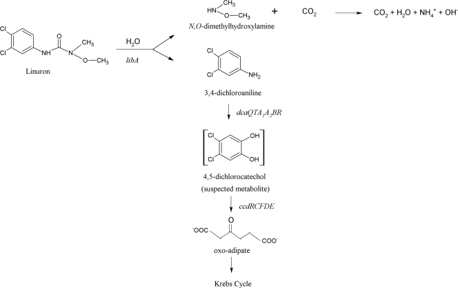
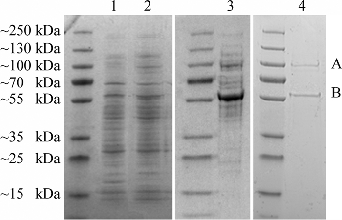
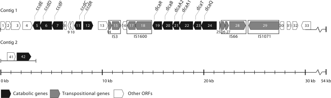
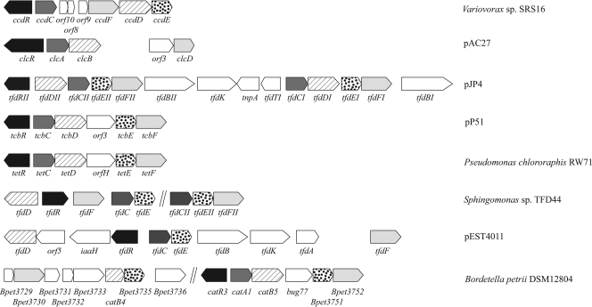
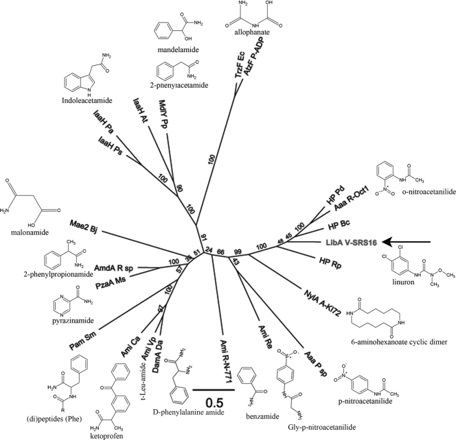
The soil bacterial isolate Variovorax sp. strain SRS16 mineralizes the phenylurea herbicide linuron. The proposed pathway initiates with hydrolysis of linuron to 3,4-dichloroaniline (DCA) and N,O-dimethylhydroxylamine, followed by conversion of DCA to Krebs cycle intermediates. Differential proteomic analysis showed a linuron-dependent upregulation of several enzymes that fit into this pathway, including an amidase (LibA), a multicomponent chloroaniline dioxygenase, and enzymes associated with a modified chlorocatechol ortho-cleavage pathway. Purified LibA is a monomeric linuron hydrolase of ∼55 kDa with a K(m) and a V(max) for linuron of 5.8 μM and 0.16 nmol min⁻¹, respectively. This novel member of the amidase signature family is unrelated to phenylurea-hydrolyzing enzymes from Gram-positive bacteria and lacks activity toward other tested phenylurea herbicides. Orthologues of libA are present in all other tested linuron-degrading Variovorax strains with the exception of Variovorax strains WDL1 and PBS-H4, suggesting divergent evolution of the linuron catabolic pathway in different Variovorax strains. The organization of the linuron degradation genes identified in the draft SRS16 genome sequence indicates that gene patchwork assembly is at the origin of the pathway. Transcription analysis suggests that a catabolic intermediate, rather than linuron itself, acts as effector in activation of the pathway. Our study provides the first report on the genetic organization of a bacterial pathway for complete mineralization of a phenylurea herbicide and the first report on a linuron hydrolase in Gram-negative bacteria.


Figures

Fig. 1.
Fig. 1.
Catabolic pathway of linuron degradation in Variovorax sp. SRS16. The catabolic steps specified by libA, dcaQTA1A2B, and ccdCFDE are indicated.
Fig. 2.
Fig. 2.
SDS-PAGE analysis of protein extracts showing linuron hydrolase activity, from linuron-amended Variovorax sp. SRS16 cultures at different purification steps. Lane 1, crude cell-free protein extract; lane 2, after 35 to 50% ammonium sulfate precipitation; lane 3, after anion-exchange chromatography; lane 4, after size exclusion chromatography. Band A was identified as transcription elongation factor G, while band B was identified as the translation product of ORF 42 (libA).
Fig. 3.
Fig. 3.
Genetic organization of contigs 1 and 2 containing the identified genes involved in linuron and DCA catabolism in Variovorax sp. SRS16. Contig 1 contains a 54-kb DNA fragment including dcaQTA1A2BR and ccdRCFDE. Upstream of ORF 33, contig 1 includes another 24-kb-long sequenced nucleotide segment. The ORFs of this part of the contig are represented in the corresponding GenBank file JN104632. Contig 2 consists of a 3.5-kb genome fragment containing the linuron hydrolase gene libA (ORF 42). Further information about the different ORFs is shown in Table 1.
Fig. 4.
Fig. 4.
Organization of the modified chlorocatechol ortho-cleavage pathway gene cluster in chloroaromatic-degrading bacterial strains (14, 18, 36, 48, 50, 55, 56). The arrows indicate the localization, size, and direction of transcription of the genes. Similar shaded patterns indicate isofunctional genes. The following enzymes are shown (the coding genes are in parentheses): 2,4-D transport protein (tfdK), chlorophenol hydroxylases (tfdB), chlorocatechol 1,2-dioxygenases (tfdC, ccdC, clcA, tcbC, tetC, and catA1), chloromuconate cycloisomerases (tfdD, ccdD, clcB, tcbD, tetD, catB4, and catB5), dienelactone hydrolases (tfdE, ccdE, tcbE, tetE, Bpet3735, and Bpet3751), maleylacetate reductases (tfdF, ccdF, clcD, tcbF, tetF, and Bpet3730), transposases (tnpA, Bpet3733, and Bpet3736), and indole acetamide hydrolase (iaaH). The ORFs Bpet3729, Bpet3731, and Bpet3732 code for hypothetical proteins. bug77 codes for a Bug family protein.
Fig. 5.
Fig. 5.
Phylogenetic analysis of LibA and selected members of the amidase signature family. A multiple amino acid alignment of the respective Pfam domains (PF01425) was used to construct a maximum-likelihood tree. The AS domain of LibA (residues 27 to 453) was aligned with the corresponding domain of the following enzymes, with their (preferred) substrates indicated: Aaa R-Oct1 (Rhodococcus sp. strain Oct1, o-nitroacetanilide [15]), Aaa P sp (Pseudomonas sp., p-nitroacetanilide [24]), AmdA R sp (Rhodococcus sp., 2-phenylpropionamide [28]), Ami Ca (Comamonas acidovorans, ketoprofen [58]), Ami Re (Rhodococcus erythropolis PR4, Gly-p-nitroacetanilide [26]), Ami R-N-771 (Rhodococcus sp. strain N-771, benzamide [32]), Ami Vp (Variovorax paradoxus, t-Leu-amide [25]), AtzF P-ADP (Pseudomonas sp. ADP, allophanate [43]), DamA Da (Delftia acidovorans, d-phenylalanine amide [22]), IaaH At (Agrobacterium tumefaciens, indole acetamide [57]), IaaH Pa (Pantoea agglomerans, indole acetamide [8]), IaaH Ps (Pseudomonas savastanoi, indole acetamide [57]), Mae2 Bj (Bradyrhizobium japonicum, malonamide [45]), MdlY Pp (Pseudomonas putida, mandelamide/2-phenylacetamide [17]), NylA (Arthrobacter sp. strain KI72, 6-aminohexanoate cyclic dimer/β-laurolactam [59]), Pam Sm (Stenotrophomonas maltophila, [di]peptides [Phe] [30]), PzaA Ms (Mycobacterium smegmatis, pyrazinamide [4]), and TrzF Ec (Enterobacter cloacae, allophanate [42]). Three hypothetical proteins most similar to LibA were included: HP Bc (Burkholderia cenocepacia J2315 [YP_002234156]), HP Pd (Pseudonocardia dioxanivorans [YP_004335959]), HP Rp (Ruegeria pomeroyi DSS-3 [YP_167742]). The scale bar represents 0.5 substitutions per site. Bootstrap values are represented at the branches.
Fig. 6.
Fig. 6.
Transcription analysis of linuron degradation genes in Variovorax sp. SRS16. RNA sampled after 30 min of growth on R2A without (lane 1) or with linuron (lane 2), DCA (lane 3) or aniline (lane 4). A “–” indicates a blank, while “+” indicates a positive control (genomic DNA of SRS16).

Similar articles

Cited by

References

    1. Altschul S. F., Gish W., Miller W., Myers E. W., Lipman D. J. 1990. Basic local alignment search tool. J. Mol. Biol. 215:403–410 - PubMed
    1. Antoine R., et al. 2005. The periplasmic binding protein of a tripartite tricarboxylate transporter is involved in signal transduction. J. Mol. Biol. 351:799–809 - PubMed
    1. Antoine R., et al. 2003. Overrepresentation of a gene family encoding extracytoplasmic solute receptors in Bordetella. J. Bacteriol. 185:1470–1474 - PMC - PubMed
    1. Boshoff H. I. M., Mizrahi V. 1998. Purification, gene cloning, targeted knockout, overexpression, and biochemical characterization of the major pyrazinamidase from Mycobacterium smegmatis. J. Bacteriol. 180:5809–5814 - PMC - PubMed
    1. Breugelmans P., D'Huys P. J., De Mot R., Springael D. 2007. Characterization of novel linuron-mineralizing bacterial consortia enriched from long-term linuron-treated agricultural soils. FEMS Microbiol. Ecol. 62:374–385 - PubMed

Publication types

LinkOut - more resources